

 PDF
PDF ePub
ePub Citation
 Print
Print


INTRODUCTION
Glucocorticoids (GCs) are very effective drugs for the treatment of inflammatory diseases and have been widely used in various diseases.[123] However, long-term use of GCs has detrimental effects on bone microstructure, leading to a decrease in bone mass and an increase in the risk of fracture.[34] It is estimated that fractures occur in 30% to 50% of patients receiving long-term GC therapy, but many patients have no symptoms of fracture because of the analgesic effects of GCs.[567] There are no definitive data on the number of patients receiving GCs for 3 months or longer in Korea, but it is estimated to be much higher than the 1% reported in other countries.[89] Therefore, GC-induced osteoporosis (GIOP) is recognized as the most common cause of secondary osteoporosis and, because there may be no specific symptoms, active management focused on prevention is needed.
The decrease of bone mass caused by GCs occurs in 2 stages. First, a rapid decrease in bone mass begins within the first 3 to 6 months of GC use, with a 6% to 12% loss of bone mass in the first year of GC use.[10] Second, long-term use of GCs can result in a 3% reduction in bone mass every year.[11] GCs affect both cortical bone and trabecular bone, but fractures occur most commonly in the vertebral body, especially in areas rich in trabecular bone, such as the lumbar spine.[3412] The risk of GC-induced fracture is already increased before a significant reduction in bone mass occurs.[3] Therefore, measurements of bone mineral density (BMD) are not sufficient to evaluate the degree of GC-induced bone loss, so it is very important to identify patients with a high risk of fracture to prevent fractures caused by GIOP.[131415] The absolute risk of an individual fracture is determined by age, gender and other risk factors for osteoporosis. Currently, fracture-risk assessment tool (FRAX) is a well-known method for assessing the risk of fracture. In addition to the risk factors included in the FRAX, low bone strength at the beginning of GC treatment and the rate of bone loss during treatment are suggested as risk factors for GC-induced fractures, the latter being determined by the dose and duration of GC treatment.[16] In a study of individual absolute fracture probability, patients receiving prednisolone at a dose greater than 30 mg/day (cumulative dose >5 g/year) showed significantly increased risk of vertebral and femoral fractures.[17] However, if GC treatment is terminated, BMD gradually increases and fracture risk decreases. Therefore, patients could benefit from continuous risk assessment with an emphasis on the appropriate duration of GC use.[91819]
As evidence of fracture risk in patients using GCs accumulates, drugs that effectively prevent fractures have been developed. However, many primary care physicians and specialists fail to recognize the severity of GIOP or determine which patients are at greatest risk for GIOP. Therefore, many patients still do not receive treatment to prevent fractures. To address this problem, guidelines for GIOP prevention and treatment have recently been developed by several countries. Notably, the guideline of the American College of Rheumatology (ACR) were revised in 2017 based on the latest evidence and applied to clinical practice.[16] In Korea, it is necessary to provide standardized clinical practical guideline (CPG) for the primary prevention and treatment of GIOP to all clinicians, to ensure that Korean patients who plan to use or use GCs receive the appropriate services for fracture prevention. The Korean Society for Bone and Mineral Research (KSBMR) and the Korean College of Rheumatology (KCR) have mutually developed guideline for the treatment of GIOP. Because of limited domestic data, this guideline was developed by adapting previously-published guidelines.
METHODS
This guideline was developed for adults over the age of 19 who plan to use or currently use GCs. Pediatric populations and people with a glomerular filtration rate of <30 mL/min were excluded. A development committee and a working committee were organized to develop guideline for the treatment of GIOP. These committees were composed of multi-disciplinary and multi-institutional organizations and included endocrinologists, rheumatologists, an orthopedist, and a methodologist (Supplementary Appendix 1). A systematic literature review was conducted, and guidelines were selected and adapted from the existing literature. The completed guideline should be revised within 5 years, and earlier revisions may be required if a new drug is approved for GIOP or if the evidence changes significantly.
1. Framework for GIOP guideline development
Methods were based on guidance for the development of CPGs version 1.0 by the National Evidence-Based Healthcare Collaborating Agency (NECA) (Supplementary Appendix 2). The process of developing this guideline included 3 major stages [20]: planning, development, and finalization. Each stage was divided into individual steps, for a total of 12 steps. The planning stage consisted of selecting topics (Step 1), assembling the development committee (Step 2), reviewing previously published guidelines (Step 3), establishing the development plan (Step 4), and selecting key questions (KQs) (Step 5). The development stage consisted of searching for, evaluating, and synthesizing evidence (Steps 6–8), making recommendations and determining the grades of recommendations (Step 9), and consensus building (Step 10). The finalization stage consisted of external reviews and publications (Steps 11 and 12).
2. Selection of KQs
To select the KQs to be addressed by the GIOP guideline, a working committee consisting of a total of nine members first reviewed six guidelines developed by the United States, France, Spain, Japan, Brazil, and the International Osteoporosis Foundation-European Calcified Tissue Society (IOF-ECTS). From these 6 guidelines, the committee selected 14 topics. After reviewing these topics, the development committee considered domestic circumstances and clinical significance to select the most relevant KQs. A final list of seven KQs was chosen, which included the patient population (P), the intervention (I), the comparator (C), and the outcome of the intervention (O) (PICO; Table 1).
3. Literature search
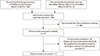
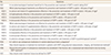
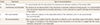
Two members of the working committee performed systematic literature searches, using the databases PubMed, OVID-EMBASE, KoreaMed, KMbase, National Guideline Clearinghouse, Guidelines International Network, and Korean Medical Guideline Information Center (KoMGI). The researchers identified a total of 309 potentially relevant articles published since 2010, excluding duplicates. By reviewing titles and abstracts, the researchers narrowed this list to the 27 most relevant articles. From these 27 articles, 7 of previously-published guidelines were identified: (1) guidelines including PICO that are consistent with KQs; (2) evidence-based guidelines, which are defined by a clear link between the recommendation and the supporting evidence, including systematic literature searches; (3) guidelines for peer review; and (4) guidelines published in Korean or English (Fig. 1, Supplementary Appendix 3 and 4).
4. Final selection process for guidelines
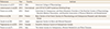
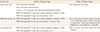
Seven guidelines were chosen based on the systematic literature review, selection criteria, and exclusion criteria (Table 2).[21222324252627] A quality assessment was performed on these seven guidelines using the Appraisal of Guidelines for Research and Evaluation II (AGREE II). In this comprehensive assessment, the scope and objectives of the guidelines, strictness of development, participation of stakeholders, clarity of expression, applicability, and editorial independence were assessed.[28] The quality assessment of guidelines using AGREE II was conducted by 3 members of the working committee as per recommendations, and the items with given divergent scores by the 3 researchers were reviewed and agreed upon. The strictness of development was specifically considered in the process of selecting guidelines. Four guidelines published in the United States (2010), IOF-ECTS (2012), Canada (2010) and France (2014) were selected.[21222324]
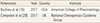
During the selection of guidelines, the National Osteoporosis Guideline Group (NOGG) and the ACR published new guidelines for GIOP treatment in 2017.[16,29] Therefore, the 2010 ACR guideline was replaced by the 2017 ACR guideline (Table 3), and the quality assessment was reevaluated for the 5 guidelines. The 2017 ACR guidelines, which ranked highly in items of “comprehensive evaluation” and “strictness of development”, were chosen and adapted (Supplementary Appendix 5). The characteristics of final 5 guidelines were summarized in Supplementary Appendix 6.
5. Writing process for guidelines
The working committee reviewed the recommendations and evidence for the final 5 guidelines, and then summarized the primary recommendations for KQs (Supplementary Appendix 7). The acceptability and applicability of the recommendations of final 5 guidelines to the KQ was assessed (Supplementary Appendix 8). After collecting the opinions of all committee members, the final recommendations were completed. If there was a lack of evidence or a need for clinical interpretation, the consensus process proceeded among the members of the working committee. The strength of evidence was divided into 5 levels (Table 4), and recommendation grade was assessed considering the level of evidence and clinical effects, patient satisfaction, quality of life, harmful reactions, and unnecessary use of resources (Table 5). The final recommendation grade was decided on the principle of agreement of more than 80%, along with the consent of the working committee.
6. Selection of eligible patients and fracture risk assessment
The development committee and working committee decided to apply the 2017 ACR guideline for the fracture risk classification and assessment, because relatively little evidence was available beyond these guidelines. Tables and figures were used with permission from the original author.
RESULTS
1. Fracture risk classification
It is important to classify patients according to fracture risk before deciding whether to use fracture-prevention treatments. To predict fracture risk, we decided to use FRAX, which is widely used in many guidelines for GIOP, including the 2017 ACR guidelines. In addition to FRAX, we also considered using history of previous fracture, BMD, and GC dose as predictors. Patients aged ≥50 years have been the focus of many studies of osteoporosis. However, FRAX applies only to patients ≥40 years old, so we divided our recommendations by adults ≥40 years for whom FRAX is applicable, and those under 40 years old who are not included in FRAX. Fracture risk was classified as high, moderate, and low (Table 6). For example, the high risk category included patients ≥40 years of age with previous osteoporotic fracture, BMD T-score ≤−2.5 in men who are ≥50 years of age or postmenopausal women, FRAX-calculated 10-year major osteoporotic fracture risk ≥20%, or FRAX-calculated 10-year hip fracture risk ≥3%. The risk of fracture is correlated with prednisolone dose. For patients taking prednisolone at a dose >7.5 mg/day, FRAX calculates an increase in fracture risk of 15% for major osteoporotic fracture and 20% for hip fracture risk.[30]
2. Fracture risk assessment
1) Initial fracture risk assessment
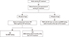
The initial fracture risk assessment should be performed as early as possible in patients with long-term GC treatment. It is appropriate to assess the fracture risk within 6 months of the initiation of long-term GC treatment, and to consider the calculated risks when selecting the specific GC treatment (Fig. 2). The most important factors in the initial assessment are the dose, duration, and method of administration of GCs, and history of previous fracture, fall, and frailty. The current nutritional status, weight loss, and the possibility of secondary osteoporosis, including thyroid disease, need to be evaluated carefully. In particular, it is necessary to investigate the variables included in FRAX (history of previous fracture, comorbidities, smoking history, alcohol consumption, family history of fracture) in patients ≥40 years of age. Adjusting FRAX for GC dose and BMD testing are also necessary. If a patient <40 years of age has risk factors for fracture (previous osteoporotic fracture, malnutrition, thyroid disease, weight loss, secondary hyperparathyroidism, hypogonadism, family history of femoral fracture, smoking, alcohol consumption, etc.), it is important to conduct a BMD early in treatment to assess fracture risk further.
2) Reassessment of fracture risk
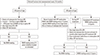
If GCs are used continuously, it is necessary to reassess the risk of fracture every 12 months (Fig. 3). For adults ≥40 years of age who continue GC treatment and are not treated with osteoporosis medications beyond calcium and vitamin D, FRAX and BMD should be performed every 1 to 3 years. FRAX and BMD are recommended every year if the initial GC dose is prednisolone ≥30 mg/day, if the cumulative dose is greater than 5 g in the previous year, or if osteoporotic fractures have occurred.
For adults ≥40 years of age who are at high risk of fracture (initial GC dose of prednisolone ≥30 mg/day, cumulative dose >5 g/year, osteoporotic fracture occurring ≥12 months after beginning osteoporosis medications, poor medication adherence or absorption, or other significant osteoporosis risk factors) who are taking osteoporosis medications with GC treatment, BMD testing should be completed every 2 to 3 years and is recommended as early as possible. BMD testing should be performed at intervals as soon as possible, even after discontinuation of osteoporosis medication.
For adults <40 years of age who have moderate or high risk and are receiving continuous GC treatment, BMD testing should be conducted every 2 to 3 years.
3. Treatment and follow-up of GIOP
1) KQ1: Is non-pharmacological treatment beneficial for the prevention and treatment of GIOP in adults taking GCs?
Non-pharmacological treatments such as exercise, good nutrition, smoking cessation, and avoiding alcohol abuse are recommended for all adults taking GCs. Because there is insufficient evidence for the effects of these treatments in GIOP, it is recommended that treatment be based on data from these treatments in postmeno-pausal osteoporosis patients [III/B].
Because there are limited data on effects of non-pharmacological treatments in the prevention and treatment of GIOP, it is recommended that treatment be based on the established non-pharmacological treatment of postmenopausal osteoporosis patients. Although the effects of these lifestyle modifications on fracture risk have not been established for patients with GIOP, non-pharmacological treatments such as weight-bearing exercise, good nutrition, smoking cessation, and avoiding alcohol abuse is recommended for all adults taking GCs [III/B].
2) KQ2: Which pharmacological treatments are effective for prevention and treatment of GIOP in adults <40 years of age?
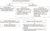
There are few randomized controlled trials comparing the effects of drugs on prevention of bone mass loss and fracture for this age group. This is because adults <40 years of age have relatively high BMD and fewer fractures than postmenopausal women. However, it is reported that long-term use of GCs in premenopausal women <40 years of age may cause changes in bone structure and weaken bone strength.[3132] Initial pharmacologic treatment for adults <40 years of age is summarized in Figure 4.
(1) KQ2-1: Is calcium and vitamin D supplementation effective in the prevention and treatment of GIOP in adults <40 years of age?
① Calcium and vitamin D are recommended for all adults taking prednisone ≥2.5 mg/day for ≥3 months [II/B].
② Sufficient calcium (1,000–1,200 mg) and vitamin D (800 IU) intake are recommended, and adequate vi-tamin D concentrations (≥20 ng/mL) should be maintained [Expert consensus/B].
③ The use of supplements should be considered when dietary intake of calcium and vitamin D is insufficient [Expert consensus/B].
④ The minimum adequate dosage should be deter-mined, because high doses of calcium and vitamin D supplementation may increase the risk of gastroin-testinal side effects and renal stones [Expert consen-sus/B].
GCs reduce intestinal absorption and renal reabsorption of calcium, and increase calcium excretion into the urine.[33] Therefore, calcium and vitamin D are recommended for adults <40 years of age who are taking prednisolone ≥2.5 mg for ≥3 months [II/B]. However, calcium alone is not effective in the prevention and treatment of GIOP,[3435] it is more effective to take calcium and vitamin D together. Patients receiving vitamin D3 (cholecalciferol) [3637] or activated vitamin D (calcitriol, alphacalcidol) [3839] in combination with calcium showed an inhibitory effect on bone loss compared to calcium alone or no treatment. Adequate intake of calcium (1,000–1,200 mg) and vitamin D (800 IU) are recommended to maintain vitamin D concentration (≥20 ng/mL) [Expert consensus/B]. The use of supplements may be considered if the intake of calcium and vitamin D through meals is insufficient [Expert consensus/B].
(2) KQ2-2: Is bisphosphonate effective in the prevention and treatment of GIOP in adults <40 years of age?
① Oral bisphosphonates are recommended for adults <40 years of age with moderate to high risk of frac-ture [II/A].
② If oral bisphosphonates are not appropriate intrave-nous bisphosphonate is recommended [II/A].
Bisphosphonates are recommended for women without childbearing potential and men <40 years of age at moderate to high fracture risk [II/A]. Alendronate was effective in prevention of bone loss and fractures in premenopausal women with high-dose GC therapy.[40] Subgroup analysis of adults <40 years of age has been done in several previously published studies on GIOP treatment. These studies have shown that bisphosphonates such as alendronate,[404142434445] risedronate,[464748] zoledronic acid [49] increased BMD and decreased fracture risk compared to placebo or calcium/vitamin D. Oral bisphosphonates, which have a relatively short half-life, are preferred. If oral bisphosphonates are not appropriate, intravenous bisphosphonate is recommended.
(3) KQ2-3: Is teriparatide effective in the prevention and treatment of GIOP in adults <40 years old?
Teriparatide is recommended for women without childbearing potential and men <40 years of age at moderate to high fracture risk [II/A]. Among patients taking prednisolone ≥5 mg/day for ≥3 months, teriparatide treatment led to significantly higher spine BMD than alendronate treatment in both postmenopausal and premenopausal women.[50]
(4) KQ2-4: Is denosumab effective in the prevention and treatment of GIOP in adults <40 years of age?
Denosumab is recommended for women without childbearing potential and men <40 years of age at moderate-to-high fracture risk [II/A]. Recently, in a randomized, double-blind, comparative study of denosumab versus risedronate in patients ≥19 years of age taking prednisolone ≥7.5 mg/day for ≥3 months, denosumab significantly increased spine and hip BMD compared to risedronate.[51]
3) KQ3: Which pharmacological treatments are effective for the prevention and treatment of GIOP in adults ≥40 years of age?
Initial pharmacologic treatment for adults ≥40 years of age is summarized in Figure 4.
(1) KQ3-1: Is calcium and vitamin D supplementation effective in the prevention and treatment of GIOP in adults ≥40 years of age?
① Calcium and vitamin D are recommended for all adults taking prednisolone ≥2.5 mg/day for ≥3 months [II/B].
② Sufficient calcium (1,000–1,200 mg) and vitamin D (800 IU) intakes are recommended, and adequate vitamin D concentrations (≥20 ng/mL) should be maintained [Expert consensus/B].
③ The use of supplements should be considered when the intake of calcium and vitamin D through meals is insufficient [Expert consensus/B].
④ The adequate dosage should be considered, because high doses of calcium and vitamin D supplementation may increase the risk of gastrointestinal side effects and renal stones [Expert consensus/B].
Recommendations for calcium and vitamin D supplementation in adults ≥40 years of age are the same as for adults <40 years of age. Calcium and vitamin D are recommended for adults ≥40 years of age who are taking prednisolone ≥2.5 mg for ≥3 months [II/B]. Sufficient calcium (1,000–1,200 mg) and vitamin D (800 IU) intake are recommended, and adequate vitamin D concentrations (≥20 ng/mL) should be maintained [Expert consensus/B]. The use of supplements should be considered when the intake of calcium and vitamin D through meals is insufficient [Expert consensus/B]. The adequate dosage should be considered, because high doses of calcium and vitamin D supplementation may increase the risk of gastrointestinal side effects and renal stones [Expert consensus/B].[52] There are concerns about increased risk of cardiovascular disease due to calcium intake, but a recent meta-analysis reported that calcium intake did not increase cardiovascular outcomes and mortality, regardless of the combination of vitamin D agents.[53]
(2) KQ3-2: Is bisphosphonate effective in the prevention and treatment of GIOP in adults ≥40 years of age?
① Oral bisphosphonates are recommended for adults ≥40 years of age with moderate-to-high risk of fracture [I/A].
② If oral bisphosphonates are not appropriate, intravenous bisphosphonate is recommended [I/A].
③ There is no evidence of increased side effects such as atypical femoral fractures or osteonecrosis of the jaw caused by bisphosphonates in patients with GIOP. However, when planning long-term bisphosphonates use in patients with GIOP, the risk-benefit ratio should be considered [Expert consensus/B].
Oral bisphosphonates are recommended for adults ≥40 years of age with moderate-to-high risk of fracture. If oral bisphosphonates are not appropriate, intravenous bisphosphonate is recommended [I/A]. Alendronate and risedronate can be used as oral bisphosphonates, and the effects of alendronate [41424344455455] and risedronate [464748565758] on the prevention and treatment of GIOP have been demonstrated through several studies. Ibandronate, an oral bisphosphonate, has been shown to increase spine and hip BMD and decrease bone turnover markers compared to placebo in postmenopausal women taking GCs for rheumatic diseases.[59] However, evidence for the use of ibandronate for GIOP prevention is still insufficient. Zoledronic acid was superior to risedronate as an intravenous bisphosphonate for the prevention and treatment of GIOP.[4960] Bisphosphonates are associated with osteonecrosis of the jaw [616263] or atypical femoral fracture,[646566676869] but there is no evidence of increased side effects caused by bisphosphonates in patients with GIOP. However, when planning long-term bisphosphonate use in patients with GIOP, the risk-benefit ratio should be considered [Expert consensus/B].
(3) KQ3-3: Is teriparatide effective in the prevention and treatment of GIOP in adults ≥40 years of age?
Teriparatide is recommended for adults ≥40 years of age with moderate-to-high risk of fracture [I/A].
Teriparatide is recommended for adults ≥40 years of age with moderate-to-high risk of fracture [I/A]. When comparing the use of teriparatide with that of estrogen for 12 months in postmenopausal women with GIOP, spine and hip BMD were significantly increased in the teriparatide group, and there was no difference in forearm BMD between the 2 groups.[70] In addition, the use of teriparatide for 18 months or 36 months led to a significant increase in the spine and hip BMD, and reduction of vertebral fracture risk, compared with the use of alendronate. However, there was no difference in the reduction effect of non-vertebral fracture risk between the 2 groups.[507172]
(4) KQ3-4: Is denosumab effective in the prevention and treatment of GIOP in adults ≥40 years of age?
Denosumab is recommended for adults ≥40 years of age with moderate-to-high risk of fracture [I/A]. For patients with rheumatoid arthritis taking methotrexate and prednisolone ≥2.5 mg/day for ≥3 months, the effects of denosumab on BMD and bone turnover were compared with placebo. Denosumab treatment increased spine and hip BMD and reduced bone turnover markers for 12 months.[73] Recently, in a randomized, double-blind, comparative study of denosumab and risedronate in patients ≥19 years of age taking prednisolone ≥7.5 mg/day for ≥3 months, denosumab significantly increased spine and femoral BMD compared to risedronate.[51]
(5) KQ3-5: Is selective estrogen receptor modulator (SERM) effective in the prevention and treatment of GIOP in postmenopausal women?
In the treatment of GIOP, SERM agents have not been sufficiently proven to be effective in the prevention of fractures. However, if bisphosphonates, teriparatide, and denosumab are not available to postmenopausal women with moderate-to-high risk of fracture, SERM should be considered [II/B].
In the treatment of GIOP, SERM has not been sufficiently proven to be effective in the prevention of fractures. However, if bisphosphonates, teriparatide, and denosumab are not available in postmenopausal women with moderate-to-high risk of fracture, SERM should be considered [II/B]. For postmenopausal women taking prednisolone ≤10 mg/day for ≥6 months, 12 months of raloxifene treatment significantly increased lumbar spine and hip BMD and decreased bone turnover markers.[74]
4) KQ4: Is it safe to use osteoporosis medications in women planning to have a pregnancy?
① Oral bisphosphonates are preferred for women with moderate-to-high risk of fracture who are planning to have a pregnancy [Expert consensus/C].
② Teriparatide is considered in women with moderate-to-high risk of fracture who are planning to have a pregnancy [Expert consensus/C].
③ Because of lack of evidence of fetal safety, intravenous bisphosphonate and denosumab are not recommended [Expert consensus/C].
④ Osteoporosis medications except calcium and vitamin D are not recommended for use during pregnancy [Expert consensus/C].
Women who plan to become pregnant should be cautious in the use of osteoporosis medication [Expert consensus/C]. Oral bisphosphonates are preferred, and teriparatide is also considered for women with moderate-to-high risk of fracture. However, intravenous bisphosphonate and denosumab are not recommended because of lack of evidence of fetal safety. Osteoporosis medications except calcium and vitamin D are not recommended for use during pregnancy. When bisphosphonates were used in pregnant rats in vivo, abnormal ossification and calcification of the offspring skeletal system were found at birth.[75] However, in pregnant women exposed to bisphosphonates before or during pregnancy, there were no differences in pregnancy outcomes or birth defects when compared to unexposed pregnant women.[7677]
5) KQ5: How should response to treatment be monitored in patients with GIOP using physical measurements, imaging, and biochemical methods?
① Annual BMD and fracture risk assessment using FRAX are recommended in patients taking continuous GCs. The simple spine x-ray examination is recommended to evaluate vertebral fractures [Expert consensus/B].
② Regular follow-up is recommended to assess compliance with osteoporosis medication [Expert consensus/B].
③ There is insufficient evidence for biochemical monitoring of treatment response in patients with GIOP [Expert consensus/I].
Annual BMD and fracture risk assessment using FRAX are recommended in patients taking continuous GCs [Expert consensus/B]. BMD is measured at the lumbar spine and hip by dual energy X-ray absorptiometry. A simple spine X-ray is recommended to evaluate vertebral fractures radiographically [Expert consensus/B]. Regular follow-up is recommended to assess compliance with osteoporosis medications [Expert consensus/B]. There is insufficient evidence for biochemical monitoring of treatment response in patients with GIOP [Expert consensus/I].
6) KQ6: Should discontinuation of osteoporosis medication be considered if the fracture risk is reassessed to be low during GIOP treatment?
When GC treatment is discontinued in patients with GIOP and low fracture risk, discontinuation of osteopo-rosis medication may be considered. Discontinuation of osteoporosis medication should be individualized, tak-ing into consideration the risk-benefit ratio [Expert con-sensus/B].
When GC treatment is discontinued in patients with GIOP and the result of fracture risk reassessment is low (low fracture risk), discontinuation of osteoporosis medication may be considered [Expert consensus/B]. Discontinuation of medication should be individualized, considering the risk-benefit ratio [Expert consensus/B]. For adults ≥40 years of age, when GC treatment was discontinued, discontinuation of osteoporosis medication was considered if the patient had a follow-up BMD T-score >−2.5, a 10-year risk of major osteoporotic fracture <10%, or a 10-year risk of hip fracture <1% after FRAX adjustment for GCs dose. For adults <40 years of age, when GC medication was discontinued, discontinuation of osteoporosis medication was considered if there were no risk factors such as low BMD (Z score <−3.0), history of previous fracture, and low body weight. When fracture risk is moderate-to-high, osteoporosis medication should be continued even if GCs are discontinued.
7) KQ7: How should initial treatment failure be defined for GIOP?
If osteoporotic fractures occur more than 2 times after
① For adults ≥40 years of age, initial treatment failure is defined as follows: Osteoporotic fractures occur more than 2 times after initiation of oral bisphosphonate treatment or osteoporotic fractures occur or there is significant BMD reduction (≥10%/year) after 12 months of treatment initiation. Switching to another osteoporosis medication is recommended [Expert consensus/B].
② When fracture risk reassessment is moderate-to-high after 5 years of oral bisphosphonate treatment, active osteoporosis treatment is recommended [Expert consensus/B].
initiation of oral bisphosphonate treatment or osteoporotic fractures occur after 12 months of treatment initiation, or if there is significant BMD reduction (≥10%/year) at follow-up, this is defined as initial treatment failure. Switching to another osteoporosis medication is recommended [Expert consensus/B].
When fracture risk reassessment is moderate-to-high after oral bisphosphonate treatment for 5 years in adults ≥40 years of age, active osteoporosis treatment is recommended [Expert consensus/B]. In this case, bisphosphonates could be used continuously without a drug holiday, and medications could be changed to intravenous bisphosphonate or other osteoporosis medications depending on the patient's drug compliance and treatment response.
DISCUSSION
The aim of developing this guideline was to promote effective treatment of GIOP by presenting standardized recommendations for prevention and treatment of GIOP for all clinicians treating patients who use or plan to use GCs. The primary implication of this guideline is that all clinicians treating patients with GCs should be aware of the risk of GIOP, identify those at high risk of fracture, and provide appropriate treatment. While osteoporosis treatment is currently given primarily to patients whose decreased bone mass was solely determined by BMD, this new guideline recommends that fracture risk should be assessed generally so that patients with moderate to high risk of fracture could receive osteoporosis treatment. Considering the pathophysiological characteristics of GIOP, it is clinically important to prevent the reduction of bone mass by glucocorticoids.
To develop this guideline, the KSBMR and the KCR jointly formed a development committee and a working committee with experts in the field. These committees used systematic literature searches and adapted previously-published guidelines, following guidelines for the development of CPGs by NECA. This new guideline includes recommendations for the assessment and monitoring of fracture risk, as well as the treatment and prevention of fractures during the period of GC administration for adults ≥19 years of age.
This guideline recommends the use of oral bisphosphonates as first line therapy for GIOP in adults ≥40 years of age [I/A]. Other guidelines for osteoporosis primarily recommend the use of intravenous bisphosphonate, teriparatide, and denosumab for high-risk groups such as GIOP.[78] However, the major published clinical studies show that oral bisphosphonates have the highest evidence level when considering fracture reduction, safety, and cost.
This guideline has several limitations and many further studies are needed. First, since domestic clinical studies on GIOP have been scarce, this guideline was developed by adapting guidelines published in other countries. Unfortunately, the guidelines published in other countries also suffer from limited clinical and epidemiological data on GIOP-induced fractures. Clinical trials assessing fracture as a primary outcome in patients taking GCs are especially needed. Notably, both GIOP and non-GIOP clinical trials show similar effects of osteoporosis medications on relative fracture risk. Therefore, results of non-GIOP clinical trials could be generalized to patients with GIOP. However, these estimates are not accurate in estimating clinical benefits from practical treatment. Second, this guideline accepts treatment criteria of FRAX, used in the USA. According to FRAX, osteoporosis treatment should be considered if the risk of major osteoporotic fractures over 10 years is >10% or hip fracture risk over 10 years is >1%. If these treatment criteria are calculated in reverse by FRAX, we can estimate the range of BMD corresponding to treatment targets according to age. For example, FRAX would recommend treatment in the following patients: Korean women with a body mass index of 25 kg/m2 when there is no other risk factor except taking GCs; and T-score of the hip BMD ≤−2.0 in all adults ≥40 years of age, ≤−1.5 in adults ≥50 years of age, ≤−1.0 in adults ≥60 years of age, and any T-score in adults ≥80 years of age. However, FRAX criteria specific to Koreans should be established to overcome the limitations of FRAX and to assess the risk of fracture more precisely. Third, there have been few studies of fractures in adults <40 years of age and, therefore, there are no means to assess fracture risk. Because of these limitations, the levels of evidence of most of the recommendations in these guidelines were moderate (II) or expert consensus.
This guideline recommends that calcium and vitamin D could be administered to patients with GIOP, as it is to postmenopausal osteoporosis patients, but the evidence is not sufficient. Although there is a concern for cardiovascular risk due to calcium and vitamin D supplementation, adequate calcium intake might be more important because GCs increase urinary calcium excretion in patients taking GCs. Hence, it is necessary to investigate the appropriate calcium and vitamin D supplementation for patients taking GCs. Additional studies in patients taking GCs are needed to investigate the differences of fracture risk according to age and gender, the role of the simple spine X-ray to assess fracture risk, fetal safety of osteoporosis medication in women with childbearing potential, and the effects of pharmacological treatment in children.
In Korea, osteoporosis medication is recognized as an insurance benefit only when the T-score is ≤−2.5 in BMD testing or when osteoporotic fracture is detected at the time of radiography. However, as described above, because the risk of GC-induced fracture increases before a significant reduction in bone mass occurs,[3] many patients' fracture risk could be underestimated if the decision to treat GIOP is made solely from the results of BMD. Therefore, gradual institutional improvement is needed to expand the insurance coverage of osteoporosis medications to patients at high risk of fracture, such as those taking GCs long-term and previous fracture history. Additionally, an efficient quality index should be developed to assess the effectiveness of this guideline in the prevention and treatment of GIOP.
 XML Download