Journal List > J Korean Soc Radiol > v.79(6) > 1108787

황, 김, 김, 강, Hwang, Kim, Kim, and Kang: 난소위체에서 발생한 장액성 경계성 종양: 증례 보고

Abstract

난소위체 낭종은 대부분 양성 단순 낭종의 형태로 나타나며 자궁부속기 종괴의 10~20%를 차지한다. 경계성 혹은 악성 난소위체 종양은 드물며, 매끈한 내부 벽에 유상돌기를 포함한다. 저자들은 2일간의 좌하복부 불편감을 주소로 내원한 19세 여성에서 발생한 난소위체 장액성 경계성 종양을 경험하여 초음파, 컴퓨터단층촬영과 자기공명영상의 소견을 보고하고자 한다.

초록

Parovarian cyst is usually simple cyst, and accounts for 10%–20% of adnexal masses. Borderline or malignant parovarian tumor is rare, and it contains papillary projection at the smooth inner wall. We report the ultrasonography, CT and MRI findings of a parovarian serous borderline tumor in 19-year-old female presenting left lower quadrant abdominal discomfort for 2 days.

서론

난소위체 낭종은 자궁부속기 종괴의 10~20%로 대부분 단순 낭종 형태의 양성 종양으로 나타난다. 경계성 혹은 악성 난소위체 종양은 드물며, 매끈한 내부 벽에 유상돌기를 포함한다(1). 저자들은 좌하복부 불편감을 주소로 내원한 19세 여성에서 발생한 난소위체 장액성 경계성 종양을 경험하여 보고하고자 한다.

증례 보고

19세 여자가 2일간 지속된 좌하복부 불편감을 주소로 내원하였다. Carbohydrate antigen 125는 29.7 U/mL(참고치: 0–35), human epididymis protein 4는 35.4 pmol/L(참고치: < 70)로 측정되었다. Serum β-human chori-onic gonadotropin는 negative로 측정되었다.
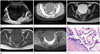
상기 증상을 주소로 실시한 하복부 초음파에서 약 12 cm 크기의 낭종이 골반 내에 있었다. 경계가 좋은 무에코의 단방성 낭종으로 내부에는 고에코의 부스러기(echogenic debris)와 벽의 내측으로 유상돌기(papillary projection)가 있었다(Fig. 1A). 또한 양측의 난소가 병변과 분리되어 정상적으로 관찰되었다. 컴퓨터단층촬영에서도 저밀도의 단방성 낭종의 벽 내측에서 약하게 조영증강되는 유상돌기가 있었다(Fig. 1B). 복강 내에 커진 림프절은 없었다. 자기공명영상에서 낭종의 내부는 T2 강조 영상에서 고신호강도, T1 강조 영상에서 저신호 강도이며, 낭종의 유상돌기는 조영증강되었다. 정상으로 보이는 양측 난소가 낭종과 구별되어 난소위체(paraovarium)에서 기원한 낭종으로 판단되었고, 내부에 조영증강되는 유상돌기가 있어 단순 낭종보다는 상피세포 종양 특히 경계성 종양이 보다 합당한 진단으로 고려되었다(Fig. 1C–E).
난소위체 종양에 대하여 적출술이 시행되었다. 적출된 낭종은 10 × 8 cm 크기였고, 대부분이 낭성이었으나, 벽 내측에서 유상돌기가 있었다.
조직검사에서 아교질성 간질과 사종체(psammoma body)를 덮고 있는 중층입방상피로 구성된 유상돌기가 관찰되었다. 중층입방상피는 뚜렷한 핵소체와 두꺼운 핵막이 관찰되었으나 간질로의 침윤은 관찰되지 않았고(Fig. 1F), 난소위체 장액성 경계성 종양(parovarian serous borderline tumor)으로 진단되었다.
환자는 합병증 없이 수술 후 2일째 퇴원하였고, 수술 3개월 후에 시행한 컴퓨터단층촬영에서 재발의 소견은 없었다.

고찰

난소위체 낭종(parovarian cyst)은 자궁부속기 종괴의 10~20%를 차지하며, 대부분 난관간막(mesosalpinx), 자궁광간막(broad ligament of uterus)에서 발생한다. 대부분 동측 난소와 구별되어 보이는 편측성 단일 단순 낭종 형태를 보이고, 드물게 양측성 혹은 다발성으로 나타나기도 한다(1). 드물게 낭종의 벽 내측으로 유상돌기(papillary projection)가 관찰되기도 한다. 이러한 경우에는 단순 낭종(simple cyst)이 아닌 상피세포성 종양일 가능성을 반드시 고려해 보아야 한다(2). 그중 난소위체 경계성 종양은 매우 드물고, 전 세계적으로 40건의 증례가 보고되었다(34).
대부분의 젊은 가임기 여성에서 발생하고, 복부통증이나 복부둘레 증가 등의 증상이 생길 수 있다(5). 낭선종(cystadenoma) 혹은 낭선암종에서 내부로 유상돌기가 관찰된다. 유상돌기는 컴퓨터단층촬영과 자기공명영상에서 조영증강을 보이며, 도플러 초음파에서 유상돌기 내부의 혈류를 관찰할 수 있다(6).
난소위체 경계성 종양의 경우 좋은 예후를 보이나 재발을 보이는 경우가 있기 때문에 지속적인 추적관찰이 필요하다(7).
결론적으로 본 증례와 같이 골반 내에 낭종이 발견된다면, 우선 난소와의 위치관계를 구분하여 낭종의 기원이 난소 또는 난소위체인지 구분하여야 할 것이다. 난소위체 낭종으로 밝혀졌다면 대부분 단순 낭종일 가능성이 높겠으나, 드물게 내부에 유상돌기가 관찰된다면 악성 종양의 가능성까지 고려하여 치료 계획을 수립하는 것과 추적 검사가 중요할 것이다.

Figures and Tables

Fig. 1

Paraovarian serous borderline tumor in a 19-year-old woman, presenting with left lower quadrant abdominal discomfort.

A. The transabdominal ultrasonography shows an approximately 12-cm-sized unilocular anechoic cyst, with papillary projection (arrow) and echogenic debris.
B. The contrast enhanced CT image shows well-defined unilocular fluid-attenuating mass, with papillary projection (arrow). The papillary projection shows subtle contrast enhancement.
C. The axial T2-weighted MR image shows a well-defined unilocular hyperintense mass with papillary projection (arrow). Both ovaries (*) are separated by the mass.
D. The axial T1-weighted MR image shows a hypointense mass and a papillary projection (arrow).
E. The axial contrast enhanced T1-weighted MR image shows an enhancing papillary projection (arrow).
F. Microscopically (hematoxylin and eosin, × 200), the thin and thick papillary projections consist of thick collagenous stroma and covered stratified cuboidal epithelium with multiple psammoma bodies (arrows). The lining cuboidal epithelial cells show slightly stratified, enlarged, hyperchromatic nuclei with prominent nucleoli and a thick nuclear membrane (arrowheads). However, there is no obvious stromal invasion or microinvasion.
jksr-79-337-g001

References

1. Zhao F, Zhang H, Ren Y, Kong F. Transvaginal sonographic characteristics of paraovarian borderline tumor. Int J Clin Exp Med. 2015; 8:2684–2688.
2. Korbin CD, Brown DL, Welch WR. Paraovarian cystadenomas and cystadenofibromas: sonographic characteristics in 14 cases. Radiology. 1998; 208:459–462.
crossref
3. Suzuki S, Furukawa S, Kyozuka H, Watanabe T, Takahashi H, Fujimori K. Two cases of paraovarian tumor of borderline malignancy. J Obstet Gynaecol Res. 2013; 39:437–441.
crossref
4. Bider D, Rabinovitch O, Schinder OM, Menczer J. Parovarian serous cystadenocarcinoma of borderline malignancy diagnosed during pregnancy. Gynecol Obstet Invest. 1988; 26:262–264.
crossref
5. Altaras MM, Jaffe R, Corduba M, Holtzinger M, Bahary C. Primary paraovarian cystadenocarcinoma: clinical and management aspects and literature review. Gynecol Oncol. 1990; 38:268–272.
crossref
6. Savelli L, Ghi T, De Iaco P, Ceccaroni M, Venturoli S, Cacciatore B. Paraovarian/paratubal cysts: comparison of transvaginal sonographic and pathological findings to establish diagnostic criteria. Ultrasound Obstet Gynecol. 2006; 28:330–334.
crossref
7. De Areia AL, Frutuoso C, Amaral N, Dias I, De Oliveira C. Paraovarian tumor of borderline malignancy—a case report. Int J Gynecol Cancer. 2004; 14:680–682.
TOOLS
Similar articles