

 PDF
PDF ePub
ePub Citation
 Print
Print


INTRODUCTION
Renal artery injury is a rare complication in blunt trauma. Renal arterial occlusion may cause devascularization of the kidney, therefore, early diagnosis and management is important. Although the rate of early diagnosis has increased substantially as the use of contrast-enhanced CT has increased, the management of renal artery injury remains controversial; emergent surgical repair, anticoagulant therapy, and endovascular treatment. Recent studies have reported the feasibility of endovascular management of renal artery injury with stent placement. However, there are difficulties in installing stents in children for several reasons. The growth of children's vessels has the possibility of developing a size discrepancy with the stent, resulting in complications such as thrombosis, stent migration. Furthermore, anticoagulation therapy is essential when installing stents. However, systemic anticoagulation to the patient with multiple organ trauma may increase risk of bleeding. To our knowledge, there are only few cases reported on renal artery injury in children, and no standard treatment strategy is determined yet. Here, we present a case of a 16-year-old girl with right renal artery occlusion associated with a grade IV liver laceration, which was treated with only balloon angioplasty.
CASE REPORT
A 16-year-old female was transferred to the emergency department complaining of abdominal pain after a traffic accident on motorcycle. Upon admission she was alert and hemodynamically stable, presenting a blood pressure of 138/71 mm Hg, 93 heart rates, 15 respiratory rate and 37.5 degrees of body temperature. Initial laboratory exams revealed hemoglobin of 10.7 g/dL, hematocrit of 32.4%, and white blood cell count (WBC) of 10.3 × 1000 µL. On urinalysis, red blood cell count and WBC were within normal range. However, microalbumin/Creatinine ratio reflecting renal function was elevated at 561.9 mg/g.
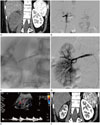
On chest posteroanterior and simple abdomen plain radiographs, there was no demonstrable abnormal finding. The patient underwent an abdomen-pelvis CT with contrast enhancement for further evaluation. CT revealed near total devascularization of the right kidney with right perirenal hematoma (Fig. 1A). There was no active contrast extravasation, but abrupt obstruction of the right renal artery was noted from 2 cm apart from the ostium with surrounding hemorrhagic fluid collection. In addition, the patient had hemorrhage at the right adrenal gland and liver laceration of grade IV injury on American Association for the Surgery of the Trauma grading scales. The patient was referred to the intervention center for the management of the right renal artery occlusion.
Under local anesthesia with 2% lidocaine (Lidocaine HCL Hydrate Injection 5 mL Huons, Seongnam, Korea), ultrasound-guided right common femoral artery was punctured using micropuncture needle (Medcomp, Harleysville, PA, USA), and a 5Fr sheath (Terumo, Tokyo, Japan) was inserted. Right renal artery angiography done with 5Fr angiographic catheter (Cobra shape, Cook medical, Bloomington, IN, USA). There was abrupt occlusion at the proximal portion of the renal artery. On selective angiography done after careful cannulation of the occluded segment using 2.0Fr microcatheter (Progreat Alpha, Terumo) and 0.016" microwire (Meister, Asahi, Seto, Japan), we could confirm that some distal branches of the renal artery was patent (Fig. 1B). The injection of 1000 IU heparin was performed as anticoagulant therapy. Then balloon angioplasty was done at the occluded main renal artery with 3 mm × 4 cm balloon catheter (Sterling, Boston Scientifics, Marlborough, MA, USA) and at the occluded proximal portion of renal artery with 5 mm × 2 cm balloon catheter (Sterling) on nominal pressure for 3 minutes, respectively (Fig. 1C). On completion angiography, although there was remained partial filling defect on the right renal artery, distal flow was patent, and the renal parenchymal perfusion showed marked recovery (Fig. 1D). The time from injury to revascularization was 12 hours, and the time from admission to revascularization was seven hours and 30 minutes.
After two days, follow-up color Doppler ultrasound was performed. Perirenal hematoma still remained around the right kidney, but vascularity was remarkably increased. The right renal artery showed normal artery waveform pattern of the sharp systolic upstroke, continuous forward diastolic flow, and low resistance waveform (Fig. 1E). Then on follow up abdomen CT with contrast enhancement after 7 days, there was a small filling defect, suspicious of dissecting flap, but parenchymal perfusion of the right kidney was patent.
The patient performed follow-up renal angiography CT after two months. Some portion of the right renal parenchyma was atrophied, but right renal artery was patent. Compared to the initial CT, there was marked improvement of parenchymal perfusion of the right kidney (Fig. 1F). The microalbumin/Creatinine ratio was normalized at 38.0 mg/g.
DISCUSSION
Renal artery injury is a rare complication in blunt trauma patients. The incidence of renal artery injury from blunt trauma ranges between 0.05% and 0.08% (12). Renovascular injuries usually occur about 2 cm from the origin of the main renal artery but may involve branch renal vessels, as was seen in our case (2). In addition, it is known that children are more susceptible to renal artery injury in blunt traumas, attributed to the relatively larger kidneys in the pediatric population, their higher mobility, and a relative lack of surrounding protective tissues (3).
Renovascular injury secondary to trauma may occur in two types - arterial spasm and arterial occlusion. Arterial spasm is considered to be secondary to contusion (4). Renal arterial occlusive injury, which may cause devascularization of the kidney, is classified as vessel avulsion, laceration, or dissection. The former two subtypes necessitate immediate surgery, such as nephrectomy or early vascular reconstruction, due to hemorrhage. Traumatic dissection of the renal artery may lead to stenosis or occlusion of the renal artery due to the presence of an intimal tear with secondary thrombus formation (5). Our case was thought to be caused by thrombotic occlusion due to flow-limiting dissection.
The kidneys are extremely vulnerable to warm ischemia. The impact of the ischemia time is even greater when complete occlusion of the renal artery occurs as there is minimal collateral renal blood flow beyond the point of occlusion. Thus, the most critical factor in preserving renal function is the reestablishment of blood flow as early as possible (2). Spirnak and Resnick (6) defined the optimal time frame for revascularization as 6–12 hours from injury. However, ischemic changes start to appear around 60–120 minutes after injury, thus it is believed that the optimal time frame is likely to be much shorter, between 3 and 4 hours, as stated by Cass (7) and Flye et al. (8) In our case, the time for revascularization was 12 hours from injury, and the mean time from admission to revascularization was 7 hours and 30 minutes.
The management of renal artery injury remains controversial in the last 30 years. In many cases, high grade renal injury accompanies other intra-abdominal organ injuries, contributing to higher morbidity and mortality. The treatment options for such cases are emergent laparotomy together with early nephrectomy, partial nephrectomy, arterial repair or renal autotransplantation (2).
Recent studies report that endovascular management for renal artery injuries in blunt trauma is safe and presents good outcome. It is believed to be an alternative treatment to surgery in cases of renal artery dissection. Most of the reported cases up to now used stents for endovascular revascularization. When installing stents, anticoagulation or antiplatelet therapy is necessary to prevent thrombus formation. However, in many cases of trauma, systemic anticoagulation is intolerable due to other visceral organ injuries. In fact, Breyer et al. (9) have failed to perform anticoagulation after renal artery stent replacement and have reported a case of failure of stent therapy due to subsequent stent thrombosis.
Until now, we have identified two cases of renal artery injury treatment with angioplasty without stenting. Both cases were pediatric patients, one with a 4.5-year-old boy and one with an 11-year-old boy. In a case reported by Abu-Gazala et al. (2), a 4.5-year-old boy had grade IV liver laceration and filling defect in right renal artery without enhancement or excretion on CT due to a military vehicle accident. The patient underwent graded renal artery angioplasty to a maximal diameter of 3.5 mm, followed by anticoagulation therapy with heparin in the pediatric intensive care unit. Time from admission to revascularization was 110 minutes. On follow up, normal flow of the right renal artery was present on ultrasound with duplex, and improvement of uptake was shown on follow up renal diethylenetriamine pentaacetic acid scan performed after 14 months. Meanwhile, Yokoyama et al. (10) reported a case of 11 year-old-boy with motor vehicle accident. CT presented grade III liver laceration and lack of contrast enhancement of the right kidney with focal extravasation of contrast media and surrounding hematoma. Recanalization and angioplasty was performed at the right kidney 4 hours after injury, but treatment was unsuccessful. Two months following the trauma, right kidney was atrophied with compensatory hypertrophy of the left kidney.
Children are more susceptible to renal artery injury in blunt traumas, but there are few cases reported so far and no standard treatment strategy. The treatment depends on the damage of other organ injuries and hemodynamic stability. In cases of renal artery occlusion, revascularization with stent placement is a recently favored treatment option; however, more risk and complications are expected in children. The stent may not fit the optimal size as children grow, and the rate of stent-related complications such as thrombosis or stent fracture may increase due to long-term stent insertion state. To this end, procedure of only balloon angioplasty can be one of the treatment options. In our case, balloon angioplasty and anticoagulation therapy was done after 12 hours of trauma. The kidney was successfully revascularized on follow up imaging studies, and the renal function showed recovery. In conclusion, endovascular treatment with stent placement is preferred as the first treatment option when there is traumatic renal artery injury; nevertheless, only balloon angioplasty may be considered as an alternative for pediatrics or patients with difficulties in anticoagulation therapy.
 XML Download