

 PDF
PDF ePub
ePub Citation
 Print
Print


INTRODUCTION
With the advance in imaging technologies and improved HCC surveillance, the incidence of early stage tumor has been improved gradually and effective treatment of early stage HCC has become increasingly important.1 Although HCC surveillance and advance in treatment technologies have led to improved patient survival, the rate of recurrence is still high. Hepatic resection (HR) and radiofrequency ablation (RFA) are now widely used to treat patients with early stage HCC. Although it remains debatable which treatment has superiority over other, HR has been regarded as the first-line treatment for patients with early stage HCC and preserved hepatic function due to its acceptable mortality, morbidity and long-term outcomes.234
Anatomical resection (AR) is defined as resection of the tumor including tumor-bearing portal vein territories.5 Theoretically, HCC has a high propensity to invade the intrahepatic vascular structures and spreads mainly through the portal venous system rather than by adjacent diffusion;678 thus, AR is regarded as effective treatment to avoid intrahepatic metastasis and recurrence. However, AR needs to sacrifice a large amount of liver parenchyma and is therefore significantly unfavorable for treating a liver that has an underlying disease. On the contrary, non-anatomical resection (NAR) is the conventional limited resection focused on achieving a non-tumoral liver parenchyma rim, without consideration of the Glisson's pedicles.910 While some studies have reported the superiority of AR over NAR,10111213 other studies have shown no prognostic difference between AR and NAR,81415 and some meta-analyses have also reported conflicting conclusions.161718 Therefore, the ideal treatment for HCC remains debatable.
A Japanese nationwide study demonstrated that AR is only beneficial for HCC of 2 to 5 cm, and not for very small HCC less than 2 cm.1920 Usually, HCC smaller than 2 cm is accepted as very early HCC and larger than 5 cm as large HCC. However, size range of 2 to 5 cm can be ambiguous and HCC of 2 to 3 cm is also accepted as early stage HCC in BCLC staging system. Therefore, we compared the outcomes following AR and NAR in patients with solitary HCC smaller than 3 cm and investigated the prognostic risk factors for HCC recurrence in these patients.
MATERIALS AND METHODS
A total of 154 patients underwent liver resection for single HCC less than 3 cm at Keimyung University Dongsan Medical Center in Daegu between January 2006 and December 2015. Among these patients, 38 patients who were treated for recurred HCC after primary treatment previously and had no clinical follow-up data after resection were excluded and 116 patients who underwent HR for primary treatment were enrolled in this study. The medical records of these patients were reviewed retrospectively and the following data were collected for each patient. AR was performed in 53 patients (45.7%) and NAR was performed in 63 patients (54.3%).
Tumor size was defined as the largest diameter of the tumor in the specimen. Single HCC was defined based on the preoperative imaging studies regardless of postoperative pathologic results including satellite nodules and/or vascular invasion. AR was defined as the systematic resection of hepatic segments according to the segmental and sectional anatomy along the hepatic vasculature based on Couinaud's classification. For the AR, Glissonean approach method and Indocyanine Green (ICG) dye injection method21 were performed in each patient. NAR was defined as liver resection without regard to the Couinaud's classification. The extent of hepatic resection (AR/NAR) was determined by surgeon's preference considering tumor location and patients' condition.
The routine follow-up program consisted of physical examination, computed tomography (CT) and laboratory tests including alpha-fetoprotein (AFP) and PIVKA-II (Protein induced vitamin K antagonist-II) level every three month for the first year, and then every six months for next five years, thereafter annually for patients who have neither recurrence nor metastasis. Recurrence was defined as the appearance of a new lesion compatible with HCC in radiologic examination during follow-up period. With regard to the type of recurrence, we considered as marginal recurrence located in same segment of liver or near the resection margin within 1 cm, and as multicentric occurrence located in another segment or more than 1 cm from resection margin.
For the investigation of risk factors predicting tumor recurrence, various cut-off points of tumor markers, which were used in previous studies, were validated using our data. However, there was no significant cut-off point to predicting tumor recurrence in this study. So, the cut-off points for AFP 500 and PIVKA-II 100, which were nearest to the statistical significance, were chosen for statistical analysis.
Statistical analysis was performed with IBM SPSS version 18.0 program (SPSS Inc., Chicago, IL, USA). The Mann-Whitney U-test was used to evaluate differences in the continuous variables. Categorical variables were compared by Fisher`s exact test. Cumulative survival curves were analyzed by using the Kaplan-Meier method, and significance was determined by log-rank test. To investigate the prognostic factors predicting tumor recurrence, univariate and stepwise multivariate regression analysis was performed using a Cox proportional hazard model with p<0.05 considered statistically significant.
RESULTS
Comparison of demographics and clinicopathologic features between resection groups
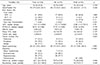
The demographics and clinical features of the 116 patients are summarized in Table 1. Among these patients 92 patients were male (79.3%) and 24 patients were female (20.7%). The mean age of patients was 56.4 years. All of these patients were classified as Child-Pugh A in preoperative liver function assessment. The median preoperative AFP and PIVKA-II were 11.9 ng/ml and 26.5 mIU/mL, and the mean preoperative ICG R-15 level was 15.2%. The mean tumor size was 2.3 cm. Among these patients, 40 patients (64%) had tumors measuring less than 2 cm and 76 patients (36%) had tumors measuring between 2 and 3 cm. Tumors were located protrusively in 27 patients (23.3%), peripherally in 69 patients (59.5%), and medially in 20 patients (17.2%). AR was performed more frequently for medial located tumor than NAR, however there was no significant difference between AR and NAR group in demographics except tumor location.
Pathological features of the patients in this study were summarized in Table 2. Mean free margin was 1.26 cm and Resection margin was grossly free from tumor in all patients, however microscopic involvement was identified in 5 patients (4.3%). Portal vein micro invasion was identified in only 1 patient (0.9%), microscopic vascular invasion (MVI) in 17 patients (14.7%), and satellite nodule was detected in 5 patients (4.3%). Underlying liver cirrhosis was identified in 82 patients (70.7%). There was no significant difference between AR and NAR group in pathologic features.
Survival & recurrence after resection
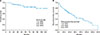
The median follow-up period was 66.65 months (range, 6.4–143.3). During a follow-up period, tumor recurrence occurred in 65 patients (56.0%) and median time to recurrence was 25.6 months after surgery, respectively. At the time of last follow-up, 19 patients (16.4%) had died of liver-related disease including tumor progression. There was no in-hospital mortality. For the entire cohort of 116 patients, 1-, 3- and 5-year overall survival rates were 99, 92 and 84% (Fig. 1A) and recurrence-free survival rates were 84, 63 and 47%, respectively (Fig. 1B).
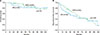
In comparison of the clinicopathologic characteristics between AR and NAR group, there was no significant differences in demographics and pathologic findings except tumor location (Table 1 and 2). The 1-, 3- and 5-year overall survival rates were 100, 88 and 81% in AR group, and 98, 95 and 86% in NAR group, respectively (Fig. 2A). There was no significant difference in overall survival between AR and NAR group (p=0.78). The 1-, 3- and 5-year recurrence-free survival rates were 75, 56 and 43% in AR group, and 90, 70 and 51% in NAR group, respectively (Fig. 2B). There was also no significant difference in recurrence-free survival between AR and NAR group (p=0.455).
Although there was no significant difference between groups in recurrence time and pattern, tumor recurrences within 1 year after resection were more frequent in AR group and after 3 years were more frequent in NAR group (Table 3).
Prognostic factor analysis for early and overall recurrence
In patients with HCC smaller than 3 cm, hepatitis B, anatomical resection, microvascular invasion, and presence of satellite nodule were associated with early recurrence (within 1 year) after resection in the univariate analysis, however high level of tumor markers, tumor size, and microscopic margin positive were not associated with early recurrence In multivariate analysis, hepatitis B (hazard ratio (HR) 8.72; p=0.035), satellite nodule (HR 3.97; p=0.029) and MVI (HR 2.79; p=0.039) were independent risk factors for early recurrence (within 1 year) after resection (Table 4). In the aspect of overall recurrence after resection for HCC smaller than 3 cm, satellite nodule (HR 6.21; p=0.005) and background liver cirrhosis (HR 1.96; p=0.032) were revealed as independent risk factors in multivariate analysis (Table 5).
Relation between safety margin, microvascular invasion and recurrence
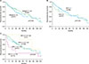
In terms of recurrence, width of safety margin (margin <1 cm) was not correlated with HCC recurrence after surgery in single HCC smaller than 3 cm (Fig. 3A). Although MVI was the independent prognostic factors for early recurrence (within 1 year), overall recurrence was not significantly affected by MVI (Fig. 3B). However, time of recurrence was significantly different in patients with MVI according to the width of safety margin (<1 cm vs ≥1 cm, p=0.049), on the other hand, there was no difference in patients without MVI according to the width of safety margin (Fig. 3C).
DISCUSSION
Liver resection has been accepted as the gold standard treatment for solitary HCC in patients with well-preserved liver function.22 However, the superiority of AR has been still controversial. Recently, with the technical improvement, laparoscopic liver resection has been widely applied for the treatment of HCC and NAR can be performed more easily than AR with laparoscopy especially in cases of small HCC which is protruded or located peripherally. For this reason, the proportion of laparoscopic resection was significantly higher in NAR group than AR group in this study.
Previously several retrospective studies reported the superiority of AR for HCC in the aspect of recurrence and survival.911121323 However, most of these studies have significant selection bias of patients, the difference of reserved liver function which is a significant postoperative prognostic factor in recurrence and survival. On the contrary, other retrospective studies which tried to control confounding factors and eliminate selection bias, reported the comparable outcomes between AR and NAR in solitary HCC less than 4 cm8 and 3 cm.15 In this study, we only included the patients with well-preserved liver function (Child-Pugh class A) and there were no differences in preoperative characteristics and postoperative pathologic results between AR group and NAR group except tumor location and the proportion of laparoscopic surgery. Our results showed that the outcomes of NAR in terms of recurrence pattern, recurrence-free survival, and overall survival were not different from those in AR (Table 3, Fig. 2). In univariate analysis for identifying prognostic factors predicting early recurrence (within 1 year), HCC recurrences within 1 year were more frequently occurred in patients underwent AR than NAR. We presumed that this result is caused by the disparity between groups in proportion of tumor size. Although there was no statistical significance between AR and NAR group in mean tumor size (2.36 vs 2.21, p=0.106) and the proportion of patients who had HCC of 2 to 3 cm (72.7% vs 60.3%, p=0.241), there were more patients who had HCC of 2 to 3 cm in AR group. And this might make the significant difference in early recurrence rate between groups in univariate analysis.
Instead of the extent of hepatic resection (AR/NAR), multivariate analysis identified that hepatitis B, the presence of satellite nodule and MVI were the independent risk factors for early (within 1 year) recurrence of tumor. There are several studies about correlation between hepatitis B viral infection and early recurrence of HCC.24252627 But the mechanism by which HBeAg positivity could possibly enhance tumor recurrence remains unclear. The possible explanation is that positive HBeAg may associate with active inflammation in liver parenchyma to promote intrahepatic metastasis by changing tumor microenvironment, finally resulting in early recurrence within a short time after hepatectomy. The other possibility is positive HBeAg may associated with increased synchronic multicentric tumor. MVI has been accepted as powerful prognostic factors predicting recurrence and survival in large and multiple HCC. However, several studies reported that MVI had no significant impact on overall survival in patients with early HCC, whereas MVI had significant impact on recurrence.192829 These data correlate with our results up to a point. Our results showed that MVI was not correlated with overall recurrence and survival, while early recurrence (within 1 year) was significantly affected by MVI. In our data, background liver cirrhosis had more significant impact on overall survival along with the presence of satellite nodule rather than MVI and viral activity. These results suggest that the prognosis after resection can be more affected by underlying liver status rather than pathobiological factors of tumor in early stage HCC, an observation supported by previous study which reported the importance of liver status as an independent prognostic factor predicting recurrence after resection.81530
The significance of safety margin in recurrence and survival after liver resection for HCC remains controversial. Several studies reported no relation between the safety margin and prognosis,8313233 while other studies suggested that safety margin less than 1 cm had an negative effect on long-term prognosis.343536 Our data suggested that the width of safety margin has no impact on survival and HCC recurrence after resection in entire cohort of patients with single HCC smaller than 3 cm. However, the impact of safety margin on tumor recurrence was different between MVI (+) and MVI (−) group when the patients were categorized according to the presence of MVI. In patients without MVI, there was no significant difference in HCC recurrence between safety margin <1 cm and safety margin ≥1 cm group. On the other hand, tumor recurrence (especially early recurrence) was more frequently observed in safety margin <1 cm group in patients with MVI (p=0.049). Based on these results, we can suggest that hepatic resection with adequate margin over 1 cm might be required for decreasing risk of early recurrence in patients with single HCC smaller than 3 cm because MVI can't be identified preoperatively. However, presence of MVI was detected in only 17 patients (14.7%) with small HCC in this study and this makes it difficult to confirm this result. Therefore, further studies with large sample size are required to confirm this result.
The limitations of this study include its retrospective nature, small volume cohort from single center and selected population. Furthermore, most of patients in this study showed favorable biologic behavior and some factors which accepted as powerful prognostic factors were observed in very few patients. this could raise the possibility of statistical error. However, similar liver function in all patients (well-preserved liver function) and no difference in clinical and pathologic findings between groups can minimize the risk of selection bias. In addition, when considering the less aggressive biologic nature of early HCC, our results might be accepted as a meaningful data.
In conclusion, the outcomes of NAR were comparable with those of AR in patients with single HCC smaller than 3 cm and well-preserved liver function. Although MVI was not correlated with overall survival and recurrence, early recurrence was significantly affected by MVI, and the outcome of resection with safety margin <1 cm was shown worse than safety margin ≥1 cm. Therefore, wide resection with adequate safety margin is recommended in patients with single HCC smaller than 3 cm. In terms of overall recurrence, back ground liver status such as cirrhosis and viral activity has more significant impact on prognosis rather than pathobiologic behavior in single HCC smaller than 3 cm.
 XML Download