

 PDF
PDF ePub
ePub Citation
 Print
Print


INTRODUCTION
Gastric ulcers, induced by several factors such as stress, smoking, acid, pepsin, ingesting nonsteroidal anti-inflammatory drugs, and most commonly by Helicobacter pylori (H. pylori) infection, affect a large portion of the population worldwide. At present, nearly 15 million people are suffering from peptic ulcer diseases, and almost 6,000 people die of it every year. Gastric ulcer healing is a complex process that involves inflammation, reepithelialization, granulation tissue formation, angiogenesis, and scar formation (123). The approaches to the treatment of gastric ulcer includes inhibition of gastric acid secretion by H2 receptor blockers or proton pump inhibitors (PPIs) as well as eradication of H. pylori (4). However, their effectiveness is compromised by side effects and the long-time continual administration. For example, 6–8 weeks after treatment, there is a problem of recurrence (5). Thus, there is a growing interest in developing new therapeutic strategies for gastric ulcer healing with reduced side effects and shortened treatment time. It would be expected to benefit millions of suffering humanity.
Hepatocyte growth factor (HGF), one of the most potent multifunctional factors, was shown to act on various epithelial and endothelial cells, promoting their proliferation, and migration, enhancing gastric epithelium proliferation, inhibiting cell apoptosis (67), reducing inflammatory infiltration, alleviating symptoms, and mucosal lesions (8), and improving local angiogenesis. Therefore, HGF may be a potential cytokine to ameliorate gastric ulcer. However, the clinical application of recombinant protein of cytokines administered orally is limited because of their instability, high dosage, and repeated administration, which increases the incidence of side effects (9).
In the current study, a new strategy was developed to treat gastric ulcer. The potential effect of HGF gene therapy mediated by the attenuated Salmonella typhimurium (S. typhimurium) on gastric ulcer was investigated. In addition, the attenuated S. typhimurium shows potent tropism for gastric mucosa. A number of trials indicated that attenuated S. typhimurium can be orally used to deliver a eukaryotic expression plasmid to gastric mucosa (1011). Therefore, a recombinant attenuated S. typhimurium strain carrying a eukaryotic expression vector encodes the human HGF gene was established (named as TPH). Its effect on repairing ulcer was investigated in vivo and ex vivo. The TPH could effectively transfer into normal gastric epithelial cells (GES-1) in vitro and highly express interest HGF mRNA and protein. The expression product could significantly stimulate proliferation of GES-1 and angiogenesis of chorioallantoic membrane (CAM). By administering TPH orally to the gastric ulcer model rats, a novel, feasible, and effective modality for gastric ulcer gene therapy was investigated.
MATERIALS AND METHODS
Construction and preparation of recombinant attenuated S. typhimurium strains TP and TPH
An attenuated S. typhimurium strain with an eukaryotic expression vector (named as TP) and with an eukaryotic expression vector encodes the human HGF gene (TPH) were prepared in Key Laboratory of Stem Cell and Gene Drug in Gansu province. Regarding the construction of TPH, the HGF cDNA was amplified from the human placental cDNA library and cloned into the eukaryotic expression vector pcK (prepared by the current investigators; this method is under review for a patent in China; the public number is CN 1869238 A), and this resulted in a eukaryotic expression plasmid pcKH. To produce TP and TPH, the pcK or pcKH was transformed into the attenuated S. typhimurium Ty21a (No.50218, from the National Institute for the Control of Pharmaceutical and Biological Products, Beijing, China) by electroporation (Multiporator 4308; Eppendorf, Hamburg, Germany). The screening of positive engineering bacteria was performed by kanamycin (Gibco, Grand Island, NY, USA) resistance, polymerase chain reaction (PCR) for amplification of cytomegalovirus (CMV) and HGF cDNA, and enzyme digestion (BamHI/ApaI, Promega Corporation, Madison, WI, USA) (12). The CMV forward primer was 5′-ccc agtacatgacct tat ggg-3′, and the reverse primer was 5′-ggagacttggaaatc ccc gt-3′. The HGF forward primer was 5′-ccatcgatgttaacatgtgggtgaccaaactc-3′, and the reverse primer was 5′-tgggatccgcggccgcctatgactgtggtacctt-3′. Thus, TP-and TPH-positive bacteria strains were obtained.
Transfection of human GES-1 cells by recombinant attenuated S. typhimurium strains
The normal gastric epithelial cells (GES-1, from the Cell Bank of Chinese Academy of Sciences in Shanghai, Shanghai, China) were cultured in the Dulbecco's Modified Eagle Medium (DMEM) with 10% fetal bovine serum (FBS; Gibco) in 5% CO2 at 37°C. After two to eight passages, the cells were harvested by trypsin (Gibco) digestion, plated at 6 × 10⁵ cells/well in a 6-well plate and incubated overnight. After washing three times with phosphate buffer solution (PBS), the medium was changed to DMEM without serum. The TP strain (3 × 10⁷ cfu/well, TP group) or TPH strain (3 × 10⁷ cfu/well, TPH group) solution in PBS and the same volume PBS/well (control group) were added into wells, respectively, and the plates were incubated at 37°C for 30 minutes. After washing three times with condition DMEM (with 50-µg/mL Gentamicin; Sigma, St. Louis, MO, USA), the medium was changed to DMEM (with 10% FBS) without antibiotics, and the plates were incubated at 37°C for 4 hours. Gentamicin (5 µg/mL) and tetracycline (5 µg/mL; Sigma) were then added to the wells. The supernatant and GES-1 cells of the three groups were harvested at 12, 24, 48, and 72 hours after transfection.
Detection of expression level of HGF mRNA by reverse transcription–-polymerase chain reaction (RT-PCR)
Total RNA from the GES-1 cells at various time points after transfection was isolated using RNAiso Plus (Po bio engineering [Dalian] Co., Ltd. [Dalian Takara], Dalian, China), according to the manufacturer's protocol. The RNA quantity was determined photometrically. Total RNA was reverse transcribed with the PrimeScript RT Reagent Kit (Po bio engineering [Dalian] Co., Ltd. [Dalian Takara]), and semi-quantitative RT-PCR was conducted with HGF-specific primers. To determine the integrity of the mRNA in each sample, β-actin mRNA was employed as an endogenous reference for human GES-1 cells. The forward primer of β-actin was 5′-ctccatcctggcctcgctgt-3′, and the reverse primer was 5′-gctgtcaccttcaccgttcc-3′. The RT-PCR products (268 bp) separated on 1.0% agarose gels were digitized by the Gel Image Analysis system and quantitated by ethidium bromide fluorescence by using the Image-Pro Plus 6.0 software (Media Cybernetics Company, Rockville, MD, USA).
Detection of expression level of HGF protein by enzyme-linked immunosorbent assay (ELISA)
The expression of HGF protein in the supernatant, from the GES-1 cells after transfection with recombinant attenuated S. typhimurium strains, was detected by the HGF ELISA Kit (R & D Systems Inc., Minneapolis, MN, USA) at various time points, according to the manufacturer's instruction. The optical density (OD) was detected at 450 nm with a microplate reader (Thermo Fisher Scientific, Pittsburgh, PA, USA). The concentration of the HGF expression product was assessed.
Evaluation of the HGF activity in GES-1 cells by MTT method
The activity of the HGF expression product for cell proliferation was monitored with a 2-(4,5-dimethyltriazol-2-yl)-2,5-diphenyl tetrazolium bromide (MTT; Sigma) assay. The GES-1 cells were cultured in 96-well plates at (5 × 10³) cells/well for 12 hours. After the medium was removed, the supernatants containing the HGF expression product (80, 160, 320, 640, and 1,280 pg) were added to each well, respectively. And supplied the total volume to 200 μL with control group supernatant. After 48 hours, the cells were incubated with 20 μL of MTT (5 mg/mL) at 37°C for 4 hours. The medium was then removed, and 150 μL of dimethylsulfoxide (DMSO) was added into each well. The plates were then incubated at 20°C for 20 minutes in dark. The OD value of each well was detected at 570 nm. The supernatants from the TP-transfected group were regarded as control stimulator under same conditions.
Effect of the HGF expression product on the angiopoiesis of chick chorioallantois
The angiogenic activity of the HGF expression product on the CAM was assayed, as described in a previous study (13). Briefly, the fertilized white Leghorn chicken eggs were incubated at 37.8°C for 13 days, and the methylcellulose (M450; Sigma) disks (5 mm in diameter, 1.5 mm in depth, prepared in the test laboratory) containing the HGF expression product (300 pg) or same volume supernatant from the TP-transfected group or only disk were, respectively, placed on CAM (6 embryos in each group). The eggs were incubated at 37.8°C for 3 days, and the mixing fixation fluid was added. After the blood vessel of allantochorion was clotted, the membrane was taken and put in a plate containing water. The vascular networks in the CAMs were blindly scored by two independent investigators for the presence or absence of an avascular zone (> 3 mm in diameter) surrounding the implant.
Induction and treatment of rat gastric ulcer model
Thirty-five male Wistar rats (aged 8–12 weeks, weighing 200–230 g, from the Animal Center of Gansu Chinese Medical College, Lanzhou, China) were housed in a cage maintained at 23°C, under 12/12-hour light/dark cycles under specific pathogen-free conditions. After 1 week of adaptation, the rats were used for the experiments. The rats were fasted, but allowed water, for 12 hours prior to the experiments. Gastric ulcers were induced with an acetic acid injection, according to a previously described method (14). with minor modifications. Briefly, the fasting rats (30 rats) were anesthetized with an intraperitoneal injection of sodium pentobarbital (30 mg/kg), and then their stomachs were exposed via a midline incision. Acetic acid (30 μL) was injected into the subserosal layer at the junction of the anterior wall of the antrum and corpus using a microsyringe. The abdominal incision was then sutured closed. From day 5 after ulcer induction, the rats were randomly divided into 3 groups: 1) TPH group, treated with 1 × 10⁹ cfu TPH; 2) TP group, treated with 1 × 10⁹ cfu TP; and 3) model control group, treated with 0.5-mL 1.19M NaHCO3. Ten rats were included in each group. For each animal, treatment was performed every other day for a total of 3 times by gavage. The other 5 rats were as normal animal group.
Gross and histological observation and scoring
The rats were sacrificed under anesthesia on day 21 after ulcer induction. The stomachs were taken from the rats and washed with cold 0.9% NaCl after being cut along the greater curvature. Among them, 5 samples from each group were immediately fixed with a 10% formalin solution for gross and histological observation and scoring. After fixed in formalin overnight, the mucosal sides of the stomachs were spread out with pins on a board and photographed. The degree of gross mucosal damage (ulcerated area) was assessed according to Kang et al. (15). The ulcerated area (mm2) was quantified using the following equation: where S represents the ulcerated area (mm2), and d1 and d2 the longest longitudinal and transverse diameters of the ulcer. Then, the ulcer and normal gastric mucosa of around 3 mm were taken and embedded in paraffin for histological analysis and immunohistochemical staining. The sections were stained with hematoxylin and eosin (H & E) for histological analysis, and a pathologist blinded to the treatment groups observed the samples under a light microscope (× 100). The crawling length of gastric epithelial cells was measured by a microscale measurement.
Immunohistochemical staining for microvascular density (MVD) with CD34 expression
To assess angiogenesis, the immunohistochemical evaluation of MVD was performed in granulation tissue at the ulcer beds. Monoclonal antibodies anti-CD34 (Abcam plc, Cambridge, UK) were used by the streptavidin-biotin-peroxidase complex (SABC) technique. Sections of 5 μm were boiled for 20 minutes in a retrieval solution and then incubated with primary antibody, secondary antibody (goat anti-mouse IgG, 1:500; Santa Cruz Biotechnology, Santa Cruz, CA, USA), and avidin-biotin-peroxidase complex (Zhongshan Biotechnology Co., Ltd., Zhongshan, China) for 45 minutes. The visualization system used the DAB kit (Zhongshan Biotechnology Co., Ltd.), and counterstaining was performed with hematoxylin. For negative control, the primary antibody was replaced with a buffer solution. MVD was measured in the ulcer base granulation tissue by the microscopical examination of sections from representative zones that had the biggest number of capillaries according to the method described previously by Weidner et al. (16). Coded specimens were evaluated quantitatively by two investigators who were unaware of the code. The microvessels were counted in zones under a × 200 objective, after initial examination with a small objective (× 100). Immunoreactive endothelial cells and nests of endothelial cells clearly separated by nearby microvessels were also counted. MVD represents the medium number of vessels counted on five microscopic fields with an objective of × 200 (BX-50; Olympus, Tokyo, Japan).
Detection of HGF and c-Met expression in gastric mucosa by ELISA
The gastric mucosa tissues (the ulcer and normal gastric mucosa of around 5 mm, weighing 500 mg) from another five samples in each group were taken and homogenized with a homogenizer in a lysis buffer containing 62.5-mmol ethylenediaminetetraacetic acid, 50-mmol Tris, pH 8.0, 0.4% deoxycholic acid, 1% Nonidet P-40, 0.5-mg/mL leupeptin, 0.5-mg/mL pepstatin, 0.5-mg/mL aprotinin, 0.2-mmol phenylmethylsulfonyl fluoride, and 0.05-mmol aminoethyl benzene sulfonyl fluoride. The homogenates were then centrifuged at 12,000 rpm for 10 minutes at 4°C. The concentration of human HGF and c-Met in the supernatant at the end time point was measured by ELISA method (same method as described earlier).
RESULTS
High expression of HGF in the GES-1 cells transfected with TPH in vitro
The TP- and TPH-positive bacteria strains were inoculated in a Lysogenybroth medium supplemented with kanamycin (50 μg/mL) and harvested 9 hours later, and the cell pellets were washed with PBS, then resuspended in 1.19M NaHCO3 solution and counted. The bacteria cells were adjusted to (3 × 10⁹) cfu/mL before administration.
To address whether the recombinant attenuated S. typhimurium genes (TP and TPH) could be transfected into the bacteria cells and effectively expressed, the human HGF mRNA level in the GES-1 cells was measured by RT-PCR, and the concentration of human HGF protein in the cell supernatant was detected by ELISA. The results showed that the HGF gene (about 2.2 kb) could be highly expressed in the GES-1 cells in the TPH group, and the highest expression appeared at 48 hours after transfection. However, no obvious expression was found in the TP and control group (Fig. 1A and 1B). The HGF protein expression was significantly higher in the TPH group than in the TP group after transfection (Fig. 1C). The maximal concentration was reached after 48 hours, which was as high as (30.8 ± 2.4) ng/(6 × 105) cells/mL. Interestingly, the HGF expression decreased gradually after 72 hours. This result could be due to an inhibition of growth induced by the high concentration of HGF or by the culturing conditions.
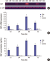 | Fig. 1HGF expression in the GES-1 cells after transfection with TPH or TP at different time points.
(A) RT-PCR products of HGF and β-actin in the agar gel electrophoresis. (B) The statistical results of RT-PCR of HGF in the GES-1 cells. TPH group: 3 × 10⁷cfu TPH strain was added into the wells of a 6-well plate; TP group: 3 × 10⁷cfu TP strain was added into the wells of a 6-well plate; control group: PBS (10 μL/well) was added into the wells of a 6-well plate; 12, 24, 48, and 72 hours indicate transfected TPH or TP or PBS for 12, 24, 48, and 72 hours, respectively. (C) Results from an ELISA of HGF concentration in cell supernatants. Cell supernatants were collected at different times after transfection, and the HGF levels were detected by ELISA. TPH group: 3 × 10⁷ cfu TPH strain was added into the wells of a 6-well plate; TP group: 3 × 10⁷ cfu TP strain was added into the wells of a 6-well plate; control group: PBS (10 μL/well) was added into the wells of a 6-well plate; 12, 24, 48, and 72 hours indicate the HGF levels from supernatant of cells transfected by TPH or TP or PBS for 12, 24, 48, and 72 hours, respectively.
GES-1 = gastric epithelial cells, TPH = attenuated Salmonella typhimurium strain carrying a eukaryotic expression vector encodes the human HGF gene, TP = attenuated Salmonella typhimurium strain with a eukaryotic expression vector, HGF = hepatocyte growth factor, RT-PCR = reverse transcription-polymerase chain reaction, PBS = phosphate buffer solution, ELISA = enzyme-linked immunosorbent assay.
*P < 0.01, TPH group vs. TP and control groups.
|
Stimulating effect of HGF on GES-1 proliferation
The result of the MTT assay showed that the HGF expression supernatant from TPH-transfected group could significantly promote GES-1 cell proliferation, with a dose-dependent manner, compared with the supernatant from the TP-transfected and control groups. And the strongest proliferation activity was observed when the expression supernatant contained 640-pg HGF protein per well; thereafter, the proliferation activity did not enhance with the increase in dosage (Fig. 2).
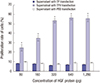 | Fig. 2TPH induced GES-1 cell proliferation.
TPH transfected the GES-1 cells. After incubation at 37°C for 48 hours, cell proliferation was measured by an MTT assay. TPH group: 3 × 10³ cells/well (transfected by TPH strain) in a 96-well plate incubated at 37°C for 48 hours; TP group: 3 × 10³ cells/well (transfected by TP strain) in a 96-well plate incubated at 37°C for 48 hours; control group: 3 × 10³ cells/well (transfected by PBS) in a 96-well plate incubated at 37°C for 48 hours.
GES-1 = gastric epithelial cells, MTT = 2-(4,5-dimethyltriazol-2-yl)-2,5-diphenyl tetrazolium bromide, TPH = attenuated Salmonella typhimurium strain carrying a eukaryotic expression vector encodes the human HGF gene, TP = attenuated Salmonella typhimurium strain with a eukaryotic expression vector, PBS = phosphate buffer solution.
*P < 0.01, TPH group vs. TP and control groups.
|
Angiogenesis activity of HGF on the chick CAM
The supernatant contains 300 pg HGF significantly promoted angiopoiesis in the periphery of a small disk. A brush-like formation of blood vessels was observed in the region of the precapillary arterioles. And the density of blood vessel was significantly higher treated by supernatant from TPH-transfected group than treated by supernatant from TP-transfected group (P < 0.05) (Fig. 3).
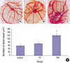 | Fig. 3Angiogenesis activity of the HGF expression product on the chick CAM.
(A) Control group: only a methylcellulose disk on CAM. (B) TP group: a disk with same volume supernatant from TP-transfected supernatant on CAM. (C) TPH group: a disk with the HGF expression product (300 pg) on CAM, from TPH-transfected supernatant. (D) The number of blood vessels was counted under a light microscope, and was found to be significantly fewer in expression supernatant from the TP-transfected and control groups than in the HGF expression product from the TPH-transfected group (P < 0.05).
HGF = hepatocyte growth factor, CAM = chorioallantoic membrane, TP = attenuated Salmonella typhimurium strain with a eukaryotic expression vector, TPH = attenuated Salmonella typhimurium strain carrying a eukaryotic expression vector encodes the human HGF gene.
*P < 0.05, TPH group vs. TP and control groups.
|
Gastric ulcer healing with TPH treatment
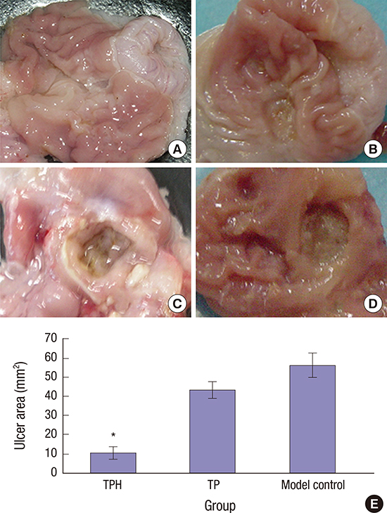
Twenty-one days after ulcer induction, the gastric mucosa of the rats in the TPH-treated group healed more rapidly than the wounds of the rats in the other two groups. In the normal control group, the gastric mucosae were smooth and intact with no ulceration (Fig. 4A). However, enlarged, deepened ulcers with severe adhesions to adjacent tissues were observed in the model control and TP-treated groups; the mucosa underwent necrosis, ulcerations, and had a dark yellowish membrane-like coating. The adjacent mucosa had obvious hyperemia and edema (Fig. 4C and 4D). In contrast, the gastric mucosae of the TPH-treated group were similar to the normal rats. The gastric mucosal lesions were smaller and shallow, and the regenerative granulation tissues were observed in ulcers (Fig. 4B). TPH treatment significantly accelerated ulcer healing. That is, compared with the TP-treated and model control groups, the mean ulcer size in the TPH-treated group was significantly smaller by day 21 after ulcer induction. Numerically, the ulcer area after 21 days of TPH treatment was 10.56 ± 3.30 mm2, which was smaller than the sizes of the TP-treated and model control groups (43.47 ± 4.18 and 56.25 ± 6.38 mm2, respectively) (Fig. 4E).
 | Fig. 4Gross observation of gastric mucosa on day 21 after induced ulcer treatment.
(A) Normal control group: the gastric mucosae from normal rats were smooth and intact with no ulceration. (B) TPH treatment group: treated with (1 × 10⁹) cfu TPH for acetic acid–induced gastric ulcer rats by gavage, the gastric mucosae of the TPH-treatment group were similar to the normal rats. The gastric mucosal lesions were smaller and shallow, and the regenerative granulation tissues were observed in ulcers. (C) Model control group: treated with 0.5-mL1.19M NaHCO3 for acetic acid–induced gastric ulcer rats by gavage. (D) TP treatment group: treated with 1 × 10⁹ cfu TP for acetic acid–induced gastric ulcer rats by gavage. In the model control and TP groups, enlarged, deepened ulcers with severe adhesions to adjacent tissues were observed, the mucosa underwent necrosis, ulcerations, and had a dark yellowish membrane-like coating. The adjacent mucosa had obvious hyperemia and edema. (E) The statistical results of the mean ulcer area size (mm2). The size of the ulcer area in 21 days after TPH treatment was smaller than that in the other two groups.
TPH = attenuated Salmonella typhimurium strain carrying a eukaryotic expression vector encodes the human HGF gene, TP = attenuated Salmonella typhimurium strain with a eukaryotic expression vector.
*P < 0.01, TPH group vs. TP and control groups.
|
Histological assessment
HE-stained sections of the three groups were made and examined separately by two experienced observers. Under a light microscope, clear glands, and mucosa with abundant submucosal vessels were observed in the normal control rats (Fig. 5A), whereas severe mucosal anabrosis and depletion with ulceration were observed in the model control group. The latter group also showed glandular necrosis in the mucosa and submucosa without obvious regeneration signs (Fig. 5E). The reepithelialization of gastric mucosa increased in the TP-treated group (Fig. 5D) than in the model control group; however, no significant histological difference was found between the TP-treated and the model control groups. After TPH treatment, the gastric mucosa significantly healed. Furthermore, increased number of glands and submucosal granulation tissue regeneration were observed in the bases of ulcers along with regenerated capillaries (Fig.5B and 5C).
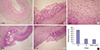 | Fig. 5Histological observations of the gastric mucosa.
(A) Normal control group: Clear glands and intact mucosa with abundant submucosal vessels were observed (H&E stain × 100). (B) TPH treatment group: Increased number of glands and submucosal granulation tissue regeneration were observed in the bases of ulcers along with regenerated capillaries. The gastric mucosa significantly healed (H&E stain × 100). (C) TPH treatment group: Increased number of glands and submucosal granulation tissue regeneration were observed in the bases of ulcers along with regenerated capillaries. The gastric mucosa significantly healed (H&E stain × 400). (D) TP-treated group: Severe mucosal anabrosis and depletion with ulceration were observed. The reepithelialization of gastric mucosa increased in the TP-treated group than in the model control group; however, no significant histological difference was found between the TP and the model control groups (H&E stain × 100). (E) Model control group: Severe mucosal anabrosis and depletion with ulceration were observed (H&E stain × 100). (F) The histological scoring for the crawling length of gastric epithelial cells from three groups. After treatment with TPH, the crawling length of gastric epithelial cells was significantly longer than in the TP and model control groups (P < 0.05). No statistical significance was observed in the mucosal epithelial crawling length between the TP and the model control groups after treatments (P > 0.05).
H&E stain = hematoxylin-eosin stain, TPH = attenuated Salmonella typhimurium strain carrying a eukaryotic expression vector encodes the human HGF gene, TP = attenuated Salmonella typhimurium strain with a eukaryotic expression vector.
*P < 0.01, TPH group vs. TP and control groups.
|
The histological scoring for three groups was summarized in Fig. 6F. After three treatments with TPH, the crawling length of gastric epithelial cells was significantly longer than in the TP-treated and model control groups (P < 0.05). No statistical significance was observed in the mucosal epithelial crawling length between the TP and the model control groups after treatments (P > 0.05).
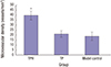 | Fig. 6Quantification of MVD on day 21 in granulation tissue from the ulcer beds by immunoreaction with CD34.
Data are presented as mean ± standard error (n = 50). The MVD in the TPH-treated group increased compared to the TP (a recombinant attenuated Salmonella strain carrying a eukaryotic expression vector)-treated and model control groups, with a significant statistical difference. The MVD in the TP-treated group was not significantly different compared with the model control group.
MVD = microvascular density, TPH = attenuated Salmonella typhimurium strain carrying a eukaryotic expression vector encodes the human HGF gene, TP = attenuated Salmonella typhimurium strain with a eukaryotic expression vector.
*P < 0.01, TPH group vs. TP and control groups.
|
Quantification of MVD in granulation tissue from the ulcer beds by immunoreaction with CD34
As shown in Fig. 6, microvessels were stained brown using the CD34 antibody. The MVD in the TPH-treated group increased compared to the TP-treated and model control groups. That is, the microvessel densities in the ulcer granulation tissues of the TPH-treated rats on day 21 were 39.9 vessels/mm2, which was greater than in the TP-treated and model control rats (21.15 and 18.9 vessels/mm2, respectively), with a significant statistical difference.
HGF and c-Met protein expression
On day 21, the expression of HGF proteins in gastric ulcer was significantly higher in the TPH-treated group than in the TP-treated and model control groups. However, the protein expression of c-Met after 21 days of TPH treatment was not significantly different from the TP-treated and model control groups (Fig. 7).
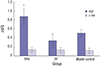 | Fig. 7HGF and c-Met protein expression in gastric mucosa by ELISA.
On day 21, the expression of HGF protein in gastric ulcer was significantly higher in the TPH-treated group than in the TP-treated and model control groups. However, the protein expression of c-Met after 21 days of TPH treatment was not significantly different from the TP-treated and model control groups.
HGF = hepatocyte growth factor, ELISA = enzyme-linked immunosorbent assay, TPH = attenuated Salmonella typhimurium strain carrying a eukaryotic expression vector encodes the human HGF gene, TP = attenuated Salmonella typhimurium strain with a eukaryotic expression vector.
*P < 0.01, TPH group vs. TP and control groups.
|
DISCUSSION
Healing of gastric ulcers occurs through multiple steps, such as formation of granulation tissue, contraction of the ulcerated tissue, angiogenesis, and reepithelialization. Neovasculature and reepithelialization of ulcer wounds play a great role for gastric ulcer healing. In this study, a novel, stable recombinant attenuated TPH was constructed, and the potential effect of the HGF gene preparation was investigated on stimulating the proliferation of GES-1 and CAM angiogenesis in vitro and promoting gastric ulcer healing in rat models. It was found that the TPH could effectively transfer into normal gastric epithelial cells in vitro and highly express interest HGF protein, which could significantly stimulate proliferation of GES-1 and angiogenesis of CAM. In vivo, the results showed that oral administration of TPH, at a dose of 1 × 10⁹ cfu for three times, promoted the crawling of gastric epithelial and accelerated the healing of the gastric mucosa in the acetic acid–induced gastric ulcer rat model.
TPH was constructed as a therapeutic agent for treating gastric ulcer, in which HGF gene was combined with attenuated S. typhimurium vector. The reasons for the combination include the following: 1) HGF is a multifunctional cytokine, which could stimulate gastric epithelium proliferation, inhibit cell apoptosis, and improve local angiogenesis (17). Early in 1984, HGF from the serum of hepatectomized rats was partially purified and described by Nakamura et al. (18). The HGF receptor encoded by the c-met proto-oncogene is a member of the cell surface receptors. As a kind of cytokine, the HGF possesses wide biological activities, including regeneration, anti-fibrosis, cytoprotection, and differentiation (19). Studies in vitro and in vivo have demonstrated that HGF binds to an upregulated c-met/HGF receptor on epithelial cells in the gastric ulcer margin to enhance cell scattering and migration and to stimulate cell proliferation. The re-epithelization of wounds plays a key role for gastric ulcer healing and in vivo healing of gastric ulcer mucosa. In this study, the crawling length of gastric epithelial cells after three treatments with TPH was significantly longer than in the TP-treated and model control groups. No statistical significance was observed in mucosal epithelial crawling length between the TP-treated and the model control groups after treatments. So it could result in promoting gastric ulcer healing (2). Considering some disadvantages for the administration of HGF protein in treating gastric ulcer, a new treatment mode of gene therapy was adopted, in which the HGF gene eukaryotic expression vector was used. Finally, gene transfer is the most critical technology in gene therapy, in which the gene delivery system is one of the key points whether gene therapy can be applied in clinic. According to the characteristics of gastric application, a new delivery system was described in this study, an attenuated S. typhimurium, which was proved to be orally used to deliver a eukaryotic expression plasmid to gastric mucosa (20). In the current study, the roles of TPH in vitro and in vivo were investigated to prove the construction idea was good.
The results of this study showed that TPH had high gene transfection efficiency to gastric epithelial cells and highly expressed interest HGF protein in vitro, which could significantly stimulate proliferation of gastric epithelial cells and angiogenesis of CAM. The proliferation of gastric epithelial cells was the base for gastric ulcer healing. The CAM of the chicken embryo was formed when the chorion and allantois fuse after around 100 hours of incubation. By day 12, the CAM extended to line the entire surface of the inner shell membrane. The expansion of the CAM capillary network was accompanied by an augmentation in chorioallantoic artery blood flow, which increased about tenfold during the period from day 9 to day 16 (21). CAM is a suitable model for studying vascular effects; it is probably the most widely used in vivo assay for studying angiogenesis. This model has several advantages over other animal models, including the fact that the CAM consists of a thin, planar vascular network, which is well adapted to observation by microscopy (2223). Furthermore, the CAM provides an easily accessible neovascular network in a transparent matrix. It allows direct visualization of blood vessels before, during, and after pharmacological intervention, and use in clinical trials is already the Food and Drug Administration (FDA) approved (24). The results of this study showed that the promoting effect on angiopoiesis was obviously observed in the periphery of the small plate in which supernatant from the TPH-transfected group was added, but the dosage effect was not obvious. The density of blood vessel was significantly fewer in HGF expression supernatant from the TP-transfected group than in the HGF expression product from the TPH-transfected group.
Since introduced in 1969 by Takagi et al. (25), the acetic acid–induced gastric ulcer model has proved useful for investigating the pathophysiology of gastric ulcer disease and the efficacy of antiulcer drugs (26). The reasons for the usefulness of this model include the following. First, the ulcer induction procedure is simple, readily resulting in ulcers of consistent size and severity with a 100% success rate. Second, the acetic acid–induced ulcers resemble human peptic ulcers both macroscopically and histologically. Indeed, the spontaneous relapse of healed ulcers is frequently observed, just as in patients with peptic ulcer disease. Finally, the ulcers respond well to various anti-ulcer drugs, such as PPI, sucralfate, and several growth factors.
Utilizing this model, the effectiveness of TPH on the healing of this type of experimental ulcer in the rat was investigated, and it was found that orally administering TPH, at a dose of 1 × 10⁹ cfu for three times, accelerated the healing of the gastric mucosa in the rat model. HGF is a multifunctional cytokine. Because the direct use of the HGF protein is limited by a variety of factors, TPH was used as a therapeutic agent for treating gastric ulcer. The attenuated S. typhimurium has a high gene transfection efficiency and reliable tropism for the intestinal mucosa. On day 21, the expression of HGF proteins in gastric ulcer was significantly higher for TPH-treated group than the TP-treated and model control groups. The attenuated S. typhimurium is also a natural immune agent that triggers an immune response after hosting in the gastrointestinal tract for a certain period of time. The results of this study also showed that the oral administration of TPH significantly improved the symptoms in the gastric ulcer rat models that were induced by acetic acid. Compared to the control groups, mucosal lesions healed and body weight increased quickly. Histologically, the ulceration size, inflammatory cell infiltration, and lesion depth were all reduced.
The regrowth of blood vessels into the ulcerated area, that is, angiogenesis, plays a pivotal role in accelerating the healing of ulcers since the neovasculature improves the local blood supply and promotes nutrient supply to the healing tissue. Angiogenesis was quantified in this study by antibody CD34 and expressed through MVD. MVD assessment is the most commonly used technique to quantify angiogenesis. An antibody against CD34 was used as a marker, staining mainly the vessels. This marker increased in the TPH-treated group than in the TP-treated and model control groups. The results showed that the mean value of MVD, quantified with anti-CD34, was higher in the TPH-treated group than in the TP-treated or model control groups.
In conclusion, the results of this study suggest that TPH accelerates the healing of acetic acid–induced gastric ulcers in rats by enhancing angiogenesis and reepithelization of wounds. As an oral agent and reliable tropism for the gastric mucosa, the TPH can be used as a therapeutic agent for treating gastric ulcer. These findings suggest that a novel, feasible, and effective modality was developed for gastric ulcer gene therapy. TPH might become an alternative treatment of gastric ulcer disease in humans sometime in the near future.
 XML Download