

 PDF
PDF ePub
ePub Citation
 Print
Print


INTRODUCTION
Total hip arthroplasty (THA) is an effective intervention for patients with severe hip disease and has been shown to lead to marked improvement in health outcomes (1). Because of the significant benefits realized with THA, utilization rates for this procedure have been increasing in most Western and Asian countries, including Korea (23). Thus, many Western epidemiological studies have examined the incidence of THA and reported osteoarthritis (OA) as the most common underlying condition for THA in Western countries (2345). However, Western epidemiological data cannot be applied to Korea, and most of these studies only determined the prevalence and trends of THA or were limited to THA for patients with OA. The pathogenesis of coxarthrosis leading to THA is complex, with many risk factors (67). Many conditions that can lead to THA include primary OA, secondary OA resulting from femoroacetabular impingement (FAI), dysplasia and peripheral joint involvement of seronegative spondylitis, such as ankylosing spondylitis (AS) and diffuse idiopathic skeletal hyperostosis, avascular necrosis (AVN), and fractures. Although the ratios of primary OA have previously been considered high, morphological deformities around the hip may cause OA over several decades (89). Recently, several studies have reported that FAI results in early OA and hip OA pathology related to FAI (101112). To our knowledge, no previous study has evaluated the epidemiology of causes leading to THA in Koreans, particularly with a focus on the incidence of FAI.
The purposes of the present study were 1) to analyze the causes leading to THA, 2) to clarify the incidence of FAI among these causes, and 3) to compare the incidence in Korea with those in other countries.
MATERIALS AND METHODS
Data selection
From January 2000 to December 2014, 1,206 hips of 818 patients who underwent primary THA at our institute were reviewed retrospectively in terms of radiographs and electronic charts. Clinical records, surgical records, and preoperative simple radiographs were evaluated to determine the causes leading to THA. The radiographs and radiographic parameters were reviewed and measured by 2 of the authors, who are orthopedic surgeons. The radiographs were also reviewed again, 4 weeks later, by the same observers. Exclusion criteria were as follows: 1) absent or insufficient radiographs or electronic charts to determine the cause, 2) revision of THA, and 3) conversion from hemiarthroplasty to THA.
Patients were categorized according to the causes leading to THA: primary OA, rheumatoid arthritis (RA), posttraumatic arthritis, post infectious arthritis, AVN of the femoral head, fracture of the femoral head or neck, AS, developmental dysplasia of the hip (DDH), Legg-Calvé-Perthes disease (LCPD), FAI, and other. The FAI group was subcategorized into cam, pincer, and mixed types.
Primary OA cases were defined as those with no systemic disease, no history of hip disease, and no remarkable deformity of the proximal femur. Atlas photographs illustrating the individual radiographic features of OA were used to standardize the readings (1314). The following radiographic parameters were also applied to determine the primary OA cases: center-edge angle > 20°, sharp angle < 45°, and acetabular roof obliquity < 15° (15).
FAI was defined by the obvious presence of a bony prominence in the anterolateral head-neck junction (cam type) and/or an acetabular abnormality, such as retroversion, coxa profunda, or acetabular protrusion (pincer type) (15). The alpha angle was considered to have been increased if > 50°, and anterior femur offset was considered to have been decreased if < 8 mm; these were classified as cam type (16). Acetabular retroversion is a form of hip dysplasia in which the alignment of the acetabulum does not face the normal anterolateral direction but is inclined more posterolaterally (17). In simple radiography, crossover of the anterior wall of acetabulum over the posterior wall (crossover sign, figure-8 sign) was considered to be acetabular retroversion, the adjoining of the acetabular fossa and ilioischial line to be coxa profunda, and the femur head passing the ilioischial to be protrusion acetabuli (18). These were classified as the pincer type. Cases having both types were classified as the mixed type. ‘Others’ were defined as cases that could not be categorized or cases with very small numbers, such as synovial chondromatosis and neurofibromatosis.
Statistical analysis
Two blinded reviewers reviewed the radiographs independently; there was no communication between the reviewers. Radiographs were presented to the reviewers 2 times in random order at intervals of 3 weeks. Intraobserver and interobserver reliabilities of the 5 measured radiological parameters (center-edge angle, sharp angle, acetabular roof obliquity, alpha angle, anterior femur offset) were assessed using intraclass correlation coefficients. And intraobserver and interobserver reliabilities of the causes leading to primary THA were assessed using Kappa coefficient. The SPSS software (ver. 19.0; SPSS Korea, Seoul, Korea) was used for statistical analysis.
RESULTS
In total, 1,206 hips in 818 patients underwent THA at out institute between January 2000 and December 2014. There were 477 male and 341 female patients. The numbers of cases undergoing THA has increased consistently since 2000 (Fig. 1). The changes of causes leading to THA showed that AVN has been decreased since the interval between 2003 and 2005 and fracture has been increased (Fig. 2).
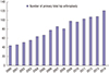 | Fig. 1The graph shows that the procedural number of total hip arthroplasties from 2000 to 2014 increased consistently.
|
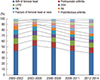 | Fig. 2The graph shows the changes of causes leading to THA.
THA = total hip arthroplasty, AVN = avascular necrosis, LCPD = Legg-Calvé-Perthes disease, DDH = developmental dysplasia of the hip, FAI = femoroacetabular impingement, RA = rheumatoid arthritis.
|
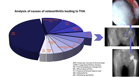
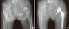
There were 32 patients (3.91%; 13 males, 19 females) in the primary OA group (Fig. 3), 41 (5.01%; 12 males, 29 females) in the RA group, 84 (10.27%; 43 males, 41 females) in the posttraumatic arthritis group, 39 (4.77%; 17 males, 22 females) in the post infectious arthritis group, 365 (44.62%; 270 males, 95 females) in the AVN group, 39 (4.77%; 17 males, 22 females) in the fracture group, 21 (2.57%; 16 males, 5 females) in the AS group, 52 (6.36%; 16 males, 36 females) in the DDH group, 71 (8.68%; 52 males, 19 females) in the LCPD group, 52 (6.36%; 13 males, 39 females) in the FAI group, and 22 (2.69%; 8 males, 14 females) in the ‘others’ group. In the FAI group, there were 36 patients (69.23%; 9 males, 27 females) of the cam type (Fig. 4), 15 (28.85%; 3 males, 12 females) of the pincer type (Fig. 5), and 1 (1.92%; 1 male) of the mixed type (Table 1).
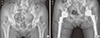 | Fig. 3Anteroposterior radiographs illustrate the case of a 62-year-old female patient who had primary OA of both hips. (A) The radiograph was taken before THA. Lateral center-edge angle, sharp angle, and acetabular roof obliquity of right hip was 37.2°, 35.8°, and 8.6°, respectively. Left hip was 43.5°, 35.2°, and 7.0°, respectively. (B) The radiograph was taken after THA to treat primary OA.
THA = total hip arthroplasty, OA = osteoarthritis.
|
 | Fig. 4Anteroposterior radiographs illustrate the case of an 85-year-old female patient who had secondary OA caused by Cam type FAI of left hip. (A) Preoperative radiograph shows typical pistol grip deformity on left hip. (B) The radiograph was taken after THA.
THA = total hip arthroplasty, FAI = femoroacetabular impingement, OA = osteoarthritis.
|
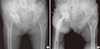 | Fig. 5Anteroposterior radiographs illustrate the case of a 76-year-old female patient who had secondary OA caused by Pincer type FAI of left hip. (A) Preoperative radiograph shows typical acetabular protrusion on right hip. (B) The radiograph was taken after THA.
THA = total hip arthroplasty, FAI = femoroacetabular impingement, OA = osteoarthritis.
|
Table 1
Causes leading to primary THA
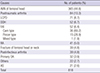
THA = total hip arthroplasty, AVN = avascular necrosis, LCPD = Legg-Calvé-Perthes disease, DDH = developmental dysplasia of the hip, FAI = femoroacetabular impingement, RA = rheumatoid arthritis, OA = osteoarthritis, AS = ankylosing spondylitis.
![]()
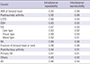
Intraobserver and interobserver correlations for causes and the combination of all measurements were found to be reproducible and reliable among observers (Tables 2 and 3).
Table 2
Intraclass intraobserver and intraobserver reliabilities of radiographic parameters
![]()
Table 3
Intraobserver and intraobserver reliabilities of the causes leading to primary THA
THA = total hip arthroplasty, AVN = avascular necrosis, LCPD = Legg-Calvé-Perthes disease, DDH = developmental dysplasia of the hip, FAI = femoroacetabular impingement, RA = rheumatoid arthritis, OA = osteoarthritis, AS = ankylosing spondylitis.
![]()
DISCUSSION
Primary THA has increased globally, including Korea. Yoon et al. (3) reported that both the number and the rate of primary THAs increased substantially between 2007 and 2011 in Korea: 7,229 hips in 2007, 7,829 hips in 2008, 8,124 hips in 2009, 8,652 hips in 2010 and 8,926 in 2011 underwent primary THAs. Our study showed similar results between 2000 and 2014 in our institution. The most common cause will be OA because OA is a leading cause of disability (2345). The 2 main categories of hip OA are primary and secondary. Although the precise proportion of each category remains controversial, primary hip OA is commonly believed to account for the majority of all hip OA cases in Western populations (19). Western epidemiological studies showed that more than 75% of primary THAs have been performed for OA (20). In the United States, hip OA affects approximately 5% of those over the age of 60 years, and approximately 360,000 hip replacement surgeries are performed annually (21). However, Korean epidemiological studies have reported that primary OA accounted for only 18% of THA cases in Korea, and the most common indication for primary THA in Korea was AVN (5), similar to our findings. This result suggests that severe hip OA is much rarer in Korea than in Western countries. This discrepancy between the Western and Korean incidence of hip OA and primary THA may be attributed partially to lifestyle factors, such as frequent kneeling or squatting, which are common in daily living in Asia and may protect against hip OA (5).
Many studies have reported that FAI could be a cause of cartilage or acetabular labrum lesions, ultimately resulting in degenerative arthritis (911). It has also become clear that so‑called primary OA of the hip is often FAI-positive. Harris (8) reported that most cases of primary OA of the hip had mild dysplasia and/or a pistol-grip deformity, with true primary OA being extremely rare. Tanzer and Noiseux (22) reported that the etiology of OA in 125 of 200 consecutive patients undergoing THA was idiopathic arthritis, but all 125 patients also had a pistol-grip deformity. Takeyama et al. (15) reported that 6 (0.6%) and 693 (73.3%) of 946 hips that underwent primary surgery for OA or other diseases of the hip were associated with FAI and DDH in Japan, respectively, and only 11 (1.2%) hips were classified as primary OA of unknown etiology. The exact reason as to why the incidence of FAI is low in Japan remains unknown, but it was suggested that the normal Japanese acetabulum may be more dysplastic than that of Caucasians, and the antero-posterior size of the proximal femur was smaller in Japanese subjects, decreasing impingement between the acetabulum and femur (15). In this study, 52 (6.36%) of 818 patients who underwent primary THA were associated with FAI and 52 (6.36%) of the patients were associated with DDH in Korea. Only 32 (3.91%) patients were associated with primary OA. The rate of FAI in Korea was higher than that in Japan. These results may be due to lifestyle. It has been suggested, for example, that frequent squatting or a crossed-legged position, particularly on the floor, common to daily life in Korea, may increase impingement.
This study has some limitations. First, there was patient bias due to geographical or institutional predominance, potentially influencing the epidemiological results. Second, this was a single center study. Thus, the sample size was small for a cohort study. However, to our knowledge, no reported study has evaluated the epidemiology of the causes leading to THA in Koreans, particularly, with a focus on the incidence of FAI.
In conclusion, the causes leading to THA in Korea differ from those in Western countries. In particular, the incidence of primary OA as a factor leading to THA in Korea is much lower than that in Western countries, whereas the incidence of AVN in Korea is much higher than that in Western countries. Also, FAI could be cause of severe secondary OA that requires THA in Korea, therefore symptomatic FAI should not be neglected.
 XML Download