Abstract
Bone marrow necrosis (BMN) is a pathologic state which is derived from various disease entities. Most commonly, it is accompanied by hematologic malignancies such as acute leukemia. The patients with marrow necrosis are generally known to have dismal prognoses but variations exist according to early diagnosis. Here we report a case of BMN in an acute lymphoblastic leukemia patient with Philadelphia chromosome at presentation.
Go to : 
REFERENCES
1.Wool GD., Deucher A. Bone marrow necrosis: ten-year retrospective review of bone marrow biopsy specimens. Am J Clin Pathol. 2015. 143:201–13.
2.Swerdlow SH., Campo E, et al. eds. WHO classifcation of tumours of haematopoietic and lymphoid tissues. 4th ed.Lyon: IARC Press;2017. p. 203–4.
3.Badar T., Shetty A., Bueso-Ramos C., Cortes J., Konopleva M., Borthakur G, et al. Bone marrow necrosis in acute leukemia: clinical characteristic and outcome. Am J Hematol. 2015. 90:769–73.

4.Kwong YL., Pollock A., Wei D., Lie AK. Philadelphia chromosome positive acute lymphoblastic leukemia masquerading as persistent asymptomatic bone marrow necrosis. Pathology. 1994. 26:183–5.

5.Petrella T., Bailly F., Mugneret F., Caillot D., Chavanet P., Guy H, et al. Bone marrow necrosis and human parvovirus associated infection preceding an Phl+ acute lymphoblastic leukemia. Leuk Lymphoma. 1992. 8:415–9.
6.Lin RF., Li JY., Lu H., Wu YJ., Qiu HR., Xiao B, et al. Bone marrow necrosis as an initial manifestation of Philadelphia chromosome and myeloid antigens positive B acute lymphoblastic leukemia—a case report. Zhong-guo Shi Yan Xue Ye Xue Za Zhi. 2006. 14:832–4.
7.Matsue K., Takeuchi M., Koseki M., Uryu H. Bone marrow necrosis associated with the use of imatinib mesylate in a patient with Philadelphia chromosome-positive acute lymphoblastic leukemia. Ann Hema-tol. 2006. 85:542–4.

8.Wade LJ., Stevenson LD. Necrosis of bone marrow with fat embolism in sickle cell anemia. Am J Path. 1941. 17:47–54.
9.Argon D., Çetiner M., Adıgüzel C., Kaygusuz I., Tuğlular TF., Tecimer T, et al. Bone marrow necrosis in a patient with non-Hodgkin lymphoma. Turk J Haematol. 2004. 21:97–100.
10.Foucar K., Reichard K., Czuchlewski D. Bone marrow pathology. 3rd ed.Chicago: American Society for Clinical Pathology;2010. p. 657–9.
11.Maisel D., Lim JY., Pollock WJ., Yatani R., Liu PI. Bone marrow necrosis: an entity often overlooked. Ann Clin Lab Sci. 1988. 18:109–15.
Go to : 
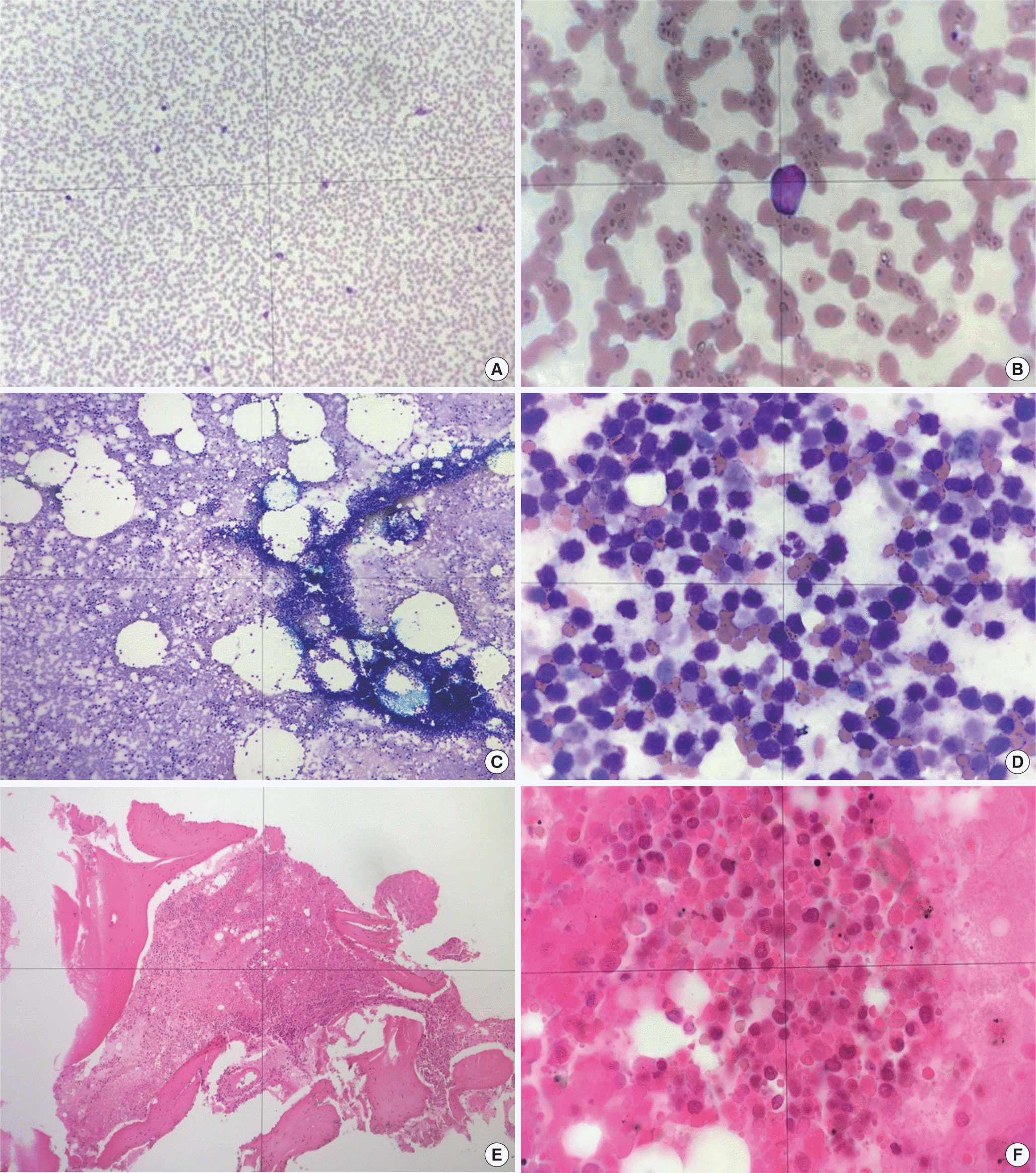
 | Fig. 1.Peripheral blood smear showing atypical lymphocytes and a few blast cells (Wright-Giemsa stain, ×200, ×1,000) (A, B). Bone marrow aspirate smear showing hypercellular marrow and coagulative necrosis (Wright-Giemsa stain, ×200) and magnified view of mostly ghost cells and one hematopoietic granulocyte (Wright-Giemsa stain, ×1,000) (C, D). Bone marrow biopsy showing almost no normal hematopoietic elements and only a few fat cells (Hematoxin and eosin stain, ×200) and magnified view of mostly amorphous, eosinophilic substance on bone marrow biopsy (Hematoxylin and eosin stain, ×1,000) (E, F). |
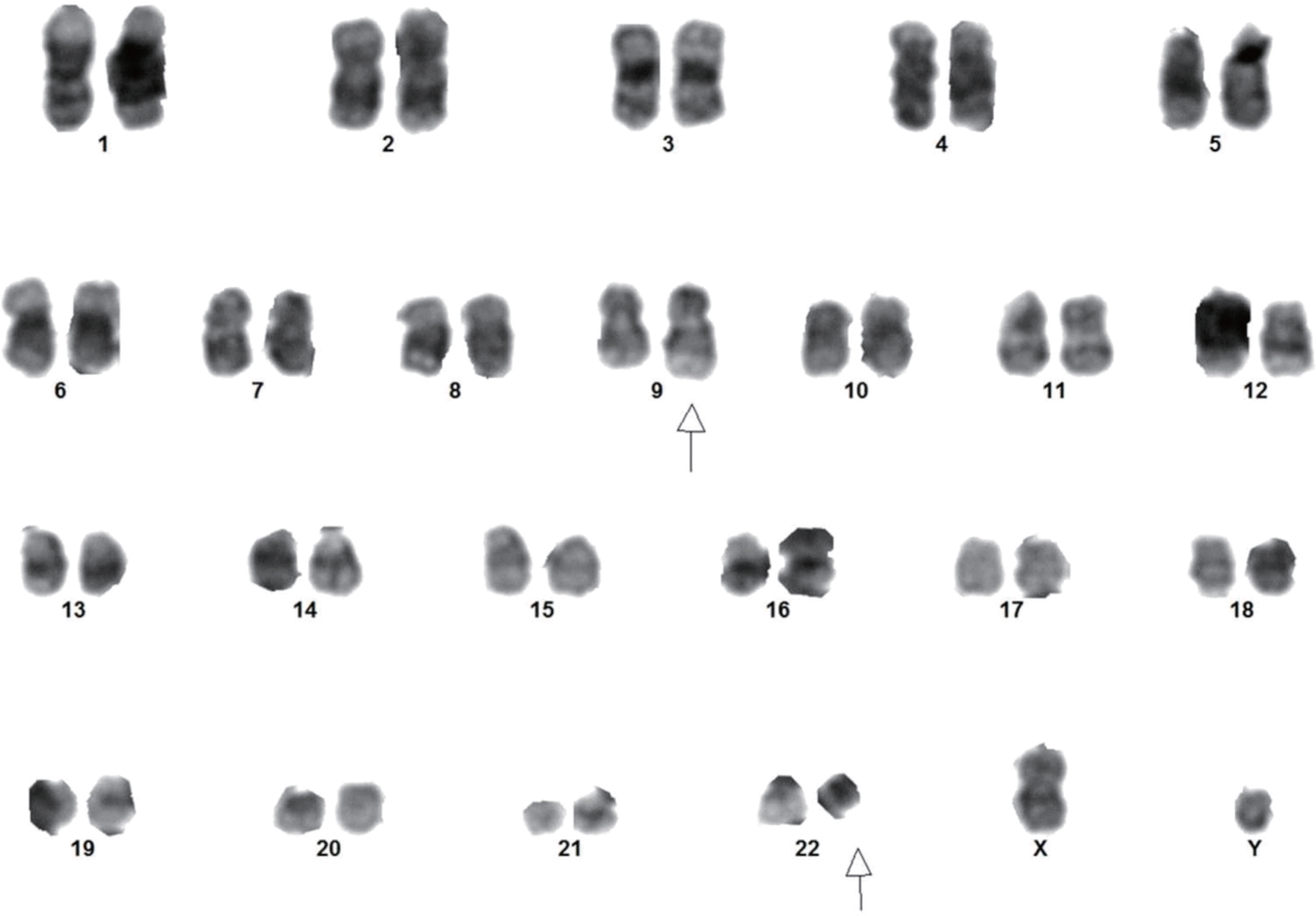
 | Fig. 2.Conventional karyotyping shows a 46,XY,t(9;22)(q34.1;q11.2)[13]/46,XY[7] (Giemsa-Leishman-Trypsin banding). |
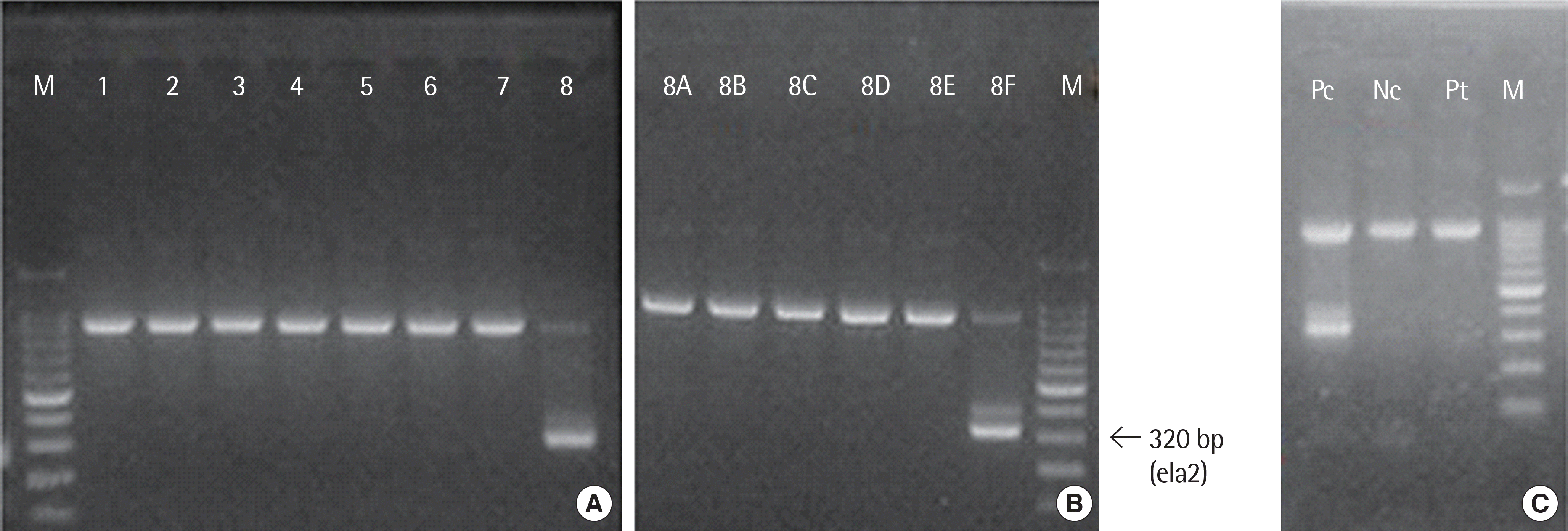
 | Fig. 3.(A) Reverse transcription polymerase chain reaction (RT-PCR) results showing band on eighth lane. (B) Split-out image showing bright band on 8F lane, corresponding to BCL-ABL1 e1a2 fusion transcript (320 base pairs). (C) Follow-up RT-PCR showing no band on third lane. Abbreviations: M, nucleic acid marker ladder; Pc, positive control; Nc, negative control; Pt, patient. |
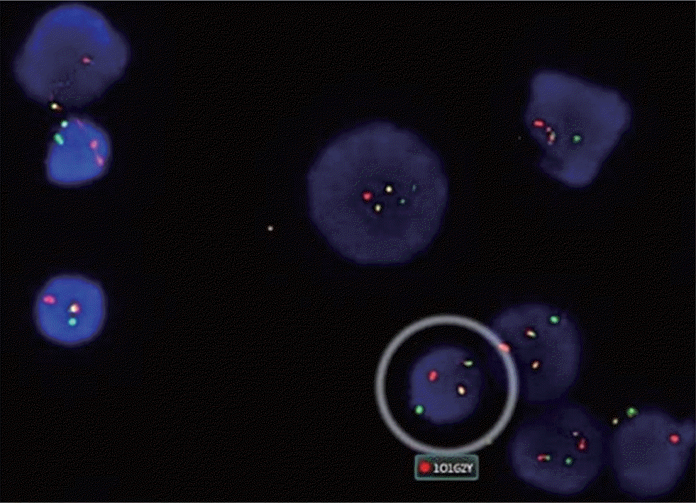
 | Fig. 4.Fluorescent in situ Hybridization study showing dual-color and dual-fusion translocation probes for ABL1 (red) and BCR (green). The circled cell shows one red signal, one green signal, and two yellow signals, respectively. |
Table 1.
Literature of Philadelphia chromosome positive acute lymphoblastic leukemia presented with bone marrow necrosis


 PDF
PDF ePub
ePub Citation
 Print
Print


 XML Download