Journal List > J Korean Med Sci > v.32(7) > 1108498

Seo, Youn, Kim, Jun, Park, Yeom, Lim, Woo, Youn, Ko, Park, Baik, Lee, Cho, and Rhee: Helicobacter pylori Antigens Inducing Early Immune Response in Infants

Abstract

To identify the Helicobacter pylori antigens operating during early infection in sera from infected infants using proteomics and immunoblot analysis. Two-dimensional (2D) large and small gel electrophoresis was performed using H. pylori strain 51. We performed 2D immunoglobulin G (IgG), immunoglobulin A (IgA), and immunoglobulin M (IgM) antibody immunoblotting using small gels on sera collected at the Gyeongsang National University Hospital from 4–11-month-old infants confirmed with H. pylori infection by pre-embedding immunoelectron microscopy. Immunoblot spots appearing to represent early infection markers in infant sera were compared to those of the large 2D gel for H. pylori strain 51. Corresponding spots were analyzed by matrix-assisted laser desorption/ionization time of flight-mass spectrometry (MALDI-TOF-MS). The peptide fingerprints obtained were searched in the National Center for Biotechnology Information (NCBI) database. Eight infant patients were confirmed with H. pylori infection based on urease tests, histopathologic examinations, and pre-embedding immunoelectron microscopy. One infant showed a 2D IgM immunoblot pattern that seemed to represent early infection. Immunoblot spots were compared with those from whole-cell extracts of H. pylori strain 51 and 18 spots were excised, digested in gel, and analyzed by MALDI-TOF-MS. Of the 10 peptide fingerprints obtained, the H. pylori proteins flagellin A (FlaA), urease β subunit (UreB), pyruvate ferredoxin oxidoreductase (POR), and translation elongation factor Ts (EF-Ts) were identified and appeared to be active during the early infection periods. These results might aid identification of serological markers for the serodiagnosis of early H. pylori infection in infants.

Graphical Abstract

jkms-32-1139-abf001

INTRODUCTION

Helicobacter pylori is an important pathogen that causes many gastric diseases such as gastirits, gastroduodenal ulcers, and gastric cancers (1). The anti-H. pylori immunoglobulin G (IgG) seropositivity rate was lowest in infants aged 7–9 months among populations from neonates to 60–69 years and increased with age in Korea (2); the most primary H. pylori infections occur in early childhood (23). The clinical diagnosis of early H. pylori infection is difficult due to the nonspecific presentations of H. pylori infection such as abdominal pain and vomiting (4), and young children cannot explain the symptoms.
Early diagnosis of H. pylori infection in young children could be helpful for investigating the disease-associated pathogenesis, and identification of the proteins that induce the early human immune response would enable early diagnosis. The primary immune response involves immunoglobulin M (IgM) antibody, and secondary exposure to the same antigen produces IgG, immunoglobulin A (IgA), and/or IgE antibodies. After H. pylori infection, IgM antibodies against early antigens of H. pylori are formed and detected transiently in the acute period. Although IgM-based serological diagnosis of H. pylori infection was observed in early infants (5), most serological tests for H. pylori infection in children are based on IgG or IgA antibodies (267). However, the sensitivity of commercial enzyme immunoassays (EIAs) was low (22.0%–33.3%) in children aged 0–6 years and varied by commercial EIA (6). This low sensitivity of commercial EIAs suggested that young children might have a different immune response to H. pylori (6). In contrast, commercial EIAs testing IgG and IgA were suitable for serodiagnosis of H. pylori infection in children aged 1–14 years, and the most recognized bands were under 40 kDa (5).
One-dimensional (1D) immunoblots show only the molecular weight (MW) of antigenic proteins that react with IgG or IgA antibodies in sera. Anti-CagA positivity on 1D immumoblots correlated with the high anti-H. pylori IgG/IgA titers in the sera of H. pylori-positive children even younger than 5 years (9). In contrast, two-dimensional (2D) polyacrylamide gel electrophoresis (PAGE) separates proteins according to both isoelectric point and MW, thereby providing superior resolution of individual proteins from a complex mixture. 2D immunoblots identified CagA as the most reactive antigen recognized by all the H. pylori-positive sera (IgG) even in children less than 3 years old (8). Furthermore, protein excision from 2D gels often allows their analysis and identification through techniques such as peptide mass fingerprinting (10).
To our knowledge, no previous study investigated anti-H. pylori IgM positivity in the sera of H. pylori-positive infants. In the present study, we identified the operating antigens of H. pylori during early infection in infants by 2D-immunoblot analysis and analyzed the proteomics of H. pylori antigens that reacted with IgM antibodies in sera from H. pylori-infected infants.

MATERIALS AND METHODS

Subjects

From 1999 to 2005, infants diagnosed with H. pylori infection using histopathologic findings or urease tests and whose serum samples and gastric biopsy specimens were preserved in the Gyeongsang National University Hospital Biobank of Korea. H. pylori infection was confirmed by immunohistochemical detection using a polyclonal antibody and immunoelectron microscopic examination (Immuno-EM) of preserved gastric biopsy specimens as reported previously (11).

Bacterial strains

The strains were isolated in previous study (12) and deposited in the H. pylori Korean Type Culture Collection (HpKTCC, http://hpktcc.knrrc.or.kr/, Gyeongsang National University School of Medicine). Frozen H. pylori strain 51 was thawed and grown on Brucella agar plates containing 10% bovine serum, vancomycin (10 μg/mL), nalidixic acid (25 μg/mL), and amphotericin B (1 μg/mL) at 37 °C under 10% CO2 and 100% humidity. After an 18-h subculture, bacterial masses were collected to prepare protein specimens for 2D electrophoresis (2DE) analysis.

2DE and image analysis

1DE and 2DE samples were prepared as described previously (13). After washing with 40 mmol/L Tris-hydrochloride (HCl) (pH 7.2) and 1 mmol/L ethylenediaminetetraacetic acid (EDTA), bacterial pellets were lysed using a buffer containing 9.5 mol/L urea, 4% 3-((3-cholamidopropyl)dimethylammonium)-1-propanesulfonate (CHAPS), and 35 mmol/L Tris-HCl (pH 7.2). The solubilized protein samples (30 μg) were mixed with the rehydration solution containing 8 mol/L urea, 4% CHAPS, 10 mmol/L dithiothreitol (DTT), and 0.2% carrier ampholytes (pH 3.0–10.0) to a final volume of 125 μL and applied to immobilized pH gradient (IPG) strips (7 cm; Bio-Rad, Hercules, CA, USA) of pH 3.0–10.0 in a re-swelling tray (Bio-Rad). After the IPG strips were rehydrated, isoelectric focusing (IEF) was performed using a Protein IEF Cell (Bio-Rad), and 3 preset programs consisting of the first conditioning step (15 minutes, 250 V), the linear voltage ramping step (3 hours, 4,000 V), and the maximum voltage ramping step of up to 30,000 Vh. The current did not exceed 50 μA per strip. Following IEF, the strips were equilibrated with 0.375 mol/L Tris buffer (pH 8.8) containing 6 mol/L urea, 2% sodium dodecyl sulfate (SDS), 20% glycerol, 2% DTT, and 0.01% bromophenol blue. The strips were equilibrated again with the same buffer supplemented with 2.5% iodoacetamide. The 2D SDS-PAGE was carried out overnight at 20 mA per gel using a 12.5% separating polyacrylamide gel (8–10 cm) without a stacking gel. The resolved protein spots on the gels were visualized by silver staining (14) and scanned using a Fluor-S MultiImager (Bio-Rad). The spot intensities of each sample were determined and analyzed using PDQUEST 2D Gel Analysis Software Version 6 (Bio-Rad) installed on a Magic Station M5660 (Samsung, Seoul, Korea).

Immunoblot analysis

Protein bands were electrophoretically transferred from the SDS-PAGE gel onto a 0.2-μm nitrocellulose membrane using a semi-dry transblot (Bio-Rad) at a constant current of 120 mA for 60 minutes. After the transfer, unbound sites on the nitrocellulose membrane were blocked for 1 hour at room temperature with blocking solution (1% bovine serum albumin, BSA-TBST solution; Tribioscience Inc., Palo Alto, CA, USA) and washed 3 times in TBS with 0.05% Tween 20 (Alfa Aesar, Haverhill, MA, USA) for 5 minutes. Thereafter, the membrane was cut into strips, and each strip was incubated with individual serum samples diluted five-fold.
Immunodetection was performed with alkaline-conjugated monoclonal rabbit anti-human IgG (1:10,000), IgA (1:500), and IgM (1:500) antibodies for 30 minutes (Bethyl Laboratories Inc., Montgomery, TX, USA). Substrate development was performed using alkaline phosphatase buffer, 0.03% nitroblue tetrazolium (Thermo Fisher Scientific Inc., Rockford, IL, USA) and 0.015% 5-bromo-4-chloro-3-indolyl phosphate (BCIP) substrate (Thermo Fisher Scientific Inc.). The MW of antigenic proteins was measured using a protein standard (Bio-Rad).

Image analysis and in-gel protein digestion

A gel image was obtained by scanning the silver-stained gels with a Fluor-S Multimager (Bio-Rad) and documented with the PDQUEST 2-D Gel Analysis Software Version 6 (Bio-Rad) installed on the Magic Station M5660 (Samsung, Korea). Silver-stained spots were excised from 2DE gels and transferred into Eppendorf tubes. The chemical reducers mixture was prepared fresh with a 1:1 ratio of 30 mmol/L potassium ferricyanide and 100 mmol/L sodium thiosulfate. One hundred microliters of the mixture was added to the Eppendorf tube and occasionally vortexed until the brownish color disappeared. After the gel pieces were rinsed three times with distilled water (DW) to stop the reaction, 500 μL 200 mmol/L ammonium bicarbonate was added to cover the gel for 20 minutes and then discarded. The gel piece was dehydrated with 100 μL acetonitrile and dried in a vacuum centrifuge. In-gel digestion was carried out using the method described by O'Connell and Stults (14). Gel pieces containing protein spots were rehydrated by adding a digestion buffer on ice. The buffer containing 12.5 ng/mL trypsin was added to cover the pieces and incubated for 45 minutes on ice. The enzyme solution was removed and replaced with 20 μL buffer without enzyme, keeping the gel pieces wet during enzymatic cleavage overnight at 37°C, and the gel pieces were subjected to vigorous vortexing for 30 minutes. Twenty microliters digested solution was transferred into a clean Eppendorf tube and dried in a vacuum, and the resulting samples were dissolved in 2 μL 0.1% trifluoroacetic acid (TFA).

Peptide mass fingerprinting

A matrix solution composed of α-cyano-4-hydroxycinamic acid (40 mg/mL) in 50% acetonitrile and 0.1% TFA was prepared for peptide mass fingerprinting. The matrix solution and sample solution were mixed at a 1:1 ratio, and 2 µL was applied onto the matrix-assisted laser desorption/ionization time of flight (MALDI-TOF)/TOF target plate, rapidly dried, and washed using deionized water. The mixture solution was dried for 10 minutes at room temperature and subjected to MALDI-TOF-mass spectrometry (MS) and MS/MS analysis using an ABI 4800 Plus TOF-TOF Mass Spectrometer (Applied Biosystems, Framingham, MA, USA). The instrument was set at 200-Hz Nd:355-nm YAG laser operation. Peaks with signal/noise ratios > 25 were selected, and the 10 most intense ions were used for MS/MS analysis in 1 kV mode, 1,000–1,250 consecutive laser exposures.
All MALDI-TOF-MS spectra were searched against the National Center for Biotechnology Information (NCBI) protein database (version 20140415; 2,845 sequences, 921,323 residues) of Helicobacter 51 proteins with a molecular mass range ± 15% of that estimated from 2DE and allowing a peptide mass accuracy of 50 ppm. If matched proteins were absent, the molecular mass window was extended. MS/MS spectral data were analyzed using Protein Pilot V.3.0 database software (with the MASCOT V.2.3.02 database search engine) at 50 ppm mass tolerance. Pyroglutamic acid modification of N-terminal glutamine, oxidation of methionine, and acrylamide modification of cysteine were considered. Individual peptide ion scores were searched using a statistically significant threshold value of P = 0.050.

Ethics statement

All of the serum and gastric biopsy specimens in paraffin blocks that we experimented had been prepared and donated by the Gyeongsang National University Hospital, a member of the Korea Biobank Network after its Institutional Review Board reviewed the research protocols (GNUHIRB-2014-09-024). The board exempted informed consent because of its retrospective nature of research.

RESULTS

Subjects


Eight infants were confirmed with H. pylori infection using urease tests, hematoxylin-eosin (H-E) staining, pre-embedding Immuno-EM, or immunohistochemistry (Table 1). The median age was 6.5 months (range, 4–11 months), and 2 were female. Upper gastrointestinal (UGI) bleeding was the most common clinical presentation. Urease test results showed all positive in body and seven positive in antrum. H-E staining revealed mild chronic gastritis in 7 infants, mild active gastritis in 1 infant, and no H. pylori infiltration in all. All 8 were confirmed with H. pylori infection by immunohistochemistry and pre-embedding immuno-EM (Fig. 1).
Table 1

Clinical data for infants enrolled in this study

jkms-32-1139-i001
No. Age, mon Sex Chief complaints Urease test, hr Histologic findings Immuno-EM
Antrum Body H-E stain Immunohisto-chemistry
1 4 Female Frequent vomiting 24 24 Mild chronic gastritis + +
2 4 Male Irritability 18 18 Mild chronic gastritis + +
3 5 Female UGI bleeding 24 36 Mild chronic gastritis + +
4 6 Male Chronic diarrhea 48 48 Mild active gastritis + +
5 7 Male HS purpura 8 8 Mild chronic gastritis + +
6 8 Male UGI bleeding Negative 24 Mild chronic gastritis + +
7 11 Male Frequent vomiting 18 12 Mild chronic gastritis + +
8 11 Male UGI bleeding 48 48 Mild chronic gastritis + +
Immuno-EM = immunoelectron microscopic examination, UGI = upper gastrointestinal, HS = Henoch-Schonlein.
Fig. 1
Histopathologic findings of the gastric antral mucosa in infant 4. (A) H-E-stained gastric mucosa (×400) showed a mild degree of chronic gastritis. (B) Immunohistochemistry with rabbit anti-H. pylori polyclonal antibody and DAB as a chromogen showed brown-colored gastric epithelial lining. (C) Immunoelectromicroscopic examination (× 12,000). Electron microscopic view of the immunohistochemical-stained area revealed several H. pylori.
DAB = 3,3′-diaminobenzidine-tetrahydrochloride-dihydrate
jkms-32-1139-g001

1D immunoblotting

1D immunoblots of IgG, IgA, and IgM antibodies for whole-cell proteins of H. pylori strain 51 were performed in 6 infants (Fig. 2). The sera of two infants were insufficient for both 1D and 2D immunoblots. IgG positivity to 120-kDa bands was observed in infants 1, 2, 3, and 5. IgA- and IgM-reactive bands were observed in infants 5, 6, and 9 and were around 60 kDa, 45 kDa, and under 30 kDA.
Fig. 2
Western blot of IgG, IgA, and IgM antibodies to whole-cell proteins of H. pylori strain 51 showed several weak IgG-reactive bands and several strong IgA- and IgM-reactive bands.
IgG = immunoglobulin G, IgA = immunoglobulin A, IgM = immunoglobulin M.
jkms-32-1139-g002

2D immunoblotting

2D immunoblotting with the IgG antibody showed reactive spots of approximately 120 kDa and pI 8–9 in 7 infants. 2D immunoblotting with the IgM antibody showed reactive spots around 43–68 kDa and pI 4–8 (Fig. 3A). The reactive spots of IgA antibodies were weaker and fewer than those of IgM antibodies (Fig. 3B).
Fig. 3
2D immunoblotting of 8 infant serum samples. (A) With IgM. Similar positive spots were observed in each membrane (circles). (B) With IgA. Similar and week positive spots were observed in each membrane (circles).
2D = two-dimensional, IgM = immunoglobulin M, IgA = immunoglobulin A.
jkms-32-1139-g003
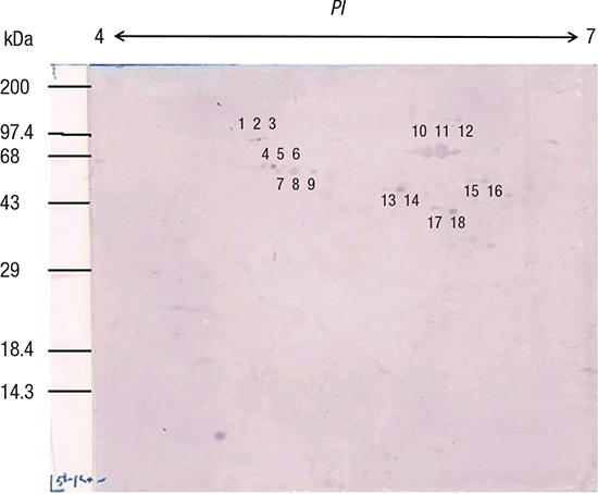
Eighteen distinctive IgM-reactive spots from the 3-month-old infant 2 were numbered (Fig. 4) and compared with the 2DE pattern of whole-cell lysates of H. pylori strain 51 (Fig. 5). These 18 IgM-immunoreactive spots were excised for MALD-TOF-MS analysis. Ten peptide fingerprints were obtained and searched against the NCBI database (Table 2). Five proteins were identified: flagellin A (FlaA), urease β subunit (UreB), pyruvate ferredoxin oxidoreductase (POR) α subunit, hemolysin secretion protein in precursor (HlyB), and translation elongation factor Ts (EF-Ts).
Fig. 4
Eighteen numbered IgM-reactive spots for the 3-month-old infant 2.
IgM = immunoglobulin M.
jkms-32-1139-g004
Fig. 5
2D PAGE of whole-cell lysates of H. pylori strain 51.
2D = two-dimensional, PAGE = polyacrylamide gel electrophoresis.
jkms-32-1139-g005
Table 2

Early IgM-reactive H. pylori antigen proteins in infant serum samples

jkms-32-1139-i002
Spot No. MW, Da/pI Protein Name KHP No. NCBI ID Unique peptides detected Sequence coverage, % Expectation value
7 53,291/6.37 FlaA KHP0717 261838156 11 30 1.8 × 10⁻⁷
10 61,833/5.64 UreB (urea amidohydrolase) KHP0075 261837530 15 26 1.4 × 10⁻¹³
11 61,833/5.64 UreB (urea amidohydrolase) KHP0075 261837530 11 21 4.7 × 10⁻⁶
12 61,833/5.64 UreB (urea amidohydrolase) KHP0075 261837530 15 28 1.8 × 10⁻¹⁸
13 45,014/5.49 POR, α subunit KHP1011 261838442 20 54 2.3 × 10⁻¹⁶
14 45,014/5.49 POR, α subunit KHP1011 261838442 20 54 2.3 × 10⁻¹⁶
15 48,660/5.89 HlyB, methyl-accepting chemotaxis protein KHP0719 261838158 15 32 2.5 × 10⁻⁶
16 48,660/5.89 HlyB, methyl-accepting chemotaxis protein KHP0719 261838158 15 32 2.5 × 10⁻⁶
17 39,980/5.77 EF-Ts KHP1407 261838826 7 27 0.96
18 39,980/5.77 EF-Ts KHP1407 261838826 7 27 0.96
IgM = immunoglobulin M, MW = molecular weight, , NCBI = National Center for Biotechnology Information, FlaA = flagellin A, UreB = urease β subunit, POR = pyruvate ferredoxin oxidoreductase, HlyB = hemolysin secretion protein in precursor, EF-Ts = translation elongation factor Ts.

DISCUSSION

In the present study, H. pylori infection was confirmed in infants by immnuohistochemistry and immuno-EM, and serum samples were tested for the anti-H. pylori IgM antibodies to identify the antigens of early-stage H. pylori infection. The 18 IgM-reactive spots on 2D immunoblots were identified as H. pylori FlaA, UreB, POR, HylB, and EF-Ts by peptide mass fingerprinting using MALDI-TOF-MS.
Several proteomics studies reported that different antigens are recognized from different gastric diseases (151617) and suggested that this might owe to persistence of strong inflammation and tissue damage leading to increased antigen presentation during ulcer development. We identified 5 IgM-reactive proteins from infants less than 1 year old that were different from the translation elongation factor EF-G (FusA), catalase (KAtA), and urease α subunit (UreA) identified in duodenal ulcers (15) or GroES (also referred to as heat shock protein A) in gastric cancer (16). In addition, H-E staining revealed mild chronic gastritis in all infants and mild active gastritis in 1 infant.
We did not observe reactive spots against CagA in the 2D IgM immunoblots although CagA has been an immunodominant antigen in children (89). In 22 H. pylori-infected Japanese children aged 2–13 years, serum IgG distinguished reactive spots identified as CagA, CagA fragments, FlaA, Omp21/AlpB, and VacA on 2D immunoblots (8). The presence of CagA bands on 1D immunoblots correlated with the high titer of anti-H. pylori IgG antibody in children aged 0–15 years (9). IgG antibodies were observed on days 63 and 857 in twin boys with acute H. pylori infection (17). Serial 1D immunoblots of IgM antibodies, using 3-, 7-, and 9-month serum samples of an infant, showed 25, 29, and 47 kDa bands for the 3-month sample; a 120 kDa (CagA) band and several 60 kDa bands for the 7-month sample; and 120, 87, 61, 56, and 54 kDa bands for the 9-month sample (18). The absence of anti-CagA IgM at 3 months was considered as acute infection, and its presence at 7 and 9 months was considered as persistent infection (18). These results suggested that CagA induces gastric mucosal inflammation 2 months after initial H. pylori infection and might not play an important role for the bacterium's survival in the gastric mucosa during early infection. Herein, the CagA-positive on the 1D IgG immunoblot was probably due to placental acquisition of maternal anti-CagA IgG given that the positive infants were younger than 7 months.
The 18 IgM-reactive spots were 33–97 kDa, consistent with previous observation of low MW bands (31 kDa and 61 kDa) in 1D IgM and IgA immunoblots for children with no IgG-positive band (5). In H. pylori-positive Australian adults, highly IgA-immunoreactive proteins were 40–80 kDa on 2D immunoblots (19); the similar MWs of immunoreactive proteins might be related to the IgA antibody, which is made in the gastric mucosa upon direct exposure to H. pylori.
In the present study, IgM-positive spots were identified as FlaA, UreB, POR, HlyB, and EF-Ts. H. pylori can actively move through mucus using its flagella, which are virulence factors. H. pylori flagella consist of two main components, FlaA and FlaB, with the majority being FlaA (20). Although a study in Japan showed that FlaA reacted with IgG in sera of uninfected children and was nonspecifically reactive in sera of infected children (8), it might play an important role for moving in the gastric mucosa during early infection. H. pylori urease comprises two structural subunits, the 26.5-kDa UreA and 60.3-kDa UreB, and 5 accessory proteins (UreC, UreD, UreE, and UreF), and its activity increases under acidic conditions (pH < 6.5) (21). POR is a housekeeping enzyme involved in energy metabolism in bacteria by converting pyruvate to acetyl-CoA in microaerophilic conditions such as the human stomach (22). HlyB belongs to one of the largest membrane protein families, the ATP-binding cassette (ABC) transporters (23). IgM antibodies against UreB, POR, and HlyB might be made through activation of these proteins during early-stage infection for survival in the acidic stomach. EF-Ts is an interacting protein involved in extending the nascent polypeptide chain during the elongation stage of bacterial translation (24). The IgM-positive antibody to EF-Ts suggested that active protein synthesis occurs in early infection.
It is important to note the following limitations in our study. First, we did not compare our patients to infants without H. pylori infection. The identified proteins were the antigens isolated from H. pylori on the NCBI database. Further studies will be needed to determine whether the identified proteins are associated with other diseases in children without H. pylori infection. In addition, we compared the results with those for H. pylori strain 51, which was isolated from a patient in our hospital, but identified the proteins using NCBI database entries from H. pylori strain J99 or 26695. H. pylori is believed to exhibit a large degree of genomic and allelic diversity (25). We did not compare the genomic sequences of H. pylori strains 51, J99, and 26695.
In summary, this is first study to evaluate the early immune response to H. pylori in infants utilizing proteomics. We identified 5 protein candidates for the essential and active proteins enabling survival and colonization in human stomach during early H. pylori infection. The identification of early immunogenic H. pylori proteins is a prerequisite for the development of serological test kits based on recombinant antigens and will support efforts for early diagnosis in infants.
In conclusion, IgM antibodies of 8 infant patients with H. pylori infection reacted with H. pylori proteins. Immunoblot spots were compared with those from whole-cell extracts of H. pylori strain 51, and 18 spots were excised, digested in gel, and analyzed by MALDI-TOF-MS. Of the ten peptide fingerprints obtained, the H. pylori proteins FlaA, UreB, POR, and EF-Ts were identified and appeared to be active during the early infection periods. These results might aid identification of serological markers for the serodiagnosis of early H. pylori infection in infants.

ACKNOWLEDGMENT

The biospecimens and data used for this study were provided by the Gyeongsang National University Hospital, a member of the Korea Biobank Network. All samples derived from the Korea Biobank Network were obtained with informed consent under Institutional Review Board-approved protocols.

Notes

Funding This study was supported by a grant from the National R & D Program for Cancer Control of the Ministry of Health and Welfare of the Republic of Korea (approval No. 0820050).

DISCLOSURE The authors have no potential conflicts of interest to disclose.

Author Contributions

  • Conceptualization: Youn HS, Cho MJ, Rhee KH.

  • Data curation: Youn JH, Kim EA, Lim JY, Ko GH, Park JS, Lee WK.

  • Investigation: Jun JS, Seo JH, Park JS, Yeom JS, Woo HO, Baik SC, Lee WK, Cho MJ.

  • Writing - original draft: Seo JH, Kim EA.

  • Writing - review & editing: Seo JH, Youn JH, Youn HS.

References

1. Blaser MJ. Helicobacter pylori and gastric diseases. BMJ. 1998; 316:1507–1510.
2. Rhee KH, Youn HS, Baik SC, Lee WK, Cho MJ, Choi HJ, Maeng KY, Ko KW. Prevalence of Helicobacter pylori infection in Korea. J Korean Soc Microbiol. 1990; 25:475–490.
3. Gologan A, Graham DY, Sepulveda AR. Molecular markers in Helicobacter pylori-associated gastric carcinogenesis. Clin Lab Med. 2005; 25:197–222.
4. Seo JH, Park JS, Yeom JS, Lim JY, Park CH, Woo HO, Baik SC, Lee WK, Cho MJ, Rhee KH, et al. Correlation between positive rate and number of biopsy samples on urease test in childhood Helicobacter pylori infection. J Korean Med Sci. 2014; 29:106–109.
5. Crabtree JE, Mahony MJ, Taylor JD, Heatley RV, Littlewood JM, Tompkins DS. Immune responses to Helicobacter pylori in children with recurrent abdominal pain. J Clin Pathol. 1991; 44:768–771.
6. Kindermann A, Konstantopoulos N, Lehn N, Demmelmair H, Koletzko S. Evaluation of two commercial enzyme immunoassays, testing immunoglobulin G (IgG) and IgA responses, for diagnosis of Helicobacter pylori infection in children. J Clin Microbiol. 2001; 39:3591–3596.
7. Oleastro M, Matos R, Cabral J, Barros R, Lopes AI, Ramalho P, Monteiro L. Evaluation of a Western blot test, Helico Blot 2.1, in the diagnosis of Helicobacter pylori infection in a pediatric population. Helicobacter. 2002; 7:210–215.
8. Akada J, Okuda M, Hiramoto N, Kitagawa T, Zhang X, Kamei S, Ito A, Nakamura M, Uchida T, Hiwatani T, et al. Proteomic characterization of Helicobacter pylori CagA antigen recognized by child serum antibodies and its epitope mapping by peptide array. PLoS One. 2014; 9:e104611.
9. Seo JH, Lim CW, Park JS, Yeom JS, Lim JY, Jun JS, Woo HO, Youn HS, Baik SC, Lee WK, et al. Correlations between the CagA antigen and serum levels of anti-Helicobacter pylori IgG and IgA in children. J Korean Med Sci. 2016; 31:417–422.
10. Haas G, Karaali G, Ebermayer K, Metzger WG, Lamer S, Zimny-Arndt U, Diescher S, Goebel UB, Vogt K, Roznowski AB, et al. Immunoproteomics of Helicobacter pylori infection and relation to gastric disease. Proteomics. 2002; 2:313–324.
11. Jung HS, Kim EJ, Kim EA, Park JH, Jun JS, Seo JH, Lim JY, Choi MB, Woo HO, Youn HS, et al. Detection of Helicobacter pylori by pre-embedding immunoelectron microscopy: comparison with immunoblotting method. J Korean Pediatr Soc. 2002; 45:862–874.
12. Cho MJ, Jeon BS, Park JW, Jung TS, Song JY, Lee WK, Choi YJ, Choi SH, Park SG, Park JU, et al. Identifying the major proteome components of Helicobacter pylori strain 26695. Electrophoresis. 2002; 23:1161–1173.
13. Jung TS, Kang SC, Choi YJ, Jeon BS, Park JW, Jung SA, Song JY, Choi SH, Park SG, Choe MY, et al. Two-dimensional gel electrophoresis of Helicobacter pylori for proteomic analysis. J Korean Soc Microbiol. 2000; 35:97–108.
14. O’Connell KL, Stults JT. Identification of mouse liver proteins on two-dimensional electrophoresis gels by matrix-assisted laser desorption/ionization mass spectrometry of in situ enzymatic digests. Electrophoresis. 1997; 18:349–359.
15. Lin YF, Chen CY, Tsai MH, Wu MS, Wang YC, Chuang EY, Lin JT, Yang PC, Chow LP. Duodenal ulcer-related antigens from Helicobacter pylori: immunoproteome and protein microarray approaches. Mol Cell Proteomics. 2007; 6:1018–1026.
16. Lin YF, Wu MS, Chang CC, Lin SW, Lin JT, Sun YJ, Chen DS, Chow LP. Comparative immunoproteomics of identification and characterization of virulence factors from Helicobacter pylori related to gastric cancer. Mol Cell Proteomics. 2006; 5:1484–1496.
17. Mitchell HM, Hazell SL, Kolesnikow T, Mitchell J, Frommer D. Antigen recognition during progression from acute to chronic infection with a cagA-positive strain of Helicobacter pylori . Infect Immun. 1996; 64:1166–1172.
18. Kim EA, Kim YO, Lim JY, Jung YS, Park CH, Woo HO, Youn HS, Ko GH, Baik SC, Lee WK, et al. Antibody response of infants to Helicobacter pylori infection. Korean J Gastroenterol. 2000; 35:704–715.
19. Lock RA, Coombs GW, McWilliams TM, Pearman JW, Grubb WB, Melrose GJ, Forbes GM. Proteome analysis of highly immunoreactive proteins of Helicobacter pylori . Helicobacter. 2002; 7:175–182.
20. Suerbaum S, Josenhans C, Labigne A. Cloning and genetic characterization of the Helicobacter pylori and Helicobacter mustelae flaB flagellin genes and construction of H. pylori flaA- and flaB-negative mutants by electroporation-mediated allelic exchange. J Bacteriol. 1993; 175:3278–3288.
21. Labigne A, Cussac V, Courcoux P. Shuttle cloning and nucleotide sequences of Helicobacter pylori genes responsible for urease activity. J Bacteriol. 1991; 173:1920–1931.
22. Narikawa S, Imai N, Yamamoto M, Suzuki T, Yanagawa A, Mizushima Y. Oxygen and carbon dioxide requirements of Helicobacter pylori . Acta Microbiol Immunol Hung. 1995; 42:367–371.
23. Schmitt L, Tampé R. Structure and mechanism of ABC transporters. Curr Opin Struct Biol. 2002; 12:754–760.
24. Wang D, Luo B, Shan W, Hao M, Sun X, Ge R. The effects of EF-Ts and bismuth on EF-Tu in Helicobacter pylori: implications for an elegant timing for the introduction of EF-Ts in the elongation and EF-Tu as a potential drug target. Metallomics. 2013; 5:888–895.
25. Alm RA, Ling LS, Moir DT, King BL, Brown ED, Doig PC, Smith DR, Noonan B, Guild BC, deJonge BL, et al. Genomic-sequence comparison of two unrelated isolates of the human gastric pathogen Helicobacter pylori. Nature. 1999; 397:176–180.
TOOLS
ORCID iDs

Ji-Hyun Seo
https://orcid.org/0000-0002-0691-3957

Jong Hyuk Youn
https://orcid.org/0000-0003-2320-6629

Eun A Kim
https://orcid.org/0000-0002-7334-9882

Jin-Su Jun
https://orcid.org/0000-0002-6382-6286

Ji-Sook Park
https://orcid.org/0000-0002-4704-2246

Jung Sook Yeom
https://orcid.org/0000-0003-0688-7493

Jae-Young Lim
https://orcid.org/0000-0001-5205-202X

Hyang-Ok Woo
https://orcid.org/0000-0001-8849-9341

Hee-Shang Youn
https://orcid.org/0000-0002-5498-838X

Gyung-Hyuck Ko
https://orcid.org/0000-0002-6721-1828

Jin-Sik Park
https://orcid.org/0000-0001-9847-4439

Seung-Chul Baik
https://orcid.org/0000-0001-6033-4078

Woo-Kon Lee
https://orcid.org/0000-0003-3913-2265

Myung-Je Cho
https://orcid.org/0000-0002-4958-9827

Kwang-Ho Rhee
https://orcid.org/0000-0002-4422-4992

Similar articles