Journal List > J Korean Med Sci > v.32(10) > 1108378

Park, Kim, and Kim: Total Occlusion of Pulmonary Arteries by Embolization of Myxoma
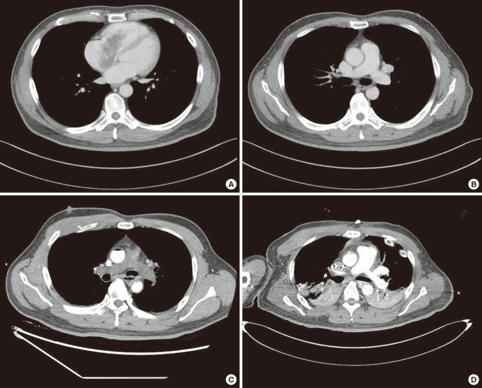
A 50-year-old male presented with acute dyspnea. Computed tomography (CT) was checked for health checkup 1 week prior and demonstrated an about 7 × 3 cm sized mass in the right atrium (RA) incidentally (Fig. 1A), without abnormalities in both pulmonary arteries and the right ventricle (RV) (Fig. 1B).
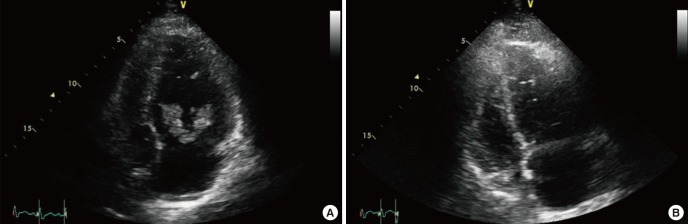
Portable echocardiography revealed RV dilatation with free wall hypokinesia, and an about 6 × 3 cm sized highly mobile, villo-papillary mass in RA with to and fro motion through the tricuspid valve (Fig. 2A and Supplementary Video 1). Sudden loss of consciousness and pulseless electrical activity were developed, and echocardiography revealed non-visualization of RA masses with total RV akinesia (Fig. 2B and Supplementary Video 2). Cardiopulmonary resuscitation and simultaneously extracorporeal membrane oxygenation (ECMO) support were performed. Follow-up chest CT revealed disappearance of the previously noted RA mass and no contrast enhancement of pulmonary arterial trees (Fig. 1C).
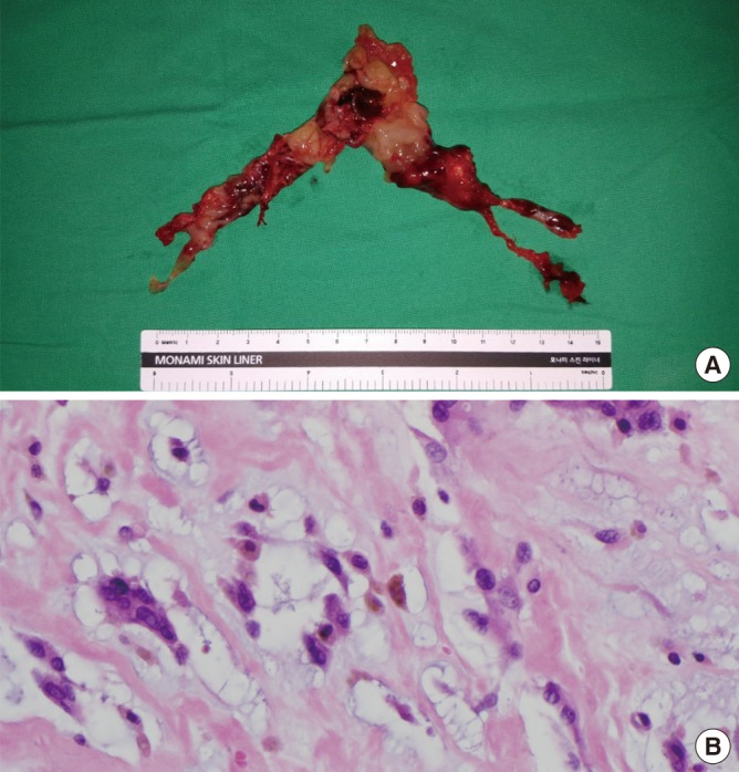
Emergent surgery revealed a small residual mass in the RA interatrial septum. Pulmonary arterial trees were totally occluded by fragile and mucoid masses which were removed by using forceps and suction (Fig. 3A). Postoperative CT revealed complete visualization of both pulmonary arteries (Fig. 1D). The masses were myxomas on histopathology (Fig. 3B). ECMO was successfully removed on the 5th hospital day.
To the best of our knowledge, this is the first case demonstrating sudden cardiac death due to total occlusion of whole pulmonary arterial trees by the embolization of huge RA myxomas during echocardiography. The present case gives several important messages. First, life-threatening complications may develop suddenly by embolization of myxomas, even in asymptomatic subjects (12). Second, prompt surgical removal should be considered in patients with high risk morphologic features of embolization (3). Third, ECMO can be a useful bridge therapy for cardiogenic shock by the embolization of myxomas before performing definitive surgical therapy (45).

Notes

DISCLOSURE: The authors have no potential conflicts of interest to disclose.

AUTHOR CONTRIBUTION: Data curation: Park H, Kim KH. Investigation: Park H, Kim KH, Kim GS. Writing - original draft: Park H, Kim GS. Writing - review & editing: Kim KH.

References

1. Fracasso T, Varchmin-Schultheiss K. Sudden death due to pulmonary embolism from right atrial myxoma. Int J Legal Med. 2009; 123:157–159. PMID: 19125270.
2. Sato H, Tanaka T, Kasai K, Kita T, Tanaka N. Sudden death due to acute pulmonary embolism from asymptomatic right atrial myxoma. J Forensic Leg Med. 2008; 15:454–456. PMID: 18761314.
3. Reynen K. Cardiac myxomas. N Engl J Med. 1995; 333:1610–1617. PMID: 7477198.
4. Chowdhury MA, Moza A, Siddiqui NS, Bonnell M, Cooper CJ. Emergent echocardiography and extracorporeal membrane oxygenation: lifesaving in massive pulmonary embolism. Heart Lung. 2015; 44:344–346. PMID: 26025763.
5. Ksela J, Knafelj R, Sostaric M, Noc M. Veno-arterial extracorporeal membrane oxygenation and embolectomy in massive pulmonary thromboembolism. Kardiol Pol. 2016; 74:393. PMID: 27098075.

Supplementary Materials

Supplementary Video 1

Portable echocardiography revealed marked dilatation of the RV with free wall hypokinesia and about 6 × 3 cm sized villo-papillary mass with irregular surface in the RA extending into the RV.

Supplementary Video 2

Echocardiography at the time of pulseless electrical activity revealed no visualization of the previously noted right atrial mass.
Fig. 1
Chest CT: a large, irregular, hypoattenuated mass in the RA (A), no abnormalities in pulmonary arteries (B). After cardiopulmonary resuscitation: no contrast enhancement of pulmonary arterial trees (C). Post-operative chest CT: complete visualization of pulmonary arteries (D).
CT = computed tomography, RA = right atrium.
jkms-32-1565-g001
Fig. 2
Portable echocardiography: marked dilatation of the RV with free wall hypokinesia and a villo-papillary mass with irregular surface in the RA (A). Echocardiography at the time of pulseless electrical activity: no visualization of the mass (B).
RV = right ventricle, RA = right atrium.
jkms-32-1565-g002
Fig. 3
Gross findings of the surgically removed masses from the pulmonary arterial trees (A). Histopathological examination confirmed the mass as myxomas (B).
jkms-32-1565-g003
TOOLS
Similar articles