

 PDF
PDF ePub
ePub Citation
 Print
Print


INTRODUCTION
Minimally invasive surgery (MIS) is gradually recognized as a safe and standard procedure for benign and early stage malignant gynecologic tumors (1). Multicenter retrospective data have been presented that MISs are increasingly applied for the management of patients with early stage uterine carcinosarcomas (UCSs), which are aggressive malignancies admixed of epithelial and stromal cells (23). With its high recurrence rate and poor overall survival for UCS, careful removal of the specimens without seeding of tumor fragments is crucial to prevent implanted metastasis during laparoscopic surgery. While port-site metastasis is one of the well-known complications of laparoscopic surgery, its incidence is reported to be very low. We present here a patient with port-site metastasis after early stage UCS surgery and review the literature.
CASE DESCRIPTION
A 53-year-old woman, nulligravida, para1-0-0-1, presented with menstrual disorders for 2 years in December 2014. On examination, in addition to erythema of vaginal mucosa and mild erosion of cervix, physical examination revealed a 3-cm bruised solitary mass with clear and smooth border prolapsed from internal cervix. Laboratory tests revealed increased cancer antigen (CA) 125 and CA199 levels. Preoperative pelvic magnetic resonance (MR) imaging showed a 12 × 9× 5-cm uterus with its cavity significant enlarged and a mass located in the left-fundus of uterus which demonstrated an isointense on T1-weighted MR image, a mixed hyperintense on T2-wighted MR image and mixed enhancing on contrast-enhanced image. A fractional curettage was initiated and potential uterine malignant tumor was diagnosed on pathology. A total laparoscopic hysterectomy (TLH) with bilateral salpingo-oophorectomy, infra-colic omentectomy, and pelvic lymphadenectomy was performed with insertion of a 10-mm trocar through a small incision in the umbilicus and placement of two 5-mm trocars 2-cm medial to the bilateral anterior superior iliac spine. There were no operative complications during the surgery. All the surgical specimens including the uterus containing the tumor and bilateral uterine adnexa had been removed through vagina. Final histology results showed a 5 × 2 cm gray brown cauliflower-like mass. Hematoxylin and eosin (H & E)-stained and immunohistochemical stained sections revealed atypical UCS penetrating the uterine superficial muscular layer without pelvic lymphatic metastasis (0/32). The final diagnosis was stage IA UCS, which was noted in patient's medical record. The patient healed without complications and underwent 4 cycles of adjuvant taxol and ifosfamide therapy.
Twelve months after the surgery, the patient presented with constant and dull right low abdominal pain in a laparoscopic incision trocar scar. The onset of pain was gradual during the 2 months and was aggravated by physical activity. Physical examination revealed a 5 × 4-cm palpable abdominal-wall incisional mass on the trocar site of right anterior superior iliac spine. Contrast-enhanced computed tomography (CT) revealed a 4.6 × 3.3-cm mass in the right low abdominal wall, with marginal moderate enhancement after contrast administration (Fig. 1). Positron emission tomography-computed tomography (PET-CT) scan revealed a 3.1×2.5-cm mass which was considered to be an isolated recurrence with high fluorodeoxyglucose (FDG) uptakes (maximum standardized uptake value [SUVmax]=12.9) in the operative region of right low abdominal wall (Fig. 2). A right low abdominal mass resection was performed and a mass measuring 6 × 3.5 × 3.5-cm was excised (Fig. 3A). The lesion was located in the muscle and surrounded by fascia. Gross tissue examination demonstrated a lobulated, pink to dusky gray-white solid tissue. H & E-stained sections revealed a metastatic squamous cell carcinoma (Fig. 3B), immunohistologic staining for CK5/6 and CD10 were positive (Fig. 3C and 3D). The patient was diagnosed with port-site metastasis without other synchronous metastases and loco-regionalinvasive recurrence of the UCS.
 | Fig. 1Contrast enhanced computed tomography scans showed a mass (arrow) in the abdominal wall near the trocar site of right anterior superior iliac spine, with marginal moderate enhancement after contrast administration. (A) Coronal section view. (B) Transverse section view.
|
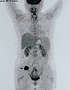 | Fig. 2PET-CT scans showed new port-site recurrence (arrow) at the right low abdominal wall with high FDG uptakes (SUVmax=12.9) in the operative trocar site.
PET-CT = positron emission tomography-computed tomography, FDG = fluorodeoxyglucose, SUVmax = maximum standardized uptake value.
|
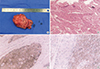 | Fig.3Pathological features of the port-site metastatic tumor. (A) Gross tissue measuring 6×3.5×3.5 cm. (B) H & E stained slides showing metastatic squamous cell carcinoma ×40. (C) Immunohistochemical stained slides showing CK5/6 was positive. (D) Immunohistochemical stained slides showing CD10 was positive.
H & E = hematoxylin and eosin.
|
DISCUSSION
UCSs are rare aggressive tumors that were considered to be sarcomas traditionally, however recently demonstrated as malignancies composed of transformation of epithelial elements (4).The worldwide annual incidence is 0.5–3.3 cases per 100,000 women (5) and the recurrence rate are reported to be over 50% despite surgery or adjuvant therapy (6). As the number of UCS patients is rare and most clinical data are retrospective, to date there is not a clear and specific guideline for the treatment of UCS. Although evidence-based treatment algorithms exist, they may be flawed due to a base on small, often retrospective studies (7).Previous studies believed UCS behaved as a sarcoma, and therefore in most cases treatment protocols for UCS followed sarcoma guideline(8). Surgery including hysterectomy with bilateral salpingo-oophorectomy and lymphadenectomy is initially recommended for patients with UCS (9).
Laparoscopic technique, as a core technique of the MIS, benefits from less postoperative complications, quicker return to normal activity and decreased postoperative pain, and it was reasonable to consider laparoscopic surgery that could be used safely in early stage UCS (4). However, complications such as inadvertent bowel injury or major vascular injury for laparoscopic surgery are related with gaining access to abdomen (10). The overall rate of major complications following laparoscopic surgery is reported to be approximately 1.4/1,000 and the incidence of port-site complication is around 21 per 100,000 cases (11).In recent years, several reports of trocar site recurrence following laparoscopic oncological surgery have been published (12). Port-site metastasis is regarded as an uncommon complication occurring in 1% of laparoscopic surgeries for gynecologic malignancies (1314). Of note, one case of port-site metastasis did occur in a patient with UCS, albeit advanced disease (4). In the present case, we report a patient diagnosed with early stage (stage IA) UCS that is recurredat the trocar-site after laparoscopic surgery.
The exact mechanisms of development of metastasis of abdominal wall are still unknown. Studies showed that recurrence of tumor at the port-site probably could be associated with several risk factors. The surgical error is a major risk factorthat leads to port-site metastasis, including rupture of tumor during surgery, direct contact between tumor and the wound for extraction of the specimen and so on. Given the surgical errors, the use of plastic bags or wound protectors and a wide abdominal incision for extraction were recommended to avoid direct contact and to allow easy passage of the specimen (12). Irrigation of the trocar site with sterile water or 5% povidone iodine is also recommended, which was not used in the present case (15).
In addition, the carbon dioxide pneumoperitoneum was considered to be another risk factor that contributes to the development of tumor implantation. Studies showed that high carbon dioxide level of pneumoperitoneum, which was likely to decrease the intracellular pH that forced the cells to translate into anaerobic metabolism, resulting in the activation of enzymes that mediated the mitosis of tumor cells, stimulated tumor growth. Meanwhile the acid environment caused by carbon dioxide to some extent might improve the ability of tumor cell adhesions and invasions, and the airflow stack effects of pneumoperitoneum might also promote the tumor adhesion on surgical instruments and trocar-site implanted metastasis (16).
Most commonly a patient with port-site metastasis presents a palpable abdominal mass at the trocar site. More published studies suggested that metastatic deposits might occur at the port-site other than the extraction site (17). The characteristics of the port-site metastasis at the early phase are supposed to be a painful solid and fixed mass with clear edge and sustained growth. CT revealed a solitary high-density shadow beneath the abdominal wall. These are difficult to distinguish from postoperative deep incisional abscess formation or local inflammatory response to suture material (18). Elevated postoperative CA125 level was noted to be associated with recurrence of the tumor, which was not confirmed by a follow-up study. Although some surgeons have suggested the additional biopsy to clarify a diagnosis, this is not a formal recommendation (4).
Nevertheless, in the present case, port-site metastasis occurred despite careful extraction vaginally of the entire specimen including the tumor, suggesting that port-site metastasis may be likely to be associated with the biology and aggressive nature of the USC. Meanwhile, although the efficiency and safety of the laparoscopic surgery for early stage gynecological malignancy had been proved by several published research studies, Gynecology Oncology Group (GOG) conducted a randomized clinical study to conclude the overall survival of patients under laparoscopic surgery with tumor recurrence of stage I ovarian cancer was as poor as with advanced ovarian cancer (19). The fact suggests that laparoscopic surgery would lead to a lower tumor stage, especially for misdiagnosis of the stage IIIC with lymphatic metastasis as early stage like the patients with UCS we present here, that might result in treatment delay and poor prognosis (19).Even worse, some have considered the port-site metastasis after adequate duration of chemotherapy might indicate the tumor recurrence and poor outcome. Therefore, when performing surgery for UCS that is thought to be an obvious high-grade malignancy, surgeons are supposed to carefully remove the specimens in case small pieces of the occult disseminated metastatic tissues are trapped between the outer surface of the trocar sleeve and the abdominal wall incisional canal. What is more, despite the low incidence, a laparotomy may be taken into consideration rather than laparoscopy to prevent port-site metastasis and more gynecological oncology clinical practices may be relevant to the management of port-site metastasis.
 XML Download