

 PDF
PDF ePub
ePub Citation
 Print
Print


INTRODUCTION
Kawasaki disease (KD) is an acute systemic vasculitis of unknown etiology that involves small- and medium-size arteries (1). The classic diagnostic criteria for KD include 5 days of fever, and at least 4 of 5 clinical features, including non-exudative bilateral conjunctival injection; erythema of the lips and oral cavity; atypical rash; edema or erythema of hands, and feet; and cervical lymphadenopathy (2). In the recently revised guidelines from the Joint Working Group in 2014, at least 5 of 6 items should be satisfied for diagnosis of KD. However, patients with 4 items of the principal symptoms can be diagnosed with KD when coronary aneurysm or dilation is recognized using two-dimensional echocardiography or coronary angiography (3). Patients with four or fewer principal symptoms indicative of KD are diagnosed with incomplete KD (4).
Prompt treatment with high-dose (2 g/kg) intravenous immunoglobulin (IVIG) and oral acetylsalicylic acid has been shown to resolve manifestations of KD, and to significantly decrease the prevalence of coronary artery abnormalities. However, most studies have indicated that 10% to 15% of patients with KD experience persistent or recurrent fever after completion of initial IVIG administration, indicating treatment resistance (5). Children with IVIG resistance are at higher risk for development of coronary artery aneurysms. Recent research has focused on identification of predictors of IVIG resistance to implement additional therapies early in the course of illness and prevent coronary lesions.
Several different risk scores are used to predict IVIG resistance in Japanese children with KD. We chose the Harada, Kobayashi, and Egami risk scores (678) (Table 1), which are most commonly used in clinical practices in Japan, and assessed their performance in Korean patients with KD. In the previous studies, there have been several trials to compare usefulness of Japanese scoring system for Korean children with KD (910). The purpose of this study was to investigate clinical risk factors to identify refractory KD, which is appropriate for Korean children.
Table 1
Baseline characteristics
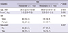
Values are presented as median (range) or number (%).
IVIG = intravenous immunoglobulin.
*Febrile days before initial dose of IVIG treatment.
![]()
MATERIALS AND METHODS
Patients and data collection
The patients with KD at Gangnam Severance Hospital (Seoul, Korea), between January 2014 and December 2015 were enrolled. Retrospective review of clinical records was performed with laboratory data regarding admission, age, sex, duration of fever (in days) at diagnosis, and the results of echocardiography were obtained.
During the study period, 6,633 patients were admitted to our pediatric department, and 350 of them (5.28%) were diagnosed with KD. KD patients were divided into IVIG responders (n = 245) and IVIG resistance (n = 105) groups. IVIG resistant patients required a second dosage of IVIG or steroid therapy because of a persistent or reappearance of fever within 36 hours after the initial IVIG treatment.
Single high-dose IVIG (2 g/kg) infused over approximately 12–24 hours to all the patients. If aspartate aminotransferase (AST) and alanine aminotransferase (ALT) were both within the reference ranges or only mildly elevated, the medium-dose aspirin (30 mg/kg/day) was administered. When body temperature presented below 37.8°C with symptomatic improvement, the low-dose aspirin therapy (5 mg/kg/day) started. IVIG-resistant patients received additional IVIG (2 g/kg) or methylprednisolone (30 mg/kg/day) as the second-line therapy.
Echocardiography
Two-dimensional echocardiography was performed at the time of diagnosis and repeated approximately 6–8 weeks after diagnosis by a pediatric cardiologist. Dimensions of the proximal left main coronary artery, proximal left anterior descending coronary artery, and proximal right coronary artery were adjusted for body surface area and presented as Z-scores based on a formula reported by Haycock et al. (11). Coronary dilatation was defined as a Z-score > 2.5 (12).
Statistical analysis
SAS version 9.4 (SAS Inc., Cary, NC, USA) and R version 3.3.0 (The R Foundation for Statistical Computing, Vienna, Austria) were used for statistical calculations. Data were expressed as the mean ± standard deviation for continuous variables, and median with range or count with percentage, as appropriate, for categorical variables. Propensity score matching was performed for the variable of age at diagnosis because it has a statistically significant difference between IVIG resistant and IVIG-responsive children. Student's t-test and Mann-Whitney U tests were used to compare groups, and the χ2 test was applied to categorical data; P < 0.05 was considered to be statistically significant. To identify independent predictors of IVIG resistance, multivariate logistic regression models were constructed using variables selected by univariate analysis. Continuous variables were converted to dichotomous variables according to the upper or lower quartile for each independent predictor identified by the multivariate logistic regression. Area under the receiver operating characteristic (ROC) curves was calculated to evaluate the capacity of the model. To identify personal risk probability that would be used in the nomogram, similar univariate and multivariate logistic analyses were performed using variables to derive a final variable formula.
RESULTS
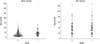
Study subjects include a total of 350 KD patients, consisting of 200 typical KD patients (57.1%) and 150 incomplete KD patients (42.9%). The number of patients with resistance was 105 (30%) and the remaining 245 were responders. Of the 105 resistant patients, 46 (43.8%) patients were incomplete KD, whereas of the 245 responder patients, 104 (42.4%) patients were incomplete KD. There was statistically significant difference in variable of age. Finally, we performed propensity score matching of 102 patients in the present study (Fig. 1).
The baseline characteristics of each group are summarized in Table 1. Age ranged from 23 to 53 months (median 38.5 months) in the IVIG-responder group, and from 23 to 51 months (median 39 months) in the IVIG-resistance group, which showed no statistical difference (P = 0.909). The total duration of fever was 2 to 17 days (mean 5.3 days) for the responder group, and 1 to 10 days (mean 5.1 days) for the resistant group. The one-day fever patient had BCG site injection, strawberry tongue, and coronary ectasia, so we diagnosed atypical Kawasaki and treated IVIG. There were no significant differences in duration of fever, sex, and incidence of recurrent KD.
Regarding laboratory parameters, IVIG-resistant individuals were found to have lower platelet levels and higher levels of C-reactive protein (CRP) and AST; these differences were statistically significant (Table 2). There were no other statistically significant differences in indicators between the two groups.
Table 2
Comparison of laboratory variables
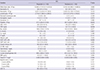
Data presented as median (range) or mean ± standard deviation, unless otherwise indicated. Bolded values indicate statistically significant differences.
IVIG = intravenous immunoglobulin, ESR = erythrocyte sedimentation rate, CRP = C-reactive protein, SGOT = serum glutamic oxaloacetic transaminase, AST = aspartate aminotransferase, SGPT = serum glutamic pyruvic transaminase, ALT = alanine aminotransferase, CK-MB = creatine kinase myocardial b fraction, BNP = brain natriuretic peptide, LMCA = left main coronary artery, LAD = left anterior descending coronary artery, RCA = right coronary artery, LVEF = left ventricular ejection fraction.
![]()
The Harada, Kobayashi, and Egami scoring systems were applied to the patients in this study, and the proportion of high-risk patients identified with each of the three risk scores were compared (Table 3). Among the three risk scores, Kobayashi showed significant differences between the IVIG-responder and IVIG-resistant groups (P = 0.014).
Table 3
Comparison of scoring systems
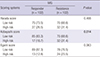
Data presented as number (%). Bolded value indicates statistically significant difference.
IVIG = intravenous immunoglobulin.
![]()
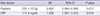
The sensitivity, specificity, positive predictive value (PPV), and negative predictive value (NPV) for each risk score system is shown in Table 4. The sensitivity of each scoring system was low (31.4%, 31.4%, and 23.5%, respectively) and the specificity was high (73.5%, 83.3%, and 87.3%, respectively). Risk scoring systems from Japan typically have good specificity but low sensitivity for predicting IVIG resistance in Korean KD patients.
Table 4
Diagnostic performance

![]()
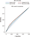
In multivariate analysis, platelet counts and CRP levels were selected as independent predictors of IVIG-resistant KD (Table 5). The cut-off values, which were identified by the ROC curve, were applied in a stepwise procedure (Fig. 2).
Table 5
Multiple conditional logistic regression analysis
| Risk factors | OR | 95% CI | P value | |
|---|---|---|---|---|
| Platelets | 278 × 103/μL | 0.997 | 0.994–0.999 | 0.012 |
| CRP | 111.4 mg/dL | 1.008 | 1.001–1.014 | 0.016 |
![]()
From the result, a nomogram was derived for personal risk probability of IVIG resistance in KD patients (Fig. 3). The underlying logistic model is given by the equation:
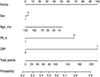 | Fig. 3Nomograms predicting the probability of IVIG resistance in patients with KD.
IVIG = intravenous immunoglobulin, KD = Kawasaki disease, Plt = platelets, CRP = C-reactive protein, mo = months, k = 103/μL
|
From this, probabilities for age, sex, platelets, and CRP variables can be estimated.
For example, if a 100-month-old female KD patient with a platelet count of 250,000/μL and CRP level of 120 mg/dL, female is linked to sex 2 and converted to 5 points and 100 months would be altered to 12.5 points. Moreover, platelets would be converted into 67.5 points, and CRP was given 100 points. The total point axis in Fig. 3 can reach a maximum of 185 points, with a predicted probability range of approximately 0.84.
DISCUSSION
In Japan, Harada (6), Kobayashi et al. (7), and Egami et al. (8) proposed scoring systems for predicting IVIG non-responders using various parameters, which is well matched in Japanese patients with complete KD. However, in a previous study, Sleeper et al. (13) concluded that these Japanese scoring systems were not suitable for the North American population. A random cohort study in a United States population found a sensitivity of 42% and a specificity of 85% for the Egami score, and a sensitivity of 42% and specificity of 87% for the Kobayashi score. Davies et al. (14) used the Kobayashi scoring system for children with KD from the United Kingdom, and found a sensitivity of 58% and a specificity of 35%. These results indicate that the Kobayashi scoring system is not useful in the British population. Fu et al. (15) predicted IVIG resistance in Chinese children with KD, and found a sensitivity of 21% and specificity of 87% for Egami scores, and a sensitivity of 49% and sensitivity of 72% for the Kobayashi score. Based on these findings, Japanese KD scoring systems were considered to have limited predictive value for IVIG response in other countries. Korea is home to the second largest KD population in the world; therefore, we have evaluated whether these scoring systems would be suitable there.
According to a recent nationwide survey in Korea in 2011, the 11.6% of patients did not respond to initial IVIG treatment and coronary artery dilation was noted in 16.4% of patients, based on the Japanese Ministry of Health and Welfare criteria (16). Despite the timely administration of IVIG, 10% to 20% of patients do not respond to treatment. These refractory patients are at increased risk of developing coronary artery complications. It is very important to identify high-risk patients for protecting against ongoing coronary arteriopathy. The significance of coronary outcomes emphasizes the importance of early prediction and timely optimal additional treatment of IVIG-resistant patients.
Previous studies have reported risk factors for refractory KD, which include high neutrophil count; low hemoglobin; thrombocytopenia; low albumin; high CRP; high lactate dehydrogenase; hyponatremia; elevated AST; abnormal findings in initial echocardiography; and recurrent KD (171819). Most risk factors are related to severe inflammatory reactions and are not suppressed by IVIG alone. In IVIG-resistance, fever represents ongoing inflammatory reactions in vessels and may reflect defects in the control of immunological stimuli (20).
Previous reports to predict IVIG resistance KD have mostly excluded incomplete KD. However, our study included patients with high rates of incomplete KD. This may have been a factor in determining whether previous studies were appropriate for our patients.
Our study tested the utility of current risk scores in Korean patients with KD. They have good specificity but low sensitivity for predicting IVIG resistance. The scoring system suggested by Kobayashi et al. (7) can help predict IVIG-resistant KD and lead to timely management and better clinical outcomes. In addition, from the results of our study, we identified a few independent predictive parameters for refractory KD — platelets and CRP level — that suggest a more focused importance of vascular inflammatory reactions in the disease process.
This study had some limitations, the first of which was its retrospective, single-center design; consequently, we could not obtain sufficient data regarding clinical characteristics and laboratory findings. Second, many patients were excluded because of a lack of adequate data or results in age matching. The incidence of coronary complications is highly reported in extreme age group, but in our study, there is a limit due to age matching. Third, we could not exclude patients with comorbidities from other febrile illnesses.
In conclusion, Korean KD patients may need an exclusive scoring system. The result of our study may be helpful in selecting patients who are at high-risk for IVIG resistance or coronary artery complications. Further multicenter studies are needed to develop a better and more comprehensive evaluation system to provide individualized treatment planning.
 XML Download