

 PDF
PDF ePub
ePub Citation
 Print
Print


Pneumatic surgical tourniquets, first introduced by Harvey Cushing in 1904, are widely used in extremity surgery to maintain a bloodless operative field. However, even when properly used and controlled, both local and systemic complications can occur1. The most common complications observed in clinical settings are neurological injuries, which, although still rare, can also be the most devasating123. We present two cases of neurological deficit of more than six months' duration following upper extremity surgery to highlight the risk and scope of such injuries.
CASE REPORT
1. Case 1
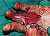
Several surgical procedures were performed on a 33-year-old male with no medical histories requiring secondary reconstruction on his left hand (Fig. 1). During the 17-hour prolonged surgery, a single tourniquet was inflated to 250 mmHg for six 2-hour periods with 30-minute breaks between each inflation, for a total ischemic time of 14 hours. Immediately after surgery, weakness of the left elbow, wrist and hand occurred, including hampered wrist and ulnar digit extension. The patient felt difficulty in maintaining elbow flexion against gravity and required an arm sling. These signs were accompanied by diminished sensation. Physiotherapy with range-ofmotion (ROM) exercises were started. Motor and sensory weakness gradually improved, and one month after surgery the patient no longer required the arm sling. However as he regained sensation, tingling and pins and needles occurred with increasing intensity. Two weeks after surgery the patient was started on gabapentin, tramadol and acetaminophen to control the tingling pain. Tingling and allodynia peaked at two months and then started to improve, with the forearm almost free of symptoms by three months.
Electromyography (EMG) and nerve conduction studies performed three months after surgery revealed ulnar, median and radial neuropathies at the upper arm with predominant involvement of sensory fibers, and median and lateral antebrachial cutaneous neuropathies. At four months the forearm and wrist had recovered most sensory functions except for cold sensation. At postoperative six months motor and sensory function had recovered sufficiently to cause no discomfort in everyday life, however allodynia and tingling pain requiring medication persisted for over three years. One year follow-up EMG studies suggested improvement of median and ulnar neuropathies but no significant interval change of radial and antebrachial cutaneous neuropathies. These findings did not match the clinical state of the patient, who had fully recovered all motor function by this time.
2. Case 2
A 45-year-old woman with a history of right nephrectomy and chronic kidney disease (CKD) required delayed coverage for a crushed index finger. A medial sural artery perforator free flap was planned. Both donor and recipient dissection proved difficult, resulting in a prolonged surgical time of 19 hours, during which a tourniquet was inflated three times (one 60-minute session and two 30-minute sessions) to 250 mmHg for a total of 120 minutes. Break time between each inflation was at least 20 minutes. Postoperative wrist-and finger-drop and cramping pain with tingling followed (Fig. 2). Splinting, physiotherapy and ROM exercises were prescribed. Nerve conduction studies performed two weeks after surgery revealed radial neuropathy at the upper arm with nearly complete axonotmesis. Five month follow-up nerve studies presented no interval changes, however the patient had significantly recovered motor function by that time. The patient, who had required oral gabapentin for six months to control the tingling and cramping pain, was almost free from these symptoms at eight months. By one year the patient had almost fully recovered motor function, and no sensory problems were observed above the wrist level; remnant sensory symptoms below the wrist were probably the result of surgical incisions and initial trauma (Fig. 3). Follow-up nerve conduction and EMG studies performed at this time suggested an improved but incomplete recovery state of the radial neuropathy.
DISCUSSION
Reports on upper limb paralysis after tourniquet use in surgery, first presented 130 years ago by Montes and Putnam, have become very rare since pneumatic tourniquets became common45. The first reference was from Bruner in 1951, and in this case, as in several others reported later, a faulty pressure gauge was presumed to have led to excessive cuff pressure45. The incidence of nerve injury varies between 0.1%–7.7% in the literature15. The reasons for this wide variation probably lies in underdiagnosis of mild injury. Postoperative limb weakness from muscle injury, rapid recovery of the nerve, and the fact that many patients are in splints or casts can easily lead to over-looked cases36. Subtle or even subclinical lesions only detectable by nerve conduction and EMG studies may also occur more frequently than anticipated7. Thankfully, prognosis is generally good, usually recovering within six months with rare permanent deficits4.
Although the exact pathophysiology of tourniquetassociated neurological injury is not yet clear, it is generally suggested that mechanical compression and shearing forces have a more important role than ischemia356. Most nerve damage occurs directly under and near the edges of the cuff, due to myelin disturbance and disruption of the nodes of Ranvier36. Both longer durations and higher pressures are associated with increased risk of nerve injury, however safe duration and pressure guidelines remain controversial2. A safe continuous inflation limit of 1.5–3 hours has generally been suggested, with a deflation break of 10–15 minutes every one to two hours for reperfusion, but concrete rules have not been definitely established23. Surgeons commonly inflate tourniquets to a fixed pressure (250 mmHg for the upper extremity) or to a fixed amount of pressure (100 mmHg) above systolic blood pressure. These practices are not ideal, and cases have been reported after using tourniquets within these guidelines, as was in both of our cases2457. Recent recommendations call for tourniquet inflation according to the limb occlusion pressure (LOP) at which the distal arterial blood flow (assessed by a Doppler probe) is occluded, and for deflation after 90 minutes for at least 10 minutes. A safety margin (according to the LOP) from 40 to 80 mmHg is added to the LOP for tourniquet pressure; for pediatric patients, a margin of 50 mmHg has been recommended23.
In case 1, although each inflation session was within two hours, the prolonged 14-hour total inflation time was suspected as the main reason for nerve injury. According to Horlocker et al.8, nerve dysfunction risk significantly increases with total tourniquet time and deflation breaks only modestly decrease this risk. In situations similar to case 1 where protracted total tourniquet time is anticipated, using two tourniquet cuffs with alternate inflation at 1-hour intervals can reduce direct pressure time at each cuff1. In case 2, despite the short tourniquet time, the patient may have been relatively vulnerable to nerve damage due to her general condition with stage 4 CKD1. Moreover, there was an intra-operative episode of accidental automatic tourniquet inflation; the possibility of multiple or prolonged malfunction events such as this during the tedious donor site dissection as the main reason for nerve damage cannot be ruled out, which highlights the importance of regularly checking equipment maintanence56. Other standard safety measures, such as using the widest contoured cuff possible, ensuring good padding beneath the cuff, applying to the proximal part of the limb with the greatest circumference (for more muscle bulk protection), and minimizing tourniquet time by making skin markings and other planning before inflation should always be implemented1237.
There were discrepancies in clinical recovery and EMG findings in both of our cases, with near-total symptomatic recovery while EMG findings suggested only partial improvement. Apart from neurological recovery, other factors such as adaptive biomechanical changes, functional compensation, muscle substitution patterns and overlapping contribution from other nerves could have also been involved in improved strength and function while the affected nerves were still healing9.
These cases demonstrate the diverse clinical presentation of tourniquet paralysis, with symptomatic progress not necessarily following EMG results. Though symptoms resolve over time, the considerable discomfort to patients warrants careful use of tourniquets with preventive measures against such neuropathies.


 XML Download