

 PDF
PDF ePub
ePub Citation
 Print
Print


Abstract
Purpose
Metacarpal fractures are common injuries of the hand. They are treated using closed reduction (CR) or open reduction (OR) techniques. The management strategy depends on fracture site characteristic and fixation methods. In this study, we evaluated pre- and postoperative fracture angulation, when metacarpal fractures bad been treated using two different techniques: CR with parallel transverse pinning and OR with plate fixation.
Methods
Forty-six patients undergoing anatomic reduction to treat extra-articular metacarpal fractures were recruited. They were included in one of two therapeutic groups: Group 1, CR with parallel transverse pinning (n=21); Group 2, OR with plate fixation (n=25). Fracture angulation values have been measured on pre- and postoperative radiologic images. Values were compared between pre- and postoperative states, and between corresponding measurements of each group.
Results
All extra-articular metacarpal fractures were successfully treated without wound related complications or the limit of joint motion. Both groups demonstrated adequate reduction at immediate postoperative period (postoperative angulation of group 1, 20°±7°; group 2, 19°±5°). During the observation at follow-up period, Group 1 exhibited slight recurrence (follow-up angulation of group 1, 24°±10°). Nonetheless, Group 2 showed adequate reduction state in both immediate postoperative and long-term follow-up periods (follow-up angulation of group 2, 18°±6°).
Figures and Tables
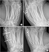 | Fig. 1Fracture angulation was analysed using uninjured contralateral metacarpal (A), injured metacarpal preoperative (B), postoperative (C), and long term follow-up (D) X-ray images. Measurements have been undergone on oblique views of hand X-ray. Measured values were utilized for intergroup comparison, and besides difference of angles were figured out (i.e., b-a, c-a, and d-a) to avoid normal angulation between metacarpal head and shaft.
|
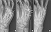 | Fig. 2Case 1. (A) A 28-year-old male patient was injured on the right 5th metacarpal bone, angulation 48°. (B) The metacarpal neck fracture was reduced using closed reduction and parallel transverse pinning method, showing 20° angulation. (C) However, heavy functional activities have led to slight recurrence (angulation 23°) at 4 week postoperative period.
|
 | Fig. 3Case 2. (A) A 30-year-old male patient was injured on the right 5th metacarpal bone, angulation 39°. (B) The metacarpal shaft fracture was reduced using open reduction and internal plate fixation method, showing 17° angulation. (C) Active exercise was followed by implant removal at 30 weeks postoperatively. The metacarpal shaft presented stability with maintenance of 15° angulation.
|
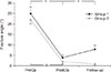 | Fig. 4Fracture angles were analysed based on the difference between angulation of injured and uninjured metacarpals. Group 1 and 2 showed adequate reduction postoperatively. However, Group 1 (closed reduction and percutaneous parallel pinning) demonstrated slight recurrence in terms of angulation (p<0.05). Nonetheless, Group 2 (open reduction and plate internal fixation) presented consistent maintenance of reduction state. PreOp: Preoperation, PostOp: Postoperation.
|
Table 1
Patient demographics: inter-group analysis did not show significant differences for number of patients, sex, age, observation period, or fracture site

![]()
Table 2
Angulation analysis in Group 1

Values are presented as mean±standard deviation. Angulation at preoperative and follow-up periods exhibited significant difference, in comparison with the average of normal side. Nonetheless, measurements at immediate postoperative state demonstrated comparable outcomes between fractured and uninjured metacarpals.
Group 1: closed reduction and percutaneous parallel pinning.
*p<0.05.
![]()
Table 3
Angulation analysis in Group 2

Values are presented as mean±standard deviation. Angulation at preoperative period exhibited significant difference, in comparison with the average of normal side. Nonetheless, postoperative and follow-up measurements showed comparable outcomes, when compared to the normal side.
Group 2: open reduction and plate internal fixation.
*p<0.05.
![]()
References
1. Kim JK, Kim DJ. Antegrade intramedullary pinning versus retrograde intramedullary pinning for displaced fifth metacarpal neck fractures. Clin Orthop Relat Res. 2015; 473:1747–1754.

2. Reformat DD, Nores GG, Lam G, et al. Outcome analysis of metacarpal and phalangeal fixation techniques at Bellevue hospital. Ann Plast Surg. 2018; 81:407–410.
3. Strauch RJ, Rosenwasser MP, Lunt JG. Metacarpal shaft fractures: the effect of shortening on the extensor tendon mechanism. J Hand Surg Am. 1998; 23:519–523.
4. Beutel BG, Ayalon O, Kennedy OD, Lendhey M, Capo JT, Melamed E. Crossed K-wires versus intramedullary headless screw fixation of unstable metacarpal neck fractures: a biomechanical study. Iowa Orthop J. 2018; 38:153–157.
5. Bao B, Zhu H, Zheng X. Fourth and fifth carpometacarpal fracture-dislocations is plate or Kirschner wire fixation superior: a retrospective cohort study. Int J Surg. 2018; 52:293–296.
6. Biz C, Iacobellis C. Comparison of percutaneous intramedullary Kirschner wire and interfragmentary screw fixation of displaced extra-articular metacarpal fractures. Acta Biomed. 2014; 85:252–264.
7. Botte MJ, Davis JL, Rose BA, et al. Complications of smooth pin fixation of fractures and dislocations in the hand and wrist. Clin Orthop Relat Res. 1992; 194–201.
8. Sletten IN, Nordsletten L, Hjorthaug GA, Hellund JC, Holme I, Kvernmo HD. Assessment of volar angulation and shortening in 5th metacarpal neck fractures: an interand intra-observer validity and reliability study. J Hand Surg Eur Vol. 2013; 38:658–666.
9. Horton TC, Hatton M, Davis TR. A prospective randomized controlled study of fixation of long oblique and spiral shaft fractures of the proximal phalanx: closed reduction and percutaneous Kirschner wiring versus open reduction and lag screw fixation. J Hand Surg Br. 2003; 28:5–9.
10. Pandey R, Soni N, Bhayana H, Malhotra R, Pankaj A, Arora SS. Hand function outcome in closed small bone fractures treated by open reduction and internal fixation by mini plate or closed crossed pinning: a randomized controlled trail. Musculoskelet Surg. 2018; DOI: 10.1007/s12306-018-0542-z. [Epub ahead of print].
11. Freeland AE, Orbay JL. Extraarticular hand fractures in adults: a review of new developments. Clin Orthop Relat Res. 2006; 445:133–145.
12. Greeven AP, Bezstarosti S, Krijnen P, Schipper IB. Open reduction and internal fixation versus percutaneous transverse Kirschner wire fixation for single, closed second to fifth metacarpal shaft fractures: a systematic review. Eur J Trauma Emerg Surg. 2016; 42:169–175.
13. Lutz M, Sailer R, Zimmermann R, Gabl M, Ulmer H, Pechlaner S. Closed reduction transarticular Kirschner wire fixation versus open reduction internal fixation in the treatment of Bennett’s fracture dislocation. J Hand Surg Br. 2003; 28:142–147.
14. Kawamura K, Chung KC. Fixation choices for closed simple unstable oblique phalangeal and metacarpal fractures. Hand Clin. 2006; 22:287–295.
15. Gülke J, Leopold B, Grözinger D, Drews B, Paschke S, Wachter NJ. Postoperative treatment of metacarpal fractures-classical physical therapy compared with a home exercise program. J Hand Ther. 2018; 31:20–28.
16. Watt AJ, Ching RP, Huang JI. Biomechanical evaluation of metacarpal fracture fixation: application of a 90° internal fixation model. Hand (N Y). 2015; 10:94–99.
17. Skirven TM, Osterman AL, Fedorczyk J, Amadio PC. Rehabilitation of the hand and upper extremity, 2-volume set E-book: expert consult. Amsterdam: Elsevier Health Sciences;2011.
18. Chiu YC, Tsai MT, Hsu CE, Hsu HC, Huang HL, Hsu JT. New fixation approach for transverse metacarpal neck fracture: a biomechanical study. J Orthop Surg Res. 2018; 13:183.
19. Lee DH, Park JW, Lee JI. Irreducible metacarpal neck fracture caused by interposition of junctura tendinum. Hand Surg. 2014; 19:441–443.
 XML Download