

 PDF
PDF ePub
ePub Citation
 Print
Print


Nerve compression caused by non-neural tumors is rare and hard to diagnose due to its symptoms mimicking the conventional nerve compression syndrome1. In the upper limb, it is known to frequently occur mainly in the forearm and hand levels. Lipomas and vascular tumors are the main causes and usually affect the medial nerve2.
Hemangioma at finger level is very rare and typically asymptomatic3, but may present with tenderness, swelling, and movement restriction, as well as peripheral nerve compression, including paraesthesia, pain, and paresis. After evaluation through an imaging study, if necessary, decompression through surgical tumor excision is the treatment of choice for pain relief. Microscopic surgery is recommended due to complex structure and sophisticated functioning arrangement of the finger.
We report a rare case in which the symptoms of nerve compression due to capillary hemangioma at the digital level were successfully resolved by microscopic surgical decompression.
CASE REPORT
A 62-year-old female presented with severe pain on radial side of the right index finger. She had no peculiar medical or family history, and had experienced no traumatic event. The pain developed since 10 months ago. Ultrasonography performed at local hospital revealed a vascular lesion and magnetic resonance imaging (MRI) was recommended. The patient had endured the pain by warm massaging, but the severe pain impeded her from sleeping. Additionally, tingling sensation had newly developed during the past few weeks, which forced her to visit our hospital.
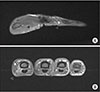
On physical examination, mild bulging of index finger on radial side at proximal phalangeal level was observed. No other abnormalities nor movement limitations were observed (Fig. 1). On MRI, a well-enhanced vascular mass of multinodular contour with about 0.7×0.5×1.0 cm size was observed on volar side of right index finger at proximal phalangeal level (Fig. 2). The space-occupying vascular lesion was thought to be compressing the radial digital nerve and surgical decompression via mass excision was planned.
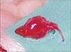
Under general anesthesia, with pneumatic tourniquet applied, a zig-zag incision was made on proximal phalanx of index finger and dissection was performed under microscopic view. A space-occupying vascular lesion was identified within the radial digital neurovascular bundle (Fig. 3). Digital artery was distorted due to the lesion, and digital nerve trunk and its branches were compressed by the mass.
Meticulous dissection was performed under microscopic view to separate the mass from surrounding tissue while preserving digital artery, digital nerve sheath, and its branches. Feeding vessel of the mass was identified, which was running up to the A2 pulley. The feeding vessel was ligated and the lesion was totally removed (Fig. 4). The final diagnosis of capillary hemangioma was made under pathologic examination. The patient's pain and tingling sensation were relieved immediately after the operation. At 6 month postoperatively, no recurrence of any abnormal symptoms was observed.
DISCUSSION
Hemangioma is benign neoplasm of endothelial cells origin, comprising 2%–6% of vascular tumors in hand region3. It is known that 70% of hemangioma in hand region spontaneously regresses before age of 7, while the remaining hemangiomas that persist in adults are due to repetitive stimulation of venous anomalies showing endothelium, polymorphism, and cell proliferation on histology4. Since hemangiomas are usually asymptomatic, conservative treatment is generally recommended3. However, surgical treatment is necessary for tumors accompanied by neuropathic symptoms such as in this case. Fingers are consisted of complex anatomical structures and sophisticated arrangement of functional tissues, hence making removal of tumor in this area challenging and requiring microscopic assist.
The 5.3%–5.5% of compression neuropathies are caused by space-occupying lesions12. In compression neuropathy caused by space-occupying lesion, pain is a common symptom but is not related to the size of the tumor5, probably due to slow growth of certain neoplasms permitting enough time for silent adaptation of the nerve to the compression1. On the other hand, certain tumors such as glomus tumor cause strong pain response in the distributed nerve territories6 by causing neuritis and irritation to the compression in pure sensory nerves. Paresis, paralysis, or atrophy caused by nerve compression are difficult to recover.
In tumor-related compressions where no mass is visible or palpable, symptoms mimicking other conventional nerve compression syndromes can lead to misdiagnosis. Thus, all nerve compression syndromes should be evaluated by diagnostic tools despite the cost ineffectiveness. MRI is the test of choice, which can define morphology and location of the tumor, allowing appropriate surgical planning1. MRI has high sensitivity in diagnosis of lipoma7 and Gadolinium-enhanced MRI has been shown to be the imaging technique of choice in the diagnosis of vascular tumors8.
Due to similarity of clinical symptoms and signs, tumor-induced compression neuropathies should be differentiated from neuromas. Painful neuromas usually occur as a sequela of nerve injury, mostly after traumatic event. After damage to the peripheral nerve, escape of the nerve fascicle to connect with distal nerve stump leads to neuroma formation9. The patient of this case had no traumatic history on right index finger, and preoperative MRI suggested vascular lesions rather than neuroma.
There are no established treatment protocols in hemangioma-induced nerve compression so far, but conservative treatment tends to fail and complete tumor removal should be the goal10. In our case with neuropathic symptom at finger level, preoperative imaging revealed a vascular lesion surrounding and compressing the adjacent digital nerve, which allowed us to establish appropriate surgical plan via meticulous dissection under microscopic view. Decompression of the digital nerve by tumor removal immediately alleviated the neuropathic pain.


 XML Download