

 PDF
PDF ePub
ePub Citation
 Print
Print


Male genital skin and soft tissue defects are usually due to trauma, fasciitis, excessive circumcision, animal bites, burns, and surgery for benign and malignant lesions1. The reconstruction of penile soft tissue defects are invariably challenging. Especially, that protruding morphology and resilient characteristics should be preserved. Various surgical techniques, including scrotal flap and pedicled anterolateral thigh flap, have been presented123. Successful reconstruction requires adequate coverage with resilience and minimal donor site morbidity. We have utilized the bilateral superficial external pudendal artery (SEPA) perforator flaps for the reconstruction of a circumferential penile shaft defect, and report the results herein.
CASE REPORT
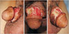
A 49-year-old man was referred from the urology department where he had received repeated debridement and repair to resolve penile abscess and soft tissue necrosis caused by a trauma. The circumferential wound presented 3×10 cm2 necrotic tissues involving superficial (Dartos) fascia (Fig. 1). We underwent debridement, preserving the deep (Buck's) facial and corpus spongiosum. Thereafter, the locations of pedicles, namely superficial external pudendal arteries (SEPAs) were traced using hand-held Doppler device on both sides of the penis and inguinal area. Then, we designed two elliptical flaps including perforators and wound margin (right, 7×3 cm; left, 6×3 cm). After incisions with regard to the design, flaps were elevated. The pedicle and vascular anatomy were visually confirmed under microscopic magnification. The diameter of the right perforator artery was 2.2 mm, and the diameter of the left perforator was 1.8 mm. The thickness of the flap was 4 mm. The flaps were transposed to the circumferential wound.
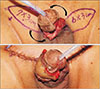
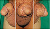
The donor sites were approximated at both subcutaneous and skin layers. Silastic drains were placed at subcutaneous layers of donor sites. Confirming the perforator wave sound on doppler device, the two transposed flaps were set in wrap-around manner. Subcutaneous layer sutures were followed by skin repair (Fig. 2, 3). The wound has healed; although there was congestion on the ventral side which resulted in a 1×1 cm sized partial flap loss. Additional debridement and repair have been performed, and flaps were maintained without complications. The patient has been observed for 27 months, showing penile resilience without deformity or wound-related problems (Fig. 4). A circumferential penile shaft defect has been successfully reconstructed using bilateral SEPA perforator flaps.
DISCUSSION
Over the past decades, penile shaft reconstruction has remained a great challenge for plastic surgeons in anatomical, functional, and aesthetical aspects. The primary goal of penile shaft reconstruction is achievement of acceptable phallus shape cosmetically as well as enough bulk with cylinder shape to guarantee sufficient rigidity for penetrative sexual intercourse. Ideally, surgical repair should be acceptable in terms of elasticity, texture, and color. When a regional local flap with genital tissue is not feasible, reconstruction can be achieved with the use of skin grafts4. Conventional regional flaps, such as various scrotal flaps show poor cosmetic results due to discrepant skin texture1. For similar reasons, split-thickness skin grafts and full-thickness skin grafts exhibit disadvantages. Resilient characteristic should be preserved in penile reconstruction. However, skin graft technique can lead to wound contracture more frequently, resulting in deformation of the penile shaft56. On the contrary, a thin subcutaneous layer of SEPA flap is advantageous for preserving resilience. In addition, this method does not require a tunneling procedure on adjacent soft tissue, which can cause morbidities. Soft tissue coverage using a free flap may be considered as the treatment of choice to overcome the above disadvantages inherent in penile reconstruction7. They are possible through various methods using radial forearm free flap, anterolateral thigh free flap, latissimus dorsi flap, scapular free flap, and so on1. Compared with the reconstruction methods mentioned above, strategic advantage of our case was well-traced SEPA perforator which existed close to the penile root showing consistent vascular anatomy.
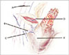
With regard to vascular anatomy, the branches from SEPA travel medially. The branch is typically originated from the inferior level of the great saphenous vein, and extended horizontally. SEPA gives rise to many willow-like branches that supply the upper perineal region. After entering the scrotum from the lateral upper side, the branch continues to run inferiorly. It supplies the upper one-third of the scrotum. The main stem anastomoses with the medial and posterior scrotal arteries8. For these reasons, surgeons can perform the operation more efficiently and confidently. In order to create a successful flap, it is important to understand detailed anatomy of the arteries that promote vascularity. There are four types of SEPA variations9. In our case, both arterial vessels presented single arteries corresponding to Type 1.
SEPA perforator flap is Type A (adipo-cutaneous course) in fasciocutaneous flap classification10. The thickness of the flap has been measured from 3 mm to 10 mm10. The flap is applicable when thin flaps are crucial, such as in penile shaft reconstruction. However, care should be taken when planning a reconstruction with limited pedicle length. Therefore, the surgical technique may be limited when the defect or penis size is large.
In conclusion, surgeons have to consider various functional and aesthetic aspects of penile reconstruction. Successful penile reconstruction can be achieved by using a flap that provides sufficient resilience with thin characteristic. The SEPA perforator flap is versatile in penile reconstruction, since the perforator exists close to the penile root and shows consistent vascular anatomy. A thin subcutaneous layer of the flap is advantageous in preserving resilience. Adequate surgical indication should be considered to accomplish favorable outcomes.
 XML Download