

 PDF
PDF ePub
ePub Citation
 Print
Print


In the past 20 years, the evolution of microsurgery has radically changed the treatment of complex lower limb wounds. However, Soft tissue reconstruction of the ankle remains a complex and challenging process despite advances in the transfer of fasciocutaneous, musculocutaneous, and composite flaps1. Clinically, there are several options of reconstructive procedures including pedicle and microsurgical free transfers of muscle and myocutaneous flaps and fascial and fasciocutaneous flaps. Compared with other flaps, peroneal artery perforator-based flap is believed that is versatile, effective and is an excellent addition to the available techniques used by reconstructive surgeons for coverage of the distal one-fourth of the leg and ankle. This flap has several advantages than other procedures2. Author planed peroneal artery perforator flap for ankle reconstruction and experienced successful result using sural neuro-lesser saphenous veno-fasciocutaneous flap due to an unexpected event.
CASE REPORT
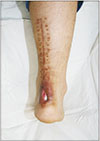
A male Asian patient, 24 years old, had a history of recurrent operation wound disruption in the ankle region with purulent secretion for about 4 months because of injury of Achilles tendon during football. Also, the patient reported repeated Achilles tendon disruption worsening. The patient reported that in the past, he had already undergone several surgical draining procedures, although he observed only immediate results as various antibiotics were administered during moments of crisis. The patient had no other medical history and did not use immunosuppressive drugs or present with any other immunosuppressive factors. Upon physical examination, the patient presented with 2×1 cm sized skin defect lesions in the ankle with exposure of Achilles tendon. In April 2017, the patient received several sharp debridements and preoperative antibiotic treatment with vancomycin 1 g, which led to the improvement of the infection (Fig. 1).
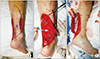
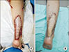
The patients were placed in a prone position under general anesthesia induced via local infiltration with a lidocaine solution containing 1% adrenalin at a 1:200,000 dilution. The perforator was identified using handheld Doppler and marked with a blue circle. Along with previously operative scar, the 17×6 cm sized fasciocutaneous flap was designed to cover skin defect with propeller technique that based on peroneal artery perforator. The dissection is continued under the fascia until the perforating branch and sural nerve, which is divided and its proximal end buried in the muscles. Peroneal perforator and saphenous vein, as well as the sural nerve branches, were identified and preserved, and the flap is raised in a retrograde position. Homeostasis is achieved using a bipolar coagulator. After careful dissection, the distal channel of the saphenous vein was ligated for flap rotation. In the process of flap rotation, an accidental division of peroneal artery perforator has occurred. It is hard to anastomosis divided perforator because of small vessel lumen diameter. Despite the division of the perforator, margin bleeding was confirmed at the flap margin and capillary refill time was also normal (Fig. 2). Based on this clinical evidence, the author assumed that flap survival was possible, and the Achilles tendon was covered by rotation flap. After flap inset, two hemovac were inserted and the flap was repaired with 4-0 Nylon. The author used 6×2 cm sized split-thickness skin graft for defect area after flap elevation. A short leg splint was applied to the extensor part. The patient experienced no complication after the surgery, and the esthetic results are considered good without any limitation to ankle movement (Fig. 3).
DISCUSSION
Various techniques have been developed for reconstruction of ankle defects. Pontén3 introduced fasciocutaneous flaps in 1981, and since then they are in use in reconstruction of soft tissue defects of the leg and foot. Inferiorly based muscle flaps have a high failure rate due to their variable vascular anatomy distally. Reversed island flaps such as the peroneal artery flap, anterior tibial artery flap and posterior tibial artery flap can be transferred to the ankle or foot. However, the need to sacrifice a major artery constitutes a potentially serious disadvantage4. At 1992, Masquelet et al.5 first described a distally based flap based on the vascular axis around the sural nerve. It was called distally based superficial sural artery flap. This flap has been shown to be ideal for reconstruction of medium and extensive soft tissue defects of the ankle and foot. The distally based superficial sural artery flap is vascularized by a median superficial sural artery with the reverse flow as this artery takes septocutaneous perforators from peroneal and tibial arteries in the distal part of the leg.
Additionally, the sural nerve has an intrinsic arterial system. These systems anastomose freely in the suprafascial plexus; combinations of these systems are used to perfuse the distally based superficial sural artery flap6.
The primary advantage of this flap is the relatively large size that can be harvested with little donor-site deformity. Dissection of the flap is easy, and blood loss is minimal. If a thin flap is required, the adipofascial flap can be used. If a neurosensorial flap is required, the lateral sural cutaneous nerve can be transferred with the flap and coapted to a recipient nerve in the defect to be reconstructed. The main advantages of these flaps are that they are well vascularized and can be neutralized, the disadvantage is that they require the necessary equipment, expertise and extended operating hours7.
In this study, peroneal artery perforator flap was the first choice for reconstruction of soft tissue defects of the ankle because of above reasons. During flap rotation, perforator was divided in an accident. However, the flap survived successfully without arterial insufficiency. There are several explanations for this phenomenon.
Sladjana et al.8 show the relationship between the elements of superficial sural artery flap neurovascular stalk, which consists not only of the lesser saphenous vein and sural nerve but also the median superficial sural artery. The lesser saphenous vein extends from the lateral border of the dorsum of the foot, passes below the lateral malleolus, and then travels along its posterior border. The terminal part of the median superficial sural artery is medial to the lesser saphenous vein. The sural nerve is located between the median superficial sural artery and the Achilles tendon8. Oberlin et al.9 recommend using a fasciocutaneous flap based on the satellite vascular networks found accompanying the sural nerve, rather than a defined artery. This flap relies on the vascular axis of the sural nerve which consists of the median superficial sural artery and the lesser saphenous vein9.
Guiraldo et al.1 reported that while investigating the vascular anatomy of the sural neurocutaneous flap in a freshly amputated limb. The accompanying arteries and the vascular plexus around the sural nerve communicate with the lower peroneal septocutaneous perforators as they emerge from the lateral crural septum. The accompanying arteries of the short saphenous vein and the lateral sural nerve contribute to this plexus from the midpoint of the leg downwards. Masquelet et al.5 developed the concept of ‘neuro-skin flaps’. They demonstrated the role of the coaxial vascular axis around the superficial sensory nerves supplying the skin.
Lee et al.10 well described the blood supply pattern of the lower leg. The posterior lower leg was evenly divided into 9 zones from zone 1 to zone 9, with zone 1 being the lateral malleolar area and zone 9 the popliteal fossa. These zones show that perforating branch of peroneal artery and sural nerve accompanying artery plays an essential role in the reverse sural flap. In reverse sural flap, reverse flow from peroneal artery perforator is the primary nutrient source. On the other hand, the antegrade flow of sural nerve accompanying artery will be used for the pedicle of the flap10. Through this anatomical concept, the survival of neuro-venous flap without peroneal artery perforator was possible. Our case has a clinical significance that the 17×6 cm sized large flap was survived by only accompanying artery of sural nerve without peroneal artery perforator.
In conclusion, sufficient blood supply was possible only with the accompanying artery of the sural nerve without peroneal perforator. The major blood supplies of this flaps are the segmental arteries of the cutaneous nerve along the sural nerve. Although the neurovascular pedicle may be able to provide some blood supply, it is essential to be careful not to injure the perforator. Also, there is the possibility of development of true anastomosis from several operations. After all, it is critical always to preserve neurovascular bundles and perforator in any circumstances.
 XML Download