

 PDF
PDF ePub
ePub Citation
 Print
Print


INTRODUCTION
Mugwort pollen is one of the most common causes of allergic rhinoconjunctivitis and asthma in late summer and autumn in China.1 In addition to seasonal respiratory symptoms, patients with mugwort pollen allergy may develop immediate allergic reactions to fruits and vegetables including oral allergy syndrome (OAS), urticaria, angioedema and anaphylaxis.23 Mugwort pollen-related food allergy is often noticed in clinical practice. Mugwort-celery-spice-syndrome, mugwort-mustard syndrome and mugwort-peach association have been reported.456 The prevalence, sensitization profiles and clinical symptoms of this form of food allergy might differ geographically. In China, systemic reactions (SR) are the main manifestation of mugwort pollen-related food allergy, and peach is the most common trigger.7
Mugwort pollen-related food allergy is considered a consequence of immunologic cross-reactivity between mugwort pollen allergens and structurally related food proteins. The cross-reactive allergen in mugwort pollen has not yet been identified. Art v 1 is the dominant allergen of mugwort pollen, and more than 95% of the mugwort pollen-allergic patients are sensitized to it.8 Recently, Art v 3, a lipid transfer protein (LTP) from mugwort, was identified as the sensitizing allergen in a Chinese peach allergic population.9 Art v 3 has 43% to 50% sequence identity with LTPs from apple, plum and peach. As a result of this sequence homology, several LTPs have high cross-reactivity. However, little is known about the importance of LTP-sensitization in mugwort pollen-related food allergy. We speculated that Art v 3 might be the sensitizer for mugwort pollen-related food allergy and that LTPs would be found to be the major food allergens in this type of food allergy.
In this study, we investigated the relevance of immunoglobulin E (IgE) reactivity to Art v 1 and Art v 3 for food allergy as well as the correlation between LTP sensitization and allergic reactions to foods.
MATERIALS AND METHODS
Study population
In total, 148 patients with mugwort pollinosis were enrolled in the allergy center of Peking Union Medical College Hospital (PUMCH) from July 2011 to May 2012. Selection of such patients was carried out on the basis of a clinical history of autumnal pollinosis, a positive skin test to mugwort extract and the presence of IgE to mugwort pollen. Exclusion criteria were sensitization to birch pollen and history of treatment with allergen-specific immunotherapy. All subjects underwent intradermal skin tests with extracts of the 8 most common pollens and serum IgE testing to mugwort, birch and culprit foods. A thorough clinical evaluation was performed by an experienced allergist that aimed to ascertain all episodes of food allergy using a standardized questionnaire adopted from Fernández-Rivas et al.10 that included 16 foods, namely, fruits/vegetables (peach, apple, mango, grape, longan, lychee, celery, and cabbage), legume/peanuts (peanuts and green bean), nuts/seeds (hazelnut, walnut, chestnut, and sunflower seeds) and spices (garlic and onion).
A diagnosis of plant-food allergy was based on a convincing history of allergic reactions immediately after food consumption, elevated food-specific IgE and/or a positive skin prick test. According to clinical symptoms after consuming allergenic foods, patients were categorized into 2 groups as follows: group 1, symptoms localized to the oral mucosa (OAS); group 2, SR including general urticaria, angioedema, laryngeal angioedema, respiratory difficulty, gastrointestinal disorders or circulatory symptoms of anaphylaxis. The diagnosis of anaphylaxis was based on the National Institute of Allergy and Infectious Diseases (NIAID) 2006 criteria11 with slight modification: patients had symptoms of at least 2 organ systems (of the skin-mucosal tissue, gastro-intestinal system, respiratory compromise and hypotension) that occurred rapidly after consuming food.
Written informed consent was obtained, and the Ethics Committee of Peking Union Medical College Hospital approved the study (No. PUMCH1103).
Determination of allergen-specific IgE
All the recruited patients were screened with ImmunoCAP (ThermoFisher Scientific, Uppsala, Sweden) for mugwort pollen, birch pollen, food allergens and the allergenic components Art v 1, Art v 3, Pru p 3 (peach LTP), Pru p 1 (Bet v 1 homolog), Pru p 4 (profilin), Ara h 9 (peanut LTP) and Cor a 8 (hazelnut LTP). Specific IgE levels > 0.35 kUA/L were considered positive.
Skin test
All patients underwent intradermal skin tests with extracts of the 8 most common pollens in China (mugwort, birch, ragweed, London plane, Japanese hop, cypress, ash, and kochia; Allergen Manufacturing and Research Center, PUMCH, Beijing, China). The pollen allergens were extracted at a ratio of 1:20 (wt/vol). Then, the extracts underwent a 1:100 dilution (v/v; except 1:1,000 for mugwort and Japanese hop) with allergen solvent for use; histamine phosphate (0.01 and 0.1 mg/mL, respectively) was used as a positive control, and allergen diluent was a negative control. Reactions were considered positive when the mean wheal diameter exceeded 5 mm.
In some suspected food-allergic patients, skin prick tests to raw vegetables, fruits and/or spices were performed. Histamine phosphate (10 mg/mL) and allergen diluent served as positive and negative controls, respectively. The test was considered positive when the mean wheal diameter was greater than 3 mm.12
Identification of peach allergens
Peach extract
Fresh peaches were obtained from a local market. To prepare peach extracts, we followed the method of Pastorello et al.13 Fresh peach peels were diluted 4:1(wt/vol) in 10 mmol/L phosphate-buffered saline (PBS; pH 7) with 2% polyvinylpolypyrrolidone, 2 mmol/L ethylenediaminetetraacetic acid (EDTA) disodium salt and 10 mmol/L sodium diethyldithiocarbamate. After homogenizing and centrifuging at 15,000 g at 4°C for 30 minutes, the supernatant was dialyzed against PBS at 4°C for 24 hours. The extracts were centrifuged for 10 minutes and frozen at −80°C.
Electrophoresis and immunoblotting
For immunoblotting, peach extracts were separated by sodium dodecyl sulfate-polyacrylamide gel electrophoresis (SDS-PAGE) and transferred to polyvinylidene difluoride membranes. The protein-binding strips were incubated with sera of patients allergic to peach and detected with horseradish peroxidase (HRPO)-labeled anti-human IgE antibody. Ten patients with mugwort pollen allergy without food allergy served as negative controls.
An immunoblotting inhibition study was done with pooled sera of 6 patients allergic to peach. Briefly, 500 μL of pooled sera was incubated for 1 hour with 500 μL of a solution containing 400 μg of mugwort, peach protein and 4 μg of purified peach LTP (Alpco Diagnostics, Windham, NH, USA), respectively. After inhibition, peach and mugwort immunoblotting was performed as described above.
Mass spectrometry
For mass spectrometry analysis, gel slices were exercised from the appropriate region (12 kD) of Coomassie-stained SDS-PAGE gels and subjected to tryptic digestion. Protein identification was performed on a MicrOTOF-QII (Bruker Daltonics Inc., Manning Park Billerica, MA, USA). The result was compared with the Mass Spectrometry Protein Sequence Database (MSDB) for protein identification using Mascot software (Matrix Science, London, UK).
Statistical analysis
Data analysis was performed using the statistical package SPSS/PC+ for statistical evaluation (SPSS, Chicago, IL, USA). The difference in allergen-specific IgE between patients with and without food allergy was compared using the Wilcoxon signed-rank test, and the Spearman rank test was used to evaluate correlation. Categorical variables were analyzed by Pearson's χ2 test. Analysis of receiver operating characteristic (ROC) curves was performed with test results presented graphically and as area under the curve (AUC) with a 95% confidence interval (CI). Differences were considered statistically significant at P < 0.05.
RESULTS
Study population
A total of 148 patients with mugwort pollen allergy (70 males and 78 females; mean age, 20.4 ± 10.9 years; range, 3–46 years) were categorized into 107 patients with food allergy and 41 food-tolerant patients. An earlier age at onset of pollinosis was observed in patients with food allergy than in food-tolerant individuals (P = 0.02, Table). Of 148 patients, 120 (81.1%) were positive for Art v 1, 108 (73.0%) were positive for Art v 3, and only 8 (5.4%) were negative for both. The levels of specific IgE to Art v 1 and Art v 3 correlated highly with the levels of IgE for mugwort pollen (r = 0.802 and r = 0.637, respectively, P < 0.01). In 148 patients with mugwort pollen allergy, sensitization to ragweed (70.3%), London plane (63.8%), humulus (61.5%) and kochia (56.8%) were most common.
Table
Clinical and demographic data of all 148 mugwort pollen-allergic individuals
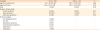
Values are presented as mean ± standard deviation (range) or number (%).
*Individuals with plant food allergy; †Individuals without plant food allergy.
![]()
Main symptoms and triggers of mugwort pollen-related food allergy
In summary, 107 of 148 (72%) patients reported allergic reactions to at least 1 of the 16 foods that included in the questionnaire. The frequency of food-induced symptoms is listed in Table. Of the 107 patients, 51 (48%) experienced at least 1 episode of food-induced anaphylaxis, and 15 (14%) reported a worsening of food-induced symptoms during mugwort pollen season.
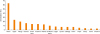
Peaches accounted for the greatest number of reported reactions (n = 68, 64%), followed by apples (n = 26, 24%), mangos (n = 21, 20%), peanuts (n = 17, 16%), and hazelnuts (n = 15, 14%, Fig. 1).
Mugwort pollen-related food allergy is associated with LTP sensitization
To evaluate the involvement of the mugwort pollen allergens in mugwort pollen-related food allergy, we compared the levels of total, mugwort pollen-specific, Art v 1-specific and Art v 3-specific IgE in patients with food allergy and food-tolerant mugwort-allergic patients (Fig. 2). No difference was found in total IgE levels between food allergy and food-tolerant mugwort-allergic patients (median, 273 kU/L; range, 19.9–2,336 kU/L and median, 343 kU/L; range, 56.8–1,437 kU/L, respectively, P > 0.05). Mugwort pollen-specific IgE levels were significantly higher in the group with food allergy (median, 35 kUA/L; range, 0.82–100 kUA/L) than in the food-tolerant group (median, 12.7 kUA/L; range, 1.0-69.5 kUA/L, P < 0.01). Similarly, Art v 3-specific IgE levels were significantly higher in patients with food allergy than in tolerant patients (median, 22.4 kUA/L; range, 0-100 kUA/L and median, 0.08 kUA/L; range, 0-85.2 kUA/L, respectively, P < 0.01), and a larger number of individuals with food allergy (87%) were positive for Art v 3 compared to tolerant individuals (37%, P < 0.01, Supplementary Fig. S1). There was no difference in the frequency or the quantity of Art v 1-specific IgE between the 2 groups.
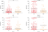 | Fig. 2Levels of total IgE and specific IgE to mugwort pollen, Art v 1 and Art v 3 were compared in FA (filled triangles, n = 107) and NFA (open circles, n = 41) patients.IgE, immunoglobulin E; FA, food-allergic; NFA, non-food-allergic.
|
ROC analysis shown in Fig. 3 described the performance of total, mugwort pollen-specific, Art v 1-specific and Art v 3-specific IgE levels in differentiating between food-allergic and food-tolerant patients. The AUC for Art v 3-specific IgE was largest with reference to that for mugwort pollen-specific and Art v 1-specific IgE, which indicated that sensitization to Art v 3 was associated with mugwort pollen-related food allergy. The best diagnostic cutoff value of Art v 3-specific IgE was 1.25 kUA/L with 70% sensitivity and 75% specificity.
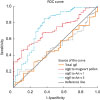 | Fig. 3Comparison of levels of total IgE and specific IgE to mugwort pollen, Art v 1 and Art v 3 in predicting mugwort pollen-related food allergy. Art v 3-specific IgE had the largest area under ROC curve (AUC, 0.757; 95% CI, 0.662–0.852; P < 0.001) with reference to total IgE (AUC, 0.479; 95% CI, 0.379–0.578; P = 0.689), specific IgE to mugwort pollen (AUC, 0.705; 95% CI, 0.620–0.789; P < 0.001) and Art v 1 (AUC, 0.495; 95% CI, 0.397–0.593; P = 0.925).IgE, immunoglobulin E; ROC, receiver operating characteristic; AUC, area under the curve; CI, confidence interval.
|
IgE reactivity to Pru p 3, Ara h 9 or Cor a 8 was more prevalent in patients with food allergy (80%, 69% or 63%, respectively) than in tolerant individuals (29%, 17% or 20%, respectively, P < 0.01, Supplementary Fig. S1). Art v 3 showed positive relationships with Pru p 3 (r = 0.608, P < 0.01), Ara h 9 (r = 0.607, P < 0.01) and Cor a 8 (r = 0.566, P < 0.01).
LTP reactivity and clinical symptoms
To analyze LTP sensitization in food allergy, we evaluated the most frequent triggers in fruits, peanut/seeds and nuts respectively. For example, 90% (61/88) of peach allergic patients were sensitized to Pru p 3; 88% (15/17) of peanut allergic patients to Ara h 9; 80% (12/15) of hazelnut allergic patients to Cor a 8. According to the severity of food-induced allergic reactions, 88 patients with peach allergy clustered into 2 main groups: OAS and SR. Participants with a history of SR showed higher levels of IgE to Pru p 3 (median, 14.05 kUA/L; range, 0.1–100 kUA/L) than OAS patients (median, 6.45 kUA/L; range, 0.03–48.6 kUA/L, P = 0.028, Fig. 4). Similarly, in peanut allergic patients, SR patients had higher values for Ara h 9 compared with OAS patients (P = 0.003, Fig. 4), while in hazelnut allergic patients, SR patients had higher values for Cor a 8 (P = 0.004, Fig. 4). The ROC curves were used to compare the performance of specific IgE to peach and its allergenic components in predicting SR to peach. Pru p 3 testing had the largest AUC (AUC, 0.77; 95% CI, 0.676–0.872; P < 0.001, Fig. 5). Measurement of IgE to Pru p 3 (cutoff, 6.63 kUA/L) had a greater predictive power for the severe allergic reaction, whereas the IgE levels of mugwort pollen (AUC, 0.57; 95% CI, 0.448-0.693; P = 0.271) and Art v 3 (AUC, 0.55; 95% CI, 0.420-0.670; P = 0.483, Fig. 5) could not predict the severity.
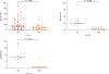 | Fig. 4IgE levels of LTP (Pru p 3, Ara h 9 and Cor a 8) were compared among food (peach, peanut and hazelnut, respectively)-allergic patients with systemic reactions and oral allergy syndrome.IgE, immunoglobulin E; LTP, lipid transfer protein.
|
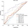 | Fig. 5ROC curve analysis for specific IgE to mugwort pollen, Art v 3, peach and its allergenic components in predicting systemic reactions to peach. Pru p 3-specific IgE had the largest area under ROC curve (AUC, 0.77; 95% CI, 0.676–0.872; P < 0.001) with reference to specific IgE to mugwort pollen (AUC, 0.57; 95% CI, 0.448–0.693; P = 0.271), Art v 3 (AUC, 0.55; 95% CI, 0.420–0.670; P = 0.483), peach (AUC, 0.73; 95% CI, 0.627–0.837; P < 0.001), Pru p 1 (AUC, 0.48; 95% CI, 0.347–0.604; P = 0.701), and Pru p 4 (AUC, 0.58; 95% CI, 0.457–0.708; P = 0.199).ROC, receiver operating characteristic; IgE, immunoglobulin E; AUC, area under the curve; CI, confidence interval.
|
Immunodetection assays
IgE reactivity profiles of peach extract were analyzed by immunoblotting with sera from 42 patients with mugwort pollen-related peach allergy (Fig. 6). Thirty-seven (88%) individuals showed IgE reactivity to the 12 kD band, and 11 (26%) to the 16 kD band. Furthermore, compared to OAS patients, a greater number of SR patients were sensitized to the 12 kD component (55% vs. 100%, P < 0.001). IgE binding to the 12 kD band of peach was fully inhibited by preincubation of pooled sera with purified peach LTP (Supplementary Fig. S2). By mass spectrometry, the 12 kD component from peach extract was identified as nonspecific LTP1. A score of 3,044 was generated using the Mascot program, and 100% coverage was obtained by means of matrix-assisted laser desorption/ionization (MALDI) analysis (Supplementary Fig. S3).
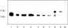 | Fig. 6Representative results of immunoblotting of peach extracts with sera from patients with mugwort pollen-related peach allergy. lane 1–6: serum from patients with systemic reactions to peach; lane 7–9: serum from patients with oral allergy syndrome; lane 10: serum from patients with mugwort pollinosis without food allergy.
|
The immunoblotting inhibition study between peach and mugwort pollen showed that mugwort pollen extract totally inhibited IgE binding to the 12 kD band of peach, whereas IgE binding to the 12 kD band of mugwort pollen was not inhibited by peach protein (Supplementary Fig. S2).
DISCUSSION
Plant food allergy is common in adolescents and adults.14 We found that 72% of this study population with mugwort pollinosis experienced food allergy. In Europe, it is reported that 47% to 73% of patients with birch pollinosis develop food allergies, while in patients with mugwort pollinosis it appears to be less common.141516 The exact prevalence of food allergy in patients sensitized to mugwort pollen is not available to date.17 Our study showed that the mugwort pollen-allergic patients with and without food allergy generally shared similar demographic and clinical characteristics of pollinosis, though patients with food allergy were younger at the onset of pollinosis than those without.
In our study population, the frequency of Art v 3-sensitized patients as well as the levels of Art v 3-specific IgE were significantly higher in food-allergic than food-tolerant patients. In contrast, neither the frequency nor the levels of Art v 1-specific IgE were associated with food-induced allergic reactions. Furthermore, Art v 3 IgE levels correlated with the IgE levels of Pru p 3, Ara h 9 and Cor a 8. In accordance with a previous study suggesting that Art v 1 was not involved in cross-reactivity to plant foods,18 our data underline the major role of Art v 3 in mugwort pollen-related food allergy. A study from China has identified Art v 3 as the sensitizing pollen allergen in a peach-allergic population.19 ImmunoCAP and enzyme-linked immunosorbent assay (ELISA) inhibition revealed that Art v 3 significantly inhibited the binding of IgE to Pru p 3, but Pru p 3 was not able to inhibit IgE binding to Art v 3,1920 which is in agreement with the results of cross-inhibition experiments in our study. In addition, a correlation has been reported between Art v 3 and Cas s 8 (chestnut LTP)21 as well as Bra o 3 (cabbage LTP).22 Together, these data suggested that Art v 3 might play a dominant role as a primary sensitizer in mugwort pollen-related food allergy. Similar amino acid sequences may provide an explanation for cross-reactivity between LTPs from different plants.2324
In this study, LTPs are major food allergens for mugwort pollen-related food allergy. Apples and hazelnuts are identified as the most frequent triggers in birch pollen-related food allergy.1525 However, both were also important offending foods, after peach, in our subjects without sensitization to birch pollen. We postulate that the 2 pollen-food syndromes might have different sensitization profiles: in birch pollen-related food allergy, the main allergens of apples and hazelnuts are Mal d 1 and Cor a 1 (Bet v 1 homolog), while in mugwort pollen-related food allergy the major allergens are Mal d 3 and Cor a 8 (LTP). A study across Europe showed the existence of 2 patterns of apple allergy: in Austria and Italy apple allergy is mild and the main allergen is Mal d 1, while in Spain apple allergy is severe and the major allergen is Mal d 3.10 Another study evaluating hazelnut allergy showed similar results.26 These findings are consistent with observations regarding the association between IgE responses to mugwort pollen and LTP sensitization.202227 However, unlike LTP-related food allergy in southern Europe in which sensitization to LTPs seems independent of any pollen hypersensitivity, in China LTP-related food allergy mainly originates from primary sensitization to mugwort pollen, and is often severe. The contrasting cause of LTP-related food allergy in China and southern Europe is unclear. A likely explanation is the dominant role of high exposure to mugwort pollen in China, which is similar to that of birch pollen exposure in Europe. Mugwort is the most common allergenic pollen in late summer and autumn in China, which is an important cause of seasonal rhinitis and asthma.2829 In our clinical practice, systemic reactions to fruits and vegetables are often reported in patients with mugwort pollen allergy. Furthermore, mugwort is the strongest allergen in LTP-related food allergy in China, and most of the allergenic epitopes of plant food LTP are also present on mugwort LTP as can be inferred from the cross-inhibition experiments.1920
Our data from ImmunoCAP showed that the levels of specific IgE to Pru p 3, Ara h 9 and Cor a 8 were significantly higher in patients with systemic reactions compared to OAS patients. Good agreement between ImmunoCAP and immunoblotting results was observed in peach-allergic subjects. Several previous studies also showed that sensitization to plant food LTP was associated with a risk of systemic allergic reactions.233031 A stable and compact secondary structure within LTPs confers their resistance to both heat treatment and gastrointestinal proteolysis, which are characteristics associated with the induction of severe symptoms.32 Therefore, high values for LTP sensitization represent a risk factor for severe allergic reactions in mugwort pollen-related food allergy.
Previous studies indicated that there were multiple components involved in the sensitization to mugwort pollen. In addition to Art v 3, other cross-reactive allergens have been reported, such as Art v 4 (profilin)33 and Art v 60 kDa.34 Sensitization patterns to cross-reactive allergens might be variable according to geographic locations and eating habits. However, LTP sensitization is prevalent in a Chinese mugwort pollen-related food-allergic patients' collective.
Currently, our study is the first cross-sectional evaluation of LTP sensitization in a mugwort pollen-related food-allergic population. This study might be limited by its single center analysis and potential bias. In addition, the diagnosis of food allergy was based on a clinical history and a skin prick test and/or specific IgE rather than double-blind placebo-controlled food challenge because of the challenging medical environment in China.
In conclusion, mugwort pollen-related food allergy is prevalent in China and is often severe. LTPs are the major food allergens for mugwort pollen-food syndrome and are associated with systemic allergic reactions. Therefore, determination of LTP sensitization is useful for risk assessment in this food allergy. In addition, immunotherapy with specific LTP, a cross-reactive allergen, existing in all the allergenic foods, might be effective for the management of food allergy in such patients.
 XML Download