Journal List > Asia Pac Allergy > v.8(4) > 1108195

Chow, Seow, Dizon, Godse, Foong, Chan, Khang, Xiang, Hidayat, Listiawan, Triwahyudi, Gondokaryono, Sutedja, Diana, Suwarsa, Dharmadji, Siswati, Danarti, Soebaryo, Budianti, and for the Asian Academy of Dermatology & Venereology: A clinician's reference guide for the management of atopic dermatitis in Asians

Abstract

Background

Atopic dermatitis (AD) is a common skin condition among Asians. Recent studies have shown that Asian AD has a unique clinical and immunologic phenotype compared with European/American AD.

Objective

The Asian Academy of Dermatology and Venereology Expert Panel on Atopic Dermatitis developed this reference guide to provide a holistic and evidence-based approach in managing AD among Asians.

Methods

Electronic searches were performed to retrieve relevant systematic reviews and guidelines on AD. Recommendations were appraised for level of evidence and strength of recommendation based on the U.K. National Institute for Health and Care Excellence and Scottish Intercollegiate Guidelines Network guidelines. These practice points were based on the consensus recommendations discussed during the Asia Pacific Meeting of Experts in Dermatology held in Bali, Indonesia in October 2016 and April 2017.

Results

The Expert Panel recommends an approach to treatment based on disease severity. The use of moisturizers is recommended across all levels of AD severity, while topical steroids are recommended only for flares not controlled by conventional skin care and moisturizers. Causes of waning efficacy must be explored before using topical corticosteroids of higher potency. Topical calcineurin inhibitors are recommended for patients who have become recalcitrant to steroid, in chronic uninterrupted use, and when there is steroid atrophy, or when there is a need to treat sensitive areas and pediatric patients. Systemic steroids have a limited role in AD treatment and should be avoided if possible. Educational programs that allow a patient-centered approach in AD management are recommended as an adjunct to conventional therapies. Recommendations on the use of phototherapy, systemic drugs, and emerging treatments are also included.

Conclusion

The management of AD among Asians requires a holistic approach, integrating evidence-based treatments while considering accessibility and cultural acceptability.

INTRODUCTION

Atopic dermatitis (AD), also referred to as atopic eczema, is a common skin condition among Asians [1]. It is a chronic inflammatory skin disease often found in patients with personal or family history of food allergy, allergic rhinitis and/or asthma [23]. Recent studies have shown that AD may have several manifestations or phenotypes, such as extrinsic vs. intrinsic AD [4], pediatric vs. adult AD [5], and European/American vs. Asian AD [67].
Asian AD clinically presents with a more clearly demarcated lesion, more prominent scaling and lichenification. Immunologic analyses have also shown that it has a unique cytokine profile that closely resembles psoriasis [89].
Challenges in AD management in Asia include variability in healthcare access in different countries, generalists' level of confidence in managing mild forms of AD, and misperceptions by patients that only dermatologists can manage AD [8]. The Asian Academy of Dermatology and Venereology Expert Panel on Atopic Dermatitis developed this reference guide to help provide a holistic and evidence-based approach in managing AD among Asians.

MATERIALS AND METHODS
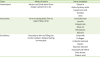
Electronic searches were performed on MEDLINE, Cochrane and Google Scholar to retrieve systematic reviews and guidelines on AD published from 2000 to 2017. The following subject headings or MeSH terms were used: ‘atopic dermatitis,’ ‘eczema,’ ‘Asian,’ ‘Chinese,’ ‘Japanese,’ ‘Korean,’ ‘Thai,’ ‘Indonesian,’ ‘Filipino,’ ‘Singaporean,’ ‘Malaysian,’ ‘Indian,’ ‘guideline,’ ‘management,’ ‘diagnosis,’ ‘treatment,’ ‘monitoring,’ ‘severity,’ ‘review,’ ‘meta-analysis,’ ‘systematic review,’ ‘evidence-based,’ ‘filaggrin,’ ‘pathophysiology,’ ‘intrinsic,’ ‘extrinsic,’ ‘pediatric,’ ‘adult,’ ‘Caucasian’ and ‘prevalence.’ Only articles in English were included.
This reference guide was based on the consensus recommendations discussed last October 2016 and April 2017 during the Asia Pacific Meeting of Experts in Dermatology held in Bali, Indonesia. The recommendations were appraised based on the U.K. National Institute for Health and Care Excellence and Scottish Intercollegiate Guidelines Network guidelines (Table 1).
Table 1

Level of evidence and strength of recommendation

apa-8-e41-i001
Level of evidence Type of evidence
1++ High-quality meta analyses, high-quality systematic reviews of clinical trials with very little risk of bias
1+ Well-conducted meta-analyses, systematic review of clinical trials or well-conducted clinical trials with low risk of bias
1- Meta-analyses, systematic reviews of clinical trials or clinical trials with high risk of bias
2++ High-quality systematic reviews of cohort or case and control studies; cohort or case and control studies with very low risk of bias and high probability of establishing a causal relationship
2+ Well-conducted cohort or case and control studies with low risk of bias and moderate probability of establishing a causal relationship
2- Cohort or case control studies with high risk of bias and significant risk that the relationship is not causal*
3 Nonanalytical studies, such as case reports and case series.
4 Expert opinion.
Strength of recommendation Evidence
A At least one meta-analysis, systematic review or clinical trial classified as 1++ and directly applicable to the target population, or a volume of scientific evidence comprising studies classified as 1+ and which are highly consistent with each other; evidence drawn from a NICE technology appraisal
B A body of scientific evidence comprising studies classified as 2++, directly applicable to the target population and highly consistent with each other, or scientific evidence extrapolated from studies classified as 1++ or 1+
C A body of scientific evidence comprising studies classified as 2+, directly applicable to the target population and highly consistent with each other, or scientific evidence extrapolated from studies classified as 2++
D Level 3 or 4 scientific evidence, or scientific evidence extrapolated from studies classified as 2+, or formal consensus
D (GPP) A good practice point (GPP) is a recommendation for best practice based on the experience of the Workgroup members
NICE, National Institute for Health and Care Excellence.

RESULTS AND DISCUSSION

Diagnosis of AD

The diagnosis of AD is clinical and is based on the morphology and distribution of the lesion, as well as the associated signs and symptoms [10]. A widely used diagnostic criteria were published by Hanifin and Rajka that consist of 4 Major and 23 Minor Criteria (Table 2). AD is diagnosed when 3 major and 3 minor criteria are met [11].
Table 2

The Hanifin and Rajka diagnostic criteria for atopic dermatitis

apa-8-e41-i002
Major criteria
• Pruritus
• Dermatitis affecting flexural surfaces in adults and the face and extensors in infants (see next page)
• Chronic or relapsing dermatitis
• Personal or family history of cutaneous or respiratory atopy
Minor criteria
• Features of the so-called “atopic facies”: facial pallor or erythema, hypopigmented patches, infraorbital darkening, infraorbital folds or wrinkles, cheilitis, recurrent conjunctivitis, and anterior neck folds
• Triggers of atopic dermatitis: foods, emotional factors, environmental factors, and skin irritants such as wool, solvents, and sweat
• Complications of atopic dermatitis: susceptibility to cutaneous viral and bacterial infections, impaired cell-mediated immunity, immediate skin-test reactivity, raised serum IgE, keratoconus, anterior subcapsular cataracts
• Others: early age of onset, dry skin, ichthyosis, hyperlinear palms, keratosis pilaris (plugged hair follicles of proximal extremities), hand and foot dermatitis, nipple eczema, white dermatographism, and perifollicular accentuation
Exclusions
Scabies
Seborrheic dermatitis
Contact dermatitis (irritant or allergic)
Ichthyoses
Cutaneous T-cell lymphoma
Psoriasis
Photosensitivity dermatoses
Immune deficiency diseases
Erythroderma of other causes
There is currently no reliable biomarker to diagnose AD. A diagnostic work-up may be performed in certain cases to help in prognostication, testing for allergic triggers or for monitoring response to treatment. These tests include serum total immunoglobulin E (IgE) levels, specific IgE levels and peripheral eosinophil count [47121314].
Total IgE is elevated in approximately 80% of AD patients classified as extrinsic type. It is not a diagnostic requirement, but it is helpful in determining prognosis or in choosing therapy. In adults, a total serum IgE level of 200 IU/mL or greater may be considered high but this may vary depending on the institution. The IgE cutoff in infants varies according to age [14]. High levels of IgE may reflect the long-term activity of AD but are often non-specific and may be seen in response to different allergens [1].
Specific IgE testing using blood serum specimens for food or inhalant allergens is also nonspecific. However, it is preferable to skin prick testing for immediate or type I hypersensitivity, especially in children. Preventing exposure to these allergens is expected to improve/prevent exacerbation of rashes [15]. Peripheral eosinophil or mast cell counts are often nondiagnostic and are not recommended for routine use [15].
Practice point: In some instances, a diagnostic work-up is done to help in prognostication, testing for allergic triggers, or for monitoring response to treatment. [Level 4, good practice point (GPP)]

Assessment of AD severity

Objective assessment of AD severity is important for appropriate management [101416]. Generally, mild disease has a more remitted course, affects less body surface area (BSA), and is associated with pruritus that is of lower intensity [17]. The SCOring Atopic Dermatitis or SCORAD Index developed by the European Task Force on Atopic Dermatitis is a comprehensive system used to assess AD severity [10131618]. Although validated, this system combines assessment of symptoms with observation of signs and is also more useful in pediatric patients [19]. The Eczema Area and Severity Index (EASI) was hence developed incorporating disease intensity and measurement of the total affected body area [19]. While the SCORAD looks at a representative site for each of the 6 signs, the EASI assesses 4 signs in various areas (4 sites) of the body and gives weight to the extent of the lesions [20]. Nonetheless, the EASI is limited by its significant emphasis on BSA measurements, which may be difficult to assess accurately and uniformly [18]. Given the complexity of these assessment methods, the Three-Item Severity (TIS) score is proposed as an alternative to the abovementioned systems for use in daily practice (Table 3). It is based on the evaluation of erythema, edema or papulation and excoriation. The TIS Score corresponds well with SCORAD and is suitable for use in routine clinical practice and for screening purposes [131821].
Table 3

The Three-Item Severity (TIS) score

apa-8-e41-i003
Symptom Score (0, none → 3, severe)
Erythema 0, 1, 2, 3
Edema/papulation 0, 1, 2, 3
Excoriation 0, 1, 2, 3
TIS score: <3, mild; 3–5, moderate; ≥6, severe.
Practice point: The TIS score correlates well with the more detailed SCORAD and can be used as a screening tool or as a monitoring tool in practice and in epidemiological studies. [Level 2+, B]

Monitoring parameters for AD

AD poses a significant burden on healthcare resources and to the quality of life (QoL) of patients [222324]. However, the measurement of QoL in infants, children and adolescents with AD remains a challenge and lacks a universally reliable tool. Hence, routine QoL assessment is not typically necessary [23].
Patient monitoring forms with a written action plan have been used in many centers and are suggested to positively affect compliance – as shown by a few small studies [25]. One such tool is the Eczema Action Plan, a patient guide that provides instructions on the control and rescue of AD. It is provided directly to patients and their caregivers [25]. More large scale prospective studies are needed to support the routine use of these tools [26].
Practice point: The use of patient monitoring forms with a written action plan may be used as an optional tool for the patient to self-monitor flares. [Level 4, GPP]

The management of AD

The goals in AD management include reduction and prevention of symptoms to improve QoL by safe and cost-effective means that are appropriate to the environment. The Expert Panel recommends a stepwise approach to treatment based on the severity of disease (e.g., mild disease warrants basic management and/or acute treatment, as needed, while moderate to severe disease may require topical anti-inflammatory and further assessment of recalcitrant lesions). Basic therapeutic recommendations integrate Dr. Thiru Thirumoorthy's “five pillars of AD management” which include education, avoidance of triggers, rebuilding barrier function, clearance of inflammatory disorders, and control and elimination of the itch-scratch cycle [16].

Education and avoidance of triggers

Education of the patient/caregiver must be communicated in lay person language and should include regular discussions on short- and long-term goals of therapy [121516272829]. Therapeutic patient education is a patient-centered approach to AD management that entails acquiring skills, such as self-management and treatment adaptation, which have been shown to lead to better disease control [283031].
The implementation of structured and multidisciplinary educational programs has led to significant improvements in subjective assessments of severity, itching and coping [29]. Educational programs differ in their type, content and organization [30]. Further studies are needed to determine the cross-applicability and cost-effectiveness of these programs in localities with different cultural norms [13].
Workshops carried out in a classroom setting or nurse-led educational sessions can improve patient awareness of their disease and compliance [2932]. The use of standardized instructional video may also be explored as a time-saving means of patient education [29].
  • Practice point:

  • · Educational programs that allow a patient-centered approach in AD management are recommended as an adjunct to conventional therapies. [Level 2+, C]

  • · Patient information leaflets presented in a local language/dialect may be considered as a cost-effective educational measure [30]. Instructional videos may also be explored. [Level 4, GPP]

  • · Specialist dermatology nurses can hold brief educational sessions, which are known to reduce AD severity. [Level 4, GPP]

  • · Specific topics may vary according to local practices; however, the following are common themes that may be discussed during these sessions:

    • · Proactive treatment (in contrast to reactive treatment) to prevent outbreak, has been strongly advocated recently.

    • · Avoidance and modification of environmental triggers is just as important as therapy. It encompasses lifestyle modification and avoidance of skin injury during flares.

    • · In the tropics, a hot and humid climate is a commonly reported cause of flare and itch. There is little information on the advice to be given regarding outdoor activities in school and the choice of material for clothing.

    • · Basic measures of itch control include keeping the nails short and wearing loose, light clothing and avoiding synthetic fabrics that dissipate heat and sweat poorly.

    • · Use of traditional medications may be a reason for flares of eczema.

  • · Food allergy in AD is debatable. The role of diet in the course and treatment of AD is controversial and is not well understood. Some literature supports the idea that an elimination diet may improve severe types of AD. However, practitioners should not recommend otherwise healthy children to be deprived of nutrition due to unnecessary food restrictions.

  • · Exposure to pets, provided that the pet is taken out of the home to get allergen exposure to the child, is recommended in recent publications.

Topical therapy

Moisturizers

Moisturizers are the mainstay in AD management and should be used liberally and frequently, especially during acute flares and in the prevention of relapse between breakouts, to moisten and protect the skin [121627]. Acceptability and availability of the moisturizer must be considered [32].
Moisturizers that attract and bind water from the deeper epidermis to the subcutaneous layer are known as humectants (Table 4). Those that form a hydrophobic film to retard transepidermal water loss (TEWL) are known as occlusives. Those that smoothen the skin by filling the cracks between desquamating corneocytes are known as emollients [3334]. Some authors classify small molecular weight proteins into the class of ‘protein rejuvenators’ [35], while ceramide-dominant moisturizers are often referred to as belonging to the class of ‘therapeutic moisturizers’ [36].
Table 4

Classification of moisturizers according to their properties

apa-8-e41-i004
Class Mode of action Some examples
Humectants Attract and bind water from deeper epidermis to SC Glycerin
Alpha hydroxy acids
Hyaluronic acid
Sorbitol
Urea
Occlusives Form a hydrophobic film to retard TEWL of SC Carnauba wax
Lanolin
Mineral oils
Olive oil
Petrolatum
Silicone
Emollients Smoothens skin by filling the cracks between desquamating corneocytes Ceramide
Collagen
Colloidal oatmeal
Elastin
Glyceryl stearate
Isopropyl palmitate
Shea butter
Stearic acid
SC, subcutaneous layer; TEWL, transepidermal water loss.
Practice point: The formulation of moisturizers must be suitable for the climate, humidity and environmental conditions of the patient to ensure compliance. It is recommended to use moisturizers across all levels of AD severity. [Level 1, A]
In Asia, traditional emollients such as virgin coconut oil are used [3738]. In patients with mild to moderate AD, camellia oil has improved itch and helped reduce the use of medicated topical ointments. Olive oil reduced the number of Staphylococcus aureus colonies but caused erythema and reduced stratum corneum integrity. Virgin coconut oil improved SCORAD, TEWL and skin capacitance scores, and reduced S. aureus colonization [3738].
There is insufficient evidence on the use of oils in bath water or the use of acidic spring water [10]. However, consistent use of moisturizers applied immediately after bathing for at least 2 to 3 times a day over affected and non-affected skin is recommended. “Double pajamas” (dry outer and moist inner layer) as a form of wet dressing enhances the efficacy of the moisturizers and this form of wet-wrap therapy with or without topical steroids can be used in moderate to severe AD [8].
New anti-inflammatory agents are added into the formulation because of their steroid-sparing effects (e.g., telmesteine, filaggrin breakdown products, Vitis vinifera, ceramide-dominant barrier repair lipids) [1337]. MAS063DP (Atopiclair) is a nonsteroidal barrier repair cream that contains glycyrrhetinic acid, V. vinifera extract and telmesteine in combination with shea butter (emollient) and hyaluronic acid (humectant) shown to be an effective monotherapy for mild to moderate AD in pediatric and adult patients [1337]. In a recent Cochrane review, MAS063DP was documented in at least four randomized trials to be four times more effective in improving AD severity and led to more reduction of itch, fewer flares, and improved patient satisfaction when compared to placebo (i.e., vehicle) [37].
Practice point: Moisturizers should be applied directly on the skin after bathing and for least 2 to 3 applications per day. [Level 1+, B]

Cleansers

There is no standard on the frequency or duration of bathing for patients with AD; however, it is recommended to carefully remove crusted skin to eliminate bacterial contaminants. The choice of cleansing products greatly influence breakout in some patients. The use of antiseptics (e.g., chlorhexidine, triclosan and potassium permanganate) while bathing has not been shown to benefit AD patients [10].
Alkaline and medicated soap removes the acid mantle of skin surface which has a normal pH of 5.5. Use of nonsoap cleansers, such as glycerin, lauryl glucoside, tocopherol-based gels (e.g., Atopiclair hydra), with low or neutral pH, hypoallergenic, and fragrance free is recommended [10].
Sodium hypochlorite bathing may be an option for some patients [39]. Strongly scrubbing with a bath towel after bathing is not recommended.
Practice point: Limited usage of neutral to low pH, hypoallergenic, and fragrance-free nonsoap cleansers is recommended. [Level 3, C]

Topical corticosteroids

Topical corticosteroids are reliable in controlling flares and are indicated for cases that have failed to respond to adequate skin care and moisturizers. They are recommended for short-term use because of potential side effects. Steroids are grouped into seven classes based on potency (Table 5) [12131427]. The availability of topical steroids may vary from country to country.
Table 5

Topical steroids grouped according to potency

apa-8-e41-i005
Class Drug Strength Dosage form
1 Clobetasol propionate 0.05 Cream, foam, ointment
Diflorasone diacetate 0.05 Ointment
2 Amcinonide 0.1 Cream, lotion, ointment
Betamethasone dipropionate 0.05 Cream, foam, ointment, solution
Fluocinonide 0.05 Cream, gel, ointment, solution
Mometasone furoate 0.1 Ointment
Triamcinolone acetonide 0.5 Cream, ointment
3–4 Betamethasone valerate 0.1 Cream, foam, lotion, ointment
Fluocinolone acetonide 0.025 Cream, ointment
Fluticasone propionate 0.05 Cream
Fluticasone propionate 0.05 Ointment
Mometasone furoate 0.1 Cream
Triamcinolone acetonide 0.1 Cream, ointment
5 Hydrocortisone butyrate 0.1 Cream, ointment, solution
Hydrocortisone probutate 0.1 Cream
Hydrocortisone valerate 0.2 Cream, ointment
6 Alclometasone dipropionate 0.05 Cream, ointment
Desonide 0.05 Cream, gel, foam, ointment
Fluocinolone acetonide 0.01 Cream, solution
7 Dexamethasone 0.1 Cream
Hydrocortisone 0.25, 0.5, 1 Cream, lotion, ointment, solution
Hydrocortisone acetate 0.5–1 Cream, ointment
Most cases of AD need only mild potency steroid. High potency topical steroid for ‘quick fixes’ and in patients who have not responded to milder steroid is not recommended. Explore other causes of waning efficacy, such as poor compliance and tachyphylaxis [1014]. Patients and caregivers should be educated on misconceptions and possible ‘steroid phobia’ [101416].
  • Practice point:

  • · Application of topical steroids is useful for flares not typically controlled by conventional skin care and moisturizers alone. [Level 1, A]

  • · It is recommended that doctors provide practical and workable instructions for patients on the use of these topical medications. Explore causes of waning efficacy before using topical corticosteroids of higher potency. [Level 1+, B]

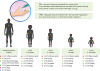
Steroid dosage and fingertip units

The right choice of topical formulation ensures better treatment outcome. Lotion and gel should be used in acute eczema with exudation and blisters, and to hairy regions. Ointment is used for thick, dry areas, and to palms and soles. Cream can be used on all areas [37].
A close approximation of adequate dosage is determined by ‘fingertip units’ (FTU; Fig. 1). FTU is the quantity of cream/ointment extruded from a tube with nozzle of 5-mm diameter covering the length of the distal phalanx of the index finger. It is about 0.45 to 0.5 g of cream and is sufficient to cover an area of 300 cm2. The quantity of cream in a FTU varies with age: adult male: 1 FTU provides 0.5 g; adult female: 1 FTU provides 0.4 g; child aged 4 years – approximately 1/3 of adult amount. A rough guide of dosage is 1 g to face and neck, 8 g to trunk (front and back), 4 g to arms, 6 g to legs, 1 g each to hands, feet and genitals [12161727].
Fig. 1

The Fingertip Unit (FTU) recommended for various age groups.

apa-8-e41-g001
Twice daily application for not more than 3 weeks is effective in most patients [1340]. Side effects, including possible hypothalamic-pituitary-adrenal axis suppression, should be monitored, particularly in children who have chronically used topical corticosteroids. Cutaneous side effects should be monitored in patients using potent steroids for longer than the recommended period. However, no specific monitoring of systemic side effects is recommended [10].
Practice point: The FTU is easily understandable as a unit of measure for both patients and clinicians. It is recommended to explain to the patient how to apply topical products using this measure and to enable clinicians to confidently advice patients without fear of overdosing. [Level 4, GPP]

Topical calcineurin inhibitors

The topical calcineurin inhibitors (TCIs) tacrolimus and pimecrolimus have comparable efficacy to topical steroid in patients with AD. TCIs are recommended for use in patients who have become recalcitrant to steroid, in cases of prolonged uninterrupted use and when there is steroid atrophy, or when there is a need to treat sensitive areas (e.g., face, anogenital area, skin folds) and pediatric patients as a steroid-sparing agent [1041]. TCIs inhibit T-lymphocyte function, which plays a central role in the development of inflammatory reactions [12].
Because tacrolimus ointment and pimecrolimus cream may cause skin discomfort, using topical corticosteroids should be considered first to minimize TCI application site reactions. The concomitant use of a topical corticosteroid with a TCI may be done for the treatment of AD [41].
Tacrolimus ointment is usually applied externally after bathing. A 0.1% tacrolimus ointment for adults should be administered at a dose of 5 g or less. A 0.03% tacrolimus ointment for children aged 2 to 5 years (<20 kg in body weight) should be given at a dose of not more than 1 g; for children aged 6 to 12 years (approximately 20 to 50 kg in body weight), at a dose of 2 to 4 g; and for children at least 13 years old (about 50 kg in body weight), a dose of up to 5 g. It should be administered at a maximum of twice per day. Pimecrolimus is available as a 1% cream and absorbed less than the tacrolimus ointment. When applied twice per day, an interval of approximately 12 hours between applications is recommended.
Occlusive dressing therapy should not be used because it may cause absorption of the drug to the bloodstream. Continuous external application of a tacrolimus ointment 2 to 3 times per week after remission induction can significantly inhibit the relapse of symptoms (proactive therapy) [12162741].
Proactive, intermittent use of TCI is recommended to help prevent relapses while reducing the need for topical corticosteroids and is more effective than the use of emollients alone [12].
The disadvantage of using TCIs is that they are expensive and may cause burning and stinging. Rare cases of skin malignancy have been reported but causal relationship has not been established. Nonetheless, clinicians should be aware of the black-box warning on the use of TCIs and discuss these as warranted [10]. TCIs do not increase the prevalence of cutaneous viral infections with use of up to 5 years; however, physicians should inform their patients of the theoretical risks. Routine blood monitoring of tacrolimus and pimecrolimus levels is not recommended at this time [10].
Practice point: TCIs are recommended for use in patients who have become recalcitrant to steroid, in chronic uninterrupted use and when there is steroid atrophy, or when there is a need to treat sensitive areas and pediatric patients. [Level 1+, B]

Phototherapy

Phototherapy may be used as second-line treatment in patients whose medical, physical, and/or psychological states are greatly affected by their disease, which may include negative impact on social or interpersonal interactions. Phototherapy can be used as maintenance therapy in chronic disease and is often used in severe AD [42]. While the mechanism of action has not been fully elucidated, ultraviolet (UV) is thought to have local anti-inflammatory and immunosuppressive action [41].
Phototherapy with narrowband ultraviolet B or UVB (UVBTL01) and UVA1 is most commonly used. Medium-dose UVA1 may be used for control of acute flares while narrowband-UVB may be used in the management of chronic AD [4143]. High dose UVA1 is useful for control of acute exacerbations. PUVA is particularly useful in AD patients with thick lichenified and keratotic palm and sole. PUVA (psoralen + UVA) lights are effective in patients with active stable disease [13404143].
Phototherapy treatment of all forms should be under the active supervision of a physician who is an expert in phototherapy techniques. The use of daylight phototherapy and home-based phototherapy are options that are currently being explored [42].
Practice point: Phototherapy is a second-line treatment, after failure of first-line treatment (emollients, topical steroids, and TCIs). [Level 2+, C]

Systemic therapy

Systemic immunomodulatory agents may be used in cases that are refractory to conventional therapy, for severe disease with large body surface involvement making topical therapy impractical, or in the event of complications of generalized exfoliative dermatitis [1216414244]. The use of these agents is off-label in most cases. Generally, any patient who requires systemic therapy should always be referred to a dermatologist [44].

Systemic steroids

Systemic steroids have limited role in AD treatment and should be avoided if possible. Judicious use should be exclusively reserved for acute, severe exacerbations and as a short-term bridge therapy to other systemic, steroid-sparing treatment [12131642].
For adults, prednisolone 30 mg daily and tapering to 5 mg within 2–3 weeks may be used in AD recalcitrant to topical therapy [13]. Other oral steroids used equivalent to prednisolone 5 mg are dexamethasone 0.75 mg, methylprednisolone 4 mg, cortisone acetate 25 mg, hydrocortisone 20 mg, betamethasone 0.75 mg, triamcinolone 4 mg, and prednisone 5 mg [45].
In children, a dosage range of 0.5 to 1.0 mg/kg of prednisone or prednisolone as tablet or oral solution for enteral administration, or triamcinolone acetonide as an intramuscular injection, may be used [42]. Clinicians are advised to educate patients about the possibility of adrenal suppression and of rebound flares upon treatment discontinuation [4142].
Practice point: Oral steroids should not be used for all cases and should be given only at a minimum dose and for the shortest duration possible in both adults and children. [Level 1++, A]

Cyclosporine

Cyclosporine is an immunosuppressant of T cells and decreases interleukin (IL)-2 production [42]. It is fast-acting and largely well tolerated by children and inpatients in crisis. It significantly improves AD in the first 1–2 months of therapy. It is especially fast in reducing pruritus in adults and children with chronic severe AD [131642].
Cyclosporine should be given at the lowest effective dose and the shortest treatment period, as toxicity is related to both high dose and prolonged treatment. A standard dose of 150 to 300 mg/day of any oral preparation (e.g., microemulsion) divided into 2 doses, and taken at the same time each day in adults may be sufficient. The initial and maintenance doses will depend on the disease severity and other comorbidities [42].
In children, the starting dose may range from 2.5 mg/kg/day and gradually increased until clinical response is adequate (up to 4–5 mg/kg/day), and a course of up to 3 months is satisfactory [122741].
Side effects are mostly predictable and dose-dependent; these include infection, nephrotoxicity, hypertension, tremor, hypertrichosis, headache, gingival hyperplasia, and an increased risk of skin cancer and lymphoma [42].
Practice point: Monitoring of patients taking cyclosporine includes checking for baseline blood pressure measurements x2, renal function testing, urinalysis with microscopy, fasting lipid profile, complete blood count with differentials (CBC), liver function, electrolytes, uric acid, and testing for tuberculosis, human immunodeficiency virus (HIV) and human chorionic gonadotropin (HCG), if indicated [Level 1++, B].

Azathioprine

Azathioprine is a purine analog that inhibits DNA synthesis. It can be used as a first-line systemic agent in older children and teenagers with chronic long-term moderate to severe AD with high total IgE when cyclosporine is neither effective nor indicated. The onset is slow, typically up to 4 weeks, and it is not typically used as a first-line medication in severe life-impacting AD [13,. 4043].
Dosing in adults is 1 to 3 mg/kg but most guidelines limit use to 2.5 mg/kg/day given as a tablet or compounded liquid, once a day. In children, 1 to 4 mg/kg/day is the allowed dose [12134244].
Myelosuppression and hypersensitivity with skin rash, hepatitis, fever, oliguria, and respiratory failure are rare but fatal adverse effects of azathioprine [27].
Practice point: Monitoring of patients taking azathioprine includes checking for baseline thiopurine methyltransferase or thiopurine S-methyltransferase (TPMT), CBC differentials and platelets, renal function, liver function, hepatitis B and C, and testing for tuberculosis, HIV and HCG, if indicated. [Level 1++, B]

Methotrexate

Methotrexate is an antifolate metabolite that negatively affects T-cell function by blocking DNA, RNA and purine synthesis. Its use in adults with AD has been demonstrated in trials to be comparable to azathioprine. It is significantly less immunosuppressive with preferable long-term safety profile [1213164144].
A standard dose is 7.5 to 10 mg of a solution given intramuscularly or subcutaneously once a week. In children, the dose of 0.2 to 0.7 mg/kg per week has been demonstrated to be effective and safe [42].
Short-term side effects include gastrointestinal upset and bone marrow suppression. Long-term side effects include liver fibrosis, and a potential effect on spermatogenesis and ovulation [42].
Practice point: Monitoring of patients taking methotrexate includes checking of CBC with differentials and platelets, renal function, liver function, hepatitis B and C, and screening for tuberculosis, HIV and HCG, and pulmonary function tests, if indicated. [Level 1++, B]

Mycophenolate mofetil

Mycophenolate mofetil blocks the purine synthesis pathway by inhibition of the inosine monophosphate dehydrogenase enzyme. It is useful for patients in whom other available systemic therapies are contraindicated or not tolerated [1213164144].
There is currently limited data to make recommendations on the dosage of this drug but a short-term oral dose of 1.0 to 1.5 g (up to 2 g) daily as monotherapy as an oral suspension, capsule or tablet, may result in clearing of skin lesion in adults resistant to other treatments [42].
In children, a dose range of 12 to 40 mg/kg daily divided into two doses titrated up to 75 mg/kg (3 g maximum) has been evaluated for pediatric AD patients. The suggested dosing in young children is 40 to 50 mg/kg/day and in young adults is 30 mg/kg/day [42].
It is well tolerated but may cause nausea, vomiting and abdominal cramping in some patients [42].
Practice point: Monitoring in mycophenolate mofetil includes checking for CBC with differentials and platelets, renal function, liver function, and testing for tuberculosis, HIV and HCG, if indicated. [Level 2+, C]

Antibiotics and antivirals

Antibiotics are needed where there is secondary bacterial infection and S. aureus is the suspected pathogen. However, there is no role for long-term oral antibiotic prophylaxis in clinically uninfected AD due to the risk of bacterial resistance and contact sensitization [1213164144]. Acyclovir and valacyclovir should be instituted without delay to patients with eczema herpeticum [1213164144].
Topical antibiotics (e.g., fusidic acid, mupirocin) may be used for focal infections. However, there is no evidence supporting their long-term use [13]. Creams are preferred for exudative skin lesions while ointments are useful for dry lesions with desquamation [43].
The recommendation is that they should be applied twice a day with bandage or 3 times a day without bandage for 7 to 10 days. Prolonged use of mupirocin may promote the emergence of resistant strains and use beyond 10 days and is thus not recommended [28].
  • Practice point:

  • · Systemic antibiotics may be used in the treatment of bacterial infections in conjunction with other standard and appropriate treatments for AD. [Level 1+, B]

  • · Systemic antivirals are indicated for eczema herpeticum. [Level 2+, B]

  • · Topical antibiotics may be used for focal skin infections for 7 to 10 days. [Level 2+, B]

Antihistamines

AD patients who suffer from dermographism, allergic rhinitis, severe itch, or other allergic illnesses may require symptomatic relief with these agents. Antihistamines generally result in partial relief [121642].
Short-term, intermittent use of sedating antihistamines may be beneficial owing to their added effect of inducing sleep. However, nonsedating oral antihistamines do not have sufficient supporting evidence to be recommended in AD. Topical antihistamines are also not recommended [41].
Practice point: Patient reliance on systemic antihistamines may indicate that the treatment plan is not sufficient to manage the symptoms. [Level 2+, C]

Complementary treatment for AD

The use of folk remedies or complementary/alternative therapies is deeply ingrained in the culture of many countries in Asia [121338404144]. These are summarized in Table 6.
Table 6

Complementary/alternative therapies used for atopic dermatitis

apa-8-e41-i006
Treatment Description Overall implications
Acupressure Use of a small titanium bead to massage an acupoint on the arm 3 times weekly to relieve pruritus and lichenification Studies limited by small number of subjects, absence of placebo and unmonitored application
Acupuncture Use of acupuncture needles to relieve allergen-induced itch intensity vs. placebo or antihistamine
Aromatherapy/massage Use of manual therapy for stress-relief is adjunctive in treatment of atopic dermatitis symptoms; improves sleep disruption Counselling and the use of relaxation therapy could have confounded any potential beneficial effects of the intervention; a much larger and better designed trial of a more representative population is needed; aromatherapy oils may be a contact allergen
Traditional herbs Use of various kinds of medicinal plants alone or in combination with others as a decoction by boiling them in water taken as a ‘tea’ or applied directly to the skin Most extensively studied in this list; clearly reported and blinded multinational trials which focus on outcomes such as quality of life and adverse events (e.g., contaminant steroid toxicity, hepatotoxicity) are necessary; quality control is a key issue
In general, there is limited evidence available that supports the routine use of these therapies. Patients should be advised that these have not been sufficiently assessed for efficacy and safety. Some traditional herbs may have unknown quantities of active contaminants (i.e., corticosteroids), potential interactions with other medications, and may contribute to flares if the patient has hypersensitivity to any one of the ingredients [38].
Practice point: Patients and their caregivers should be advised that complementary/alternative therapies have not undergone sufficient efficacy and safety evaluation. They should be encouraged to share the use of such options during their visits. [Level 4, GPP]

Future research directions

There is emerging evidence on adjunctive therapies for AD. More information regarding the safety and efficacy of these agents will be available in the future.
  • · Naltrexone is an opiate receptor antagonist that may be used in difficult-to-treat AD cases [13].

  • · Aprepitant, a new neurokinin-receptor antagonist, has been shown to be effective in resolving chronic pruritus. More studies are needed to establish the role of these agents in AD [46].

  • · A new generation of moisturizers with antioxidants, such as vitamins, polyphenols, furfuryl palmitate and grape seed oil with antipruritic agents, have been shown to significantly improve AD symptomatically at the same level as topical corticosteroids. More trials will be available showing its effects on epidermal permeability barrier function in the future [46].

  • · Nutrient supplementation may be of benefit in preventing AD development and in reducing the severity of flares. The overall result of a meta-analysis suggests that probiotics could be an option for the treatment of AD, especially for moderate to severe AD in children and adults [47]. A recent systematic review identified Lactobacillus rhamnosus GG as the most frequently studied probiotic strain for AD [48]. Further study is needed to better understand the mechanism of these agents.

  • · Dupilumab, a biologic with IL-4 and IL-13-blocking activities, provided as 2 injections a month has been shown to be highly effective. This option is useful for adults with moderate-to-severe AD that has failed systemic treatment and may be available for adolescents in the future. Consult your pharmacies and local formularies regarding approval of this option [1349].

  • · Omalizumab, an anti-IgE antibody, is effective in patients with AD and asthma with blood IgE of 30–700 IU. It is currently available for use for asthma and chronic spontaneous urticaria [13].

  • · Intravenous immunoglobulin has not been shown to be effective in recent studies and is not recommended for use in AD [40].

  • · To date, the role of vitamin D supplementation in AD remains to be unclear. It is known that cathelicidins in the skin are relatively deficient in individuals with AD, and that vitamin D may mediate the expression of innate cathelicidins in the skin. Interventional studies have yielded mixed results [132940].

  • · A phosphodiesterase 4 inhibitor, roflumilast, is one of the latest nonsteroidal topical therapy options in the armamentarium of AD management [50].

  • · Novel targeting agents used to treat recalcitrant pruritus have been evaluated in a small number of studies. Specific mediators and pathways, such as the protease-activated receptor 2, the H4 histamine pathway and the transient receptor potential vanilloid (TRPV) ion channels, are continually being studied in the development of topical antipruritic options [51]. Apart from their antipruritic properties, TRPV1 antagonists appear to play a role in maintaining epidermal barrier function and may be available as an option in the near future [52].

ACKNOWLEDGEMENTS

The Asian Academy of Dermatology & Venereology has received publication support and funding from A. Menarini Pte Ltd but has maintained editorial independence. Dr Dennis Malvin H. Malgapo of MIMS Pte Ltd provided editorial and technical writing support. The authors have no competing interests to disclose.

Notes

Author Contributions

  • Conceptualization: Steven KW Chow, Chew Swee Seow, Maria Victoria Dizon, Kiran Godse, Henry Foong, Vicheth Chan, Tran Hau Khang, Leihong Xiang, Syarief Hidayat, M. Yulianto Listiawan, Danang Triwahyudi, Srie Prihianti Gondokaryono, Endang Sutedja, Inne Arline Diana, Oki Suwarsa, Hartati Purbo Dharmadji, Agnes Sri Siswati, Retno Danarti, Retno Soebaryo, Windy Keumala Budianti.

  • Data curation: Seow Chew Swee, Maria Victoria Dizon, Kiran Godse, Henry Foong, Vicheth Chan, Tran Hau Khang, Leihong Xiang, Syarief Hidayat, M. Yulianto Listiawan, Danang Triwahyudi, Srie Prihianti Gondokaryono, Endang Sutedja, Inne Arline Diana, Oki Suwarsa, Hartati Purbo Dharmadji, Agnes Sri Siswati, Retno Danarti, Retno Soebaryo, Windy Keumala Budianti.

  • Funding acquisition: Syarief Hidayat.

  • Investigation: Steven KW Chow, Chew Swee Seow, Kiran Godse, Henry Foong, Tran Hau Khang, Inne Arline Diana, Retno Danarti, Retno Soebaryo, Windy Keumala Budianti.

  • Project administration: Srie Prihianti Gondokaryono.

  • Resources: Srie Prihianti Gondokaryono.

  • Supervision: Chew Swee Seow.

  • Validation: Steven KW Chow.

  • Writing - original draft: Steven KW Chow, Chew Swee Seow, Kiran Godse, Henry Foong, Tran Hau Khang, Inne Arline Diana, Retno Danarti, Retno Soebaryo, Windy Keumala Budianti.

  • Writing - review & editing: Chew Swee Seow, Maria Victoria Dizon, Kiran Godse, Henry Foong, Vicheth Chan, Tran Hau Khang, Leihong Xiang, Syarief Hidayat, M. Yulianto Listiawan, Danang Triwahyudi, Srie Prihianti Gondokaryono, Endang Sutedja, Inne Arline Diana, Oki Suwarsa, Hartati Purbo Dharmadji, Agnes Sri Siswati, Retno Danarti, Retno Soebaryo, Windy Keumala Budianti.

References

1. Nutten S. Atopic dermatitis: global epidemiology and risk factors. Ann Nutr Metab. 2015; 66:Suppl 1. 8–16.
crossref
2. Leung DY, Boguniewicz M, Howell MD, Nomura I, Hamid QA. New insights into atopic dermatitis. J Clin Invest. 2004; 113:651–657.
crossref
3. Leung DY. New insights into atopic dermatitis: role of skin barrier and immune dysregulation. Allergol Int. 2013; 62:151–161.
crossref
4. Suárez-Fariñas M, Dhingra N, Gittler J, Shemer A, Cardinale I, de Guzman Strong C, Krueger JG, Guttman-Yassky E. Intrinsic atopic dermatitis shows similar TH2 and higher TH17 immune activation compared with extrinsic atopic dermatitis. J Allergy Clin Immunol. 2013; 132:361–370.
crossref
5. Kim KH. Overview of atopic dermatitis. Asia Pac Allergy. 2013; 3:79–87.
crossref
6. Chen H, Common JE, Haines RL, Balakrishnan A, Brown SJ, Goh CS, Cordell HJ, Sandilands A, Campbell LE, Kroboth K, Irvine AD, Goh DL, Tang MB, van Bever HP, Giam YC, McLean WH, Lane EB. Wide spectrum of filaggrin-null mutations in atopic dermatitis highlights differences between Singaporean Chinese and European populations. Br J Dermatol. 2011; 165:106–114.
crossref
7. Noda S, Suárez-Fariñas M, Ungar B, Kim SJ, de Guzman Strong C, Xu H, Peng X, Estrada YD, Nakajima S, Honda T, Shin JU, Lee H, Krueger JG, Lee KH, Kabashima K, Guttman-Yassky E. The Asian atopic dermatitis phenotype combines features of atopic dermatitis and psoriasis with increased TH17 polarization. J Allergy Clin Immunol. 2015; 136:1254–1264.
crossref
8. Chan YC, Tay YK, Sugito TL, Boediardja SA, Chau DD, Nguyen KV, Yee KC, Alias M, Hussein S, Dizon MV, Roa F, Chan YH, Wananukul S, Kullavanijaya P, Singalavanija S, Cheong WK. A study on the knowledge, attitudes and practices of Southeast Asian dermatologists in the management of atopic dermatitis. Ann Acad Med Singapore. 2006; 35:794–803.
9. Ho SG, Chan HH. The Asian dermatologic patient: review of common pigmentary disorders and cutaneous diseases. Am J Clin Dermatol. 2009; 10:153–168.
10. Eichenfield LF, Tom WL, Berger TG, Krol A, Paller AS, Schwarzenberger K, Bergman JN, Chamlin SL, Cohen DE, Cooper KD, Cordoro KM, Davis DM, Feldman SR, Hanifin JM, Margolis DJ, Silverman RA, Simpson EL, Williams HC, Elmets CA, Block J, Harrod CG, Smith Begolka W, Sidbury R. Guidelines of care for the management of atopic dermatitis: section 2. Management and treatment of atopic dermatitis with topical therapies. J Am Acad Dermatol. 2014; 71:116–132.
11. Hanifin JM, Rajka G. Diagnostic features of atopic eczema. Acta Derm Venereol (Stockh). 1980; 92:44–47.
12. Katayama I, Aihara M, Ohya Y, Saeki H, Shimojo N, Shoji S, Taniguchi M, Yamada H. Japanese Society of Allergology. Japanese guidelines for atopic dermatitis 2017. Allergol Int. 2017; 66:230–247.
crossref
13. Nankervis H, Thomas KS, Delamere FM, Barbarot S, Rogers NK, Williams HC. Scoping systematic review of treatments for eczema. Southampton (UK): NIHR Journals Library;2016.
14. Saeki H, Nakahara T, Tanaka A, Kabashima K, Sugaya M, Murota H, Ebihara T, Kataoka Y, Aihara M, Etoh T, Katoh N. Committee for Clinical Practice Guidelines for the Management of Atopic Dermatitis of Japanese Dermatological Association. Clinical Practice Guidelines for the Management of Atopic Dermatitis 2016. J Dermatol. 2016; 43:1117–1145.
crossref
15. Eichenfield LF, Tom WL, Chamlin SL, Feldman SR, Hanifin JM, Simpson EL, Berger TG, Bergman JN, Cohen DE, Cooper KD, Cordoro KM, Davis DM, Krol A, Margolis DJ, Paller AS, Schwarzenberger K, Silverman RA, Williams HC, Elmets CA, Block J, Harrod CG, Smith Begolka W, Sidbury R. Guidelines of care for the management of atopic dermatitis: section 1. Diagnosis and assessment of atopic dermatitis. J Am Acad Dermatol. 2014; 70:338–351.
16. Rubel D, Thirumoorthy T, Soebaryo RW, Weng SC, Gabriel TM, Villafuerte LL, Chu CY, Dhar S, Parikh D, Wong LC, Lo KK. Asia-Pacific Consensus Group for Atopic Dermatitis. Consensus guidelines for the management of atopic dermatitis: an Asia-Pacific perspective. J Dermatol. 2013; 40:160–171.
crossref
17. Eichenfield LF, Boguniewicz M, Simpson EL, Russell JJ, Block JK, Feldman SR, Clark AR, Tofte S, Dunn JD, Paller AS. Translating atopic dermatitis management guidelines into practice for primary care providers. Pediatrics. 2015; 136:554–565.
crossref
18. Oranje AP. Practical issues on interpretation of scoring atopic dermatitis: SCORAD Index, objective SCORAD, patient-oriented SCORAD and Three-Item Severity score. Curr Probl Dermatol. 2011; 41:149–155.
crossref
19. Hanifin JM, Thurston M, Omoto M, Cherill R, Tofte SJ, Graeber M. The eczema area and severity index (EASI): assessment of reliability in atopic dermatitis. EASI Evaluator Group. Exp Dermatol. 2001; 10:11–18.
20. Grinich EE, Schmitt J, Küster D, Spuls PI, Williams HC, Chalmers JR, Thomas KS, Apfelbacher C, Prinsen CAC, Furue M, Stuart B, Carter B, Simpson EL. Standardized reporting of the Eczema Area and Severity Index (EASI) and the Patient-Oriented Eczema Measure (POEM): a recommendation by the Harmonising Outcome Measures for Eczema (HOME) Initiative. Br J Dermatol. 2018; 179:540–541.
crossref
21. Wolkerstorfer A, de Waard van der Spek FB, Glazenburg EJ, Mulder PG, Oranje AP. Scoring the severity of atopic dermatitis: three item severity score as a rough system for daily practice and as a pre-screening tool for studies. Acta Derm Venereol. 1999; 79:356–359.
22. Chu CY. Measuring quality of life in infants, children and adolescents with eczema. Br J Dermatol. 2017; 176:848–849.
crossref
23. Heinl D, Prinsen CA, Deckert S, Chalmers JR, Drucker AM, Ofenloch R, Humphreys R, Sach T, Chamlin SL, Schmitt J, Apfelbacher C. Measurement properties of adult quality-of-life measurement instruments for eczema: a systematic review. Allergy. 2016; 71:358–370.
crossref
24. Leung DY, Guttman-Yassky E. Assessing the current treatment of atopic dermatitis: Unmet needs. J Allergy Clin Immunol. 2017; 139:4S. S47–S48.
crossref
25. Leong KF. My Eczema Action Plan [Internet]. cited 2018 Oct 25. Available from: https://www.kairos2.com/my_eczema_action_plan.pdf/.
26. Ridd MJ, King AJL, Le Roux E, Waldecker A, Huntley AL. Systematic review of self-management interventions for people with eczema. Br J Dermatol. 2017; 177:719–734.
crossref
27. Kim JE, Kim HJ, Lew BL, Lee KH, Hong SP, Jang YH, Park KY, Seo SJ, Bae JM, Choi EH, Suhr KB, Lee SC, Ko HC, Park YL, Son SW, Seo YJ, Lee YW, Cho SH, Park CW, Roh JY. Consensus Guidelines for the Treatment of Atopic Dermatitis in Korea (Part I): General Management and Topical Treatment. Ann Dermatol. 2015; 27:563–577.
crossref
28. Stalder JF, Bernier C, Ball A, De Raeve L, Gieler U, Deleuran M, Marcoux D, Eichenfield LF, Lio P, Lewis-Jones S, Gelmetti C, Takaoka R, Chiaverini C, Misery L, Barbarot S. Oriented Patient-Education Network in Dermatology (OPENED). Therapeutic patient education in atopic dermatitis: worldwide experiences. Pediatr Dermatol. 2013; 30:329–334.
crossref
29. Sidbury R, Tom WL, Bergman JN, Cooper KD, Silverman RA, Berger TG, Chamlin SL, Cohen DE, Cordoro KM, Davis DM, Feldman SR, Hanifin JM, Krol A, Margolis DJ, Paller AS, Schwarzenberger K, Simpson EL, Williams HC, Elmets CA, Block J, Harrod CG, Smith Begolka W, Eichenfield LF. Guidelines of care for the management of atopic dermatitis: Section 4. Prevention of disease flares and use of adjunctive therapies and approaches. J Am Acad Dermatol. 2014; 71:1218–1233.
30. Barbarot S, Bernier C, Deleuran M, De Raeve L, Eichenfield L, El Hachem M, Gelmetti C, Gieler U, Lio P, Marcoux D, Morren MA, Torrelo A, Stalder JF. Oriented Patient-Education Network in Dermatology. Therapeutic patient education in children with atopic dermatitis: position paper on objectives and recommendations. Pediatr Dermatol. 2013; 30:199–206.
crossref
31. Gonzales F, Ramdane N, Delebarre-Sauvage C, Modiano P, Duhamel A, Lasek A. Monitoring of topical corticosteroid phobia in a population of parents with children with atopic dermatitis using the TOPICOP® scale: prevalence, risk factors and the impact of therapeutic patient education. J Eur Acad Dermatol Venereol. 2017; 31:e172–4.
32. Giam YC, Hebert AA, Dizon MV, Van Bever H, Tiongco-Recto M, Kim KH, Soebono H, Munasir Z, Diana IA, Luk DC. A review on the role of moisturizers for atopic dermatitis. Asia Pac Allergy. 2016; 6:120–128.
crossref
33. Arkwright PD, Motala C, Subramanian H, Spergel J, Schneider LC, Wollenberg A. Atopic Dermatitis Working Group of the Allergic Skin Diseases Committee of the AAAAI. Management of difficult-to-treat atopic dermatitis. J Allergy Clin Immunol Pract. 2013; 1:142–151.
crossref
34. Varothai S, Nitayavardhana S, Kulthanan K. Moisturizers for patients with atopic dermatitis. Asian Pac J Allergy Immunol. 2013; 31:91–98.
35. Sethi A, Kaur T, Malhotra SK, Gambhir ML. Moisturizers: the slippery road. Indian J Dermatol. 2016; 61:279–287.
crossref
36. Koh MJ, Giam YC, Liew HM, Foong AY, Chong JH, Wong SM, Tang MB, Ho MS, Tan LS, Mason JM, Cork MJ. Comparison of the Simple Patient-Centric Atopic Dermatitis Scoring System PEST with SCORAD in young children using a ceramide dominant therapeutic moisturizer. Dermatol Ther (Heidelb). 2017; 7:383–393.
crossref
37. van Zuuren EJ, Fedorowicz Z, Christensen R, Lavrijsen A, Arents BW. Emollients and moisturisers for eczema. Cochrane Database Syst Rev. 2017; 2:CD012119.
crossref
38. Vieira BL, Lim NR, Lohman ME, Lio PA. Complementary and alternative medicine for atopic dermatitis: an evidence-based review. Am J Clin Dermatol. 2016; 17:557–581.
crossref
39. Wollenberg A, Oranje A, Deleuran M, Simon D, Szalai Z, Kunz B, Svensson A, Barbarot S, von Kobyletzki L, Taieb A, de Bruin-Weller M, Werfel T, Trzeciak M, Vestergard C, Ring J, Darsow U. European Task Force on Atopic Dermatitis/EADV Eczema Task Force. ETFAD/EADV Eczema task force 2015 position paper on diagnosis and treatment of atopic dermatitis in adult and paediatric patients. J Eur Acad Dermatol Venereol. 2016; 30:729–747.
crossref
40. Hoare C, Li Wan Po A, Williams H. Systematic review of treatments for atopic eczema. Health Technol Assess. 2000; 4:1–191.
crossref
41. Chu CY, Lee CH, Shih IH, Chen HC, Huang PH, Yang CY, Wang WJ, Chen YJ, Sheu HM, Wang WM, Lee WR, Lo YH, Dai YS, Wang LF, Tsai TF, Yang CH. Taiwanese Dermatological Association consensus for the management of atopic dermatitis. Dermatol Sin. 2015; 33:220–230.
crossref
42. Sidbury R, Davis DM, Cohen DE, Cordoro KM, Berger TG, Bergman JN, Chamlin SL, Cooper KD, Feldman SR, Hanifin JM, Krol A, Margolis DJ, Paller AS, Schwarzenberger K, Silverman RA, Simpson EL, Tom WL, Williams HC, Elmets CA, Block J, Harrod CG, Begolka WS, Eichenfield LF. American Academy of Dermatology. Guidelines of care for the management of atopic dermatitis: section 3. Management and treatment with phototherapy and systemic agents. J Am Acad Dermatol. 2014; 71:327–349.
43. Galli E, Neri I, Ricci G, Baldo E, Barone M, Belloni Fortina A, Bernardini R, Berti I, Caffarelli C, Calamelli E, Capra L, Carello R, Cipriani F, Comberiati P, Diociaiuti A, El Hachem M, Fontana E, Gruber M, Haddock E, Maiello N, Meglio P, Patrizi A, Peroni D, Scarponi D, Wielander I, Eichenfield LF. Consensus conference on clinical management of pediatric atopic dermatitis. Ital J Pediatr. 2016; 42:26.
crossref
44. Kim JE, Kim HJ, Lew BL, Lee KH, Hong SP, Jang YH, Park KY, Seo SJ, Bae JM, Choi EH, Suhr KB, Lee SC, Ko HC, Park YL, Son SW, Seo YJ, Lee YW, Cho SH, Park CW, Roh JY. Consensus Guidelines for the Treatment of Atopic Dermatitis in Korea (Part II): Systemic Treatment. Ann Dermatol. 2015; 27:578–592.
crossref
45. Liu D, Ahmet A, Ward L, Krishnamoorthy P, Mandelcorn ED, Leigh R, Brown JP, Cohen A, Kim H. A practical guide to the monitoring and management of the complications of systemic corticosteroid therapy. Allergy Asthma Clin Immunol. 2013; 9:30.
crossref
46. Man G, Elias PM, Man MQ. Therapeutic benefits of enhancing permeability barrier for atopic eczema. Dermatologica Sin. 2015; 33:84–89.
crossref
47. Foolad N, Brezinski EA, Chase EP, Armstrong AW. Effect of nutrient supplementation on atopic dermatitis in children: a systematic review of probiotics, prebiotics, formula, and fatty acids. JAMA Dermatol. 2013; 149:350–355.
48. Kim SO, Ah YM, Yu YM, Choi KH, Shin WG, Lee JY. Effects of probiotics for the treatment of atopic dermatitis: a meta-analysis of randomized controlled trials. Ann Allergy Asthma Immunol. 2014; 113:217–226.
crossref
49. Ariëns LFM, Bakker DS, van der Schaft J, Garritsen FM, Thijs JL, de Bruin-Weller MS. Dupilumab in atopic dermatitis: rationale, latest evidence and place in therapy. Ther Adv Chronic Dis. 2018; 9:159–170.
crossref
50. Sakkas LI, Mavropoulos A, Bogdanos DP. Phosphodiesterase 4 inhibitors in immune-mediated diseases: mode of action, clinical applications, current and future perspectives. Curr Med Chem. 2017; 24:3054–3067.
crossref
51. Tey HL, Yosipovitch G. Targeted treatment of pruritus: a look into the future. Br J Dermatol. 2011; 165:5–17.
crossref
52. Bonchak JG, Swerlick RA. Emerging therapies for atopic dermatitis: TRPV1 antagonists. J Am Acad Dermatol. 2018; 78:3S1. S63–S66.
crossref
TOOLS
ORCID iDs

Steven Chow
https://orcid.org/0000-0002-9094-2653

Similar articles