Journal List > Immune Netw > v.18(4) > 1108116

Ignacio, Lee, Wilson, Beeghly-Fadiel, Whalen, and Son: Chemokine Network and Overall Survival in TP53 Wild-Type and Mutant Ovarian Cancer

Abstract

Ovarian cancer (OC) has the highest mortality rate among gynecological malignancies. Because chemokine network is involved in OC progression, we evaluated associations between chemokine expression and survival in tumor suppressor protein p53 (TP53) wild-type (TP53WT) and mutant (TP53m) OC datasets. TP53 was highly mutated in OC compared to other cancer types. Among OC subtypes, CXCL14 was predominantly expressed in clear cell OC, and CCL15 and CCL20 in mucinous OC. TP53WT endometrioid OC highly expressed CXCL14 compared to TP53m, showing better progression-free survival but no difference in overall survival (OS). TP53m serous OC highly expressed CCL8, CCL20, CXCL10 and CXCL11 compared to TP53WT. CXCL12 and CCL21 were associated with poor OS in TP53WT serous OC. CXCR2 was associated with poor OS in TP53m serous OC, while CXCL9, CCL5, CXCR4, CXCL11, and CXCL13 were associated with better OS. Taken together, specific chemokine signatures may differentially influence OS in TP53WT and TP53m OC.

Abbreviations

CI

confidence interval

EOC

epithelial ovarian cancer

GEO

Gene Expression Omnibus

HGSOC

high-grade serous ovarian cancer

HR

hazard ratio

LGSOC

low-grade serous ovarian cancer

NCBI

National Center for Biotechnology Information

OC

ovarian cancer

OS

overall survival

PFS

progression-free survival

RFS

recurrence-free survival

TCGA

The Cancer Genome Atlas

TP53

tumor suppressor protein p53

TP53m

tumor suppressor protein p53 mutant

TP53WT

tumor suppressor protein p53 wild-type

INTRODUCTION

Ovarian cancer (OC) is the fifth leading cause of cancer deaths among women with 22,240 new cases and 14,070 deaths estimated in the US in 2018 (1). OC alone accounts for 5% of cancer deaths, which is due to the fact that early-stage OC is asymptomatic, thus when diagnosed it has frequently already spread throughout the abdominal cavity (2). Although the 5-year survival rate of OC is 92% for women diagnosed at an early-localized stage, it has low survival rate, only 17% to 28% for those with advanced-diseased stage (3). OC has been classified based on cell of origin, such as epithelial ovarian cancer (EOC) which arises in the epithelium and accounts for up to 90% of cases, and non-EOCs from germ cells for 3% and sex cord-stromal cells for 2% (4). Furthermore, EOC is histologically subdivided into 4 subtypes such as serous (up to 70%), endometrioid (10%), mucinous (6%), and clear cell (6%) (45) that differ in their epidemiologic, genetic changes, tumor markers, and response to therapy (6). In particular, high-grade serous ovarian cancer (HGSOC) is highly-aggressive, is diagnosed at advanced-stage, and has a poor prognosis (7). A frequent molecular alteration that accounts to over 95% of cases of HGSOC is the mutation in the tumor suppressor protein p53 (TP53) (89). In contrast, low-grade serous ovarian cancer (LGSOC) as well as other subtypes, such as mucinous, endometrioid and clear cells frequently do not carry TP53 mutations (1011).
To increase the survival rate of advanced OC, we need to identify molecular drivers that can serve as prognostic markers. Chemokines and their receptors have recently been receiving attention due to their possible involvement in OC progression and metastasis (1213). Chemokines, chemotactic cytokines interacting with G-protein-coupled receptors, contribute to cell proliferation, inflammation, metastasis, and tumorigenesis (141516). TP53 mutations play a significant role in shifting the effects of inflammation toward oncogenic outcomes, making cancer cells more aggressive in response to inflammatory cytokines (17). We have shown that the frequent mutation of TP53 in OC enhances proinflammatory chemokines, leading to inflammatory tumor microenvironment (18). In addition, TP53 inactivation during carcinogenesis affects immune surveillance by interfering with chemokine expression (19).
To date, there have been no prior reports on patient survival based on TP53 status and chemokines in OC. Here, we investigated the chemokine network in different subtypes of human EOC, focusing on whether there is association in TP53 status and chemokine network and how this correlates with OC survival.

MATERIALS AND METHODS

Data analysis

Data analysis was performed on publicly available microarray data-sets that were deposited in the National Center for Biotechnology Information (NCBI) Gene Expression Omnibus (GEO) (http://www.ncbi.nlm.nih.gov/geo/) database under accession numbers GSE6008 and GSE63885. For GSE6008, RNA expression data were from 99 individual OC cases (37 endometrioid, 41 serous, 13 mucinous, and 8 clear cell carcinomas) and 4 individual normal ovarian samples. GSE63885 is gene expression data from 101 OC surgical samples, including 71 serous carcinomas, for which somatic TP53 gene mutation status was available.
We utilized Gitools 2.3.1 (http://www.gitools.org) based on Oracle Java 7, an open-source tool to perform Genomic Data Analysis and Visualization as interactive heat-maps (20). Kaplan-Meier plotter database (http://kmplot.com/analysis/index.php?p=service&cancer=ovar) was utilized to evaluate progression-free survival (PFS) and overall survival (OS) using proportional hazards regression to estimate hazard ratios (HRs) and 95% confidence intervals (CIs) based on gene expression profile of chemokines among 1,656 OC patients from GEO and The Cancer Genome Atlas (TCGA); chemokines were specified with probe sets (Affymetrix HG-U133A, HG-U133A 2.0, and HG-U133 Plus 2.0 microarrays) (21). TP53 alteration frequency profile across many types of cancers was acquired from cBioPortal (www.cbioportal.org) (2223); studies with <25% of altered samples were not included. Data on expression levels were analyzed using Student's t-test and 1-way ANOVA as appropriate. If statistical significance (p≤0.05) was indicated by ANOVA, then data were further analyzed using Tukey's pairwise comparisons to detect specific group differences.

RESULTS AND DISCUSSION

TP53 is the most frequently altered gene in many types of malignancies, with mutations in at least 50% of human cancers. First, we checked the alteration frequency in TP53 across different types of cancers. TP53 mutation was common across many malignancies, while deletion, amplification and multiple alterations in TP53 occurred in only a few types of cancers (Fig. 1). OC in the published TCGA dataset, which contains only HGSOC (9), had the highest percentage of TP53 mutation (Fig. 1). Even all of de novo HGSOC are supposed to contain TP53 somatic mutations or deletions, except for the rare HGSOC that develop from a low-grade serous tumor precursor (24).
Figure 1

Analysis of alteration frequency (%) in TP53 across many cancer types. The 150 studies deposited in cBioPortal (www.cbioportal.org) were assessed to analyze the TP53% alteration frequency profile. The 68 studies with <25% alteration frequency were filtered out to create this graph. Green, blue, red and gray bars indicate mutation, deletion, amplification and multiple alterations in TP53, respectively. The dot line indicates 80%.

in-18-e29-g001
OC is associated with chronic inflammation (25). Previously, we reported that loss or mutation of TP53 may promote tumor progression by enhancing proinflammatory chemokines in OC (18). Chemokines and their receptors are differentially expressed in tumors, influencing cancer progression. We then evaluated whether the different subtypes of EOC would show different patterns of chemokine expression profiles based on publicly available GEO data (GSE6008). Clear cell OC has high CXCL14 expression, while mucinous OC had high CCL15 and CCL20 expression (Fig. 2). Functional roles of these chemokines in clear cell and mucinous OC are largely unknown. On the other hand, normal ovarian samples highly expressed CCL18 and CXCL12 compared to other EOC subtypes (Fig. 2). Consistently, normal ovarian stroma expressed a higher level of CXCL12 compared to EOC (26). Although serum CCL18 was elevated in patients with EOC (27), patients with higher CCL18 in tumor samples had better OS in OC (HR=0.88; 95% CI=0.77–1.00; n=1,656) based on our analysis. Data of TP53 mutation in clear cell and mucinous OC subtypes are not available for statistical analysis because of limited data. Next, we classified endometrioid EOC into TP53 wild-type (TP53WT) and TP53 mutant (TP53m) and assessed chemokine signatures. CXCL14 was highly expressed in TP53WT endometrioid OC compared to TP53m (Fig. 3A). Because of limited data to identify TP53WT and TP53m endometrioid OC patients for OS measurement, we used total endometrioid OC to investigate OS and PFS based on the expression levels of CXCL14. Patients with high expression of CXCL14 has better PFS (HR=0.22; 95% CI=0.08–0.63) but unchanged OS in endometrioid OC (Fig. 3B). CXCL14 protein level is positively correlated to OS of breast cancer patients (28). Moreover, CXCL14 acts as a tumor suppressor gene that is epigenetically silenced during lung tumorigenesis, and the re-expression of CXCL14 leads to increased cell death and reduced growth of lung tumor xenografts (29). CXCL14 is a potent angiostatic chemokine which prevents chemokine- and growth factor-induced angiogenesis (30). High expression levels of CXCL14 in clear cell and endometrioid OC may be one reason why these subtypes have better prognosis than serous OC. Interestingly, CXCL14 was reported to be expressed in normal tissue, such as the epithelia (31), and OC stroma (26).
Figure 2

Chemokine and chemokine receptor signatures in EOC subtypes. Heatmap of chemokine expression profiles in EOC subtypes, including clear cell (n=8, yellow), endometrioid (n=37, blue), mucinous (n=13, green), and serous (n=41, red), and normal ovarian samples (n=4, black) from the NCBI GEO (http://www.ncbi.nlm.nih.gov/geo/) database (GSE6008) using Gitools 2.3.1. Bold yellow, green and black letters indicate dominant chemokines in clear cell, mucinous and normal samples, respectively. The right panel indicates statistical analysis of chemokine expression intensities using ANOVA and Tukey's pairwise comparisons.

in-18-e29-g002
Figure 3

Chemokine and chemokine receptor signatures in TP53WT and TP53m endometrioid OC. (A) Heatmap of chemokine and chemokine receptor expression profiles in TP53WT (n=22) and TP53m (n=15) endometrioid OC from NCBI GEO (http://www.ncbi.nlm.nih.gov/geo/) database (GSE6008) using Gitools 2.3.1. Blue letter indicates the dominant chemokine in TP53WT endometrioid OC. Right panel indicates statistical analysis of chemokine expression intensities using Student's t-test. (B) Kaplan-Meier plots for OS and PFS based on expression levels of CXCL14 in endometrioid OC patients from GEO and TCGA (probes; Affymetrix HG-U133A, HG-U133A 2.0, and HG-U133 Plus 2.0 microarrays). Black and red letters indicate low and high expression of CXCL14, respectively.

in-18-e29-g003
We determined the chemokine signature in TP53WT and TP53m serous EOC subtype from GEO dataset (GSE63885). Chemokines, such as CCL8, CCL20, CXCL10 and CXCL11, are highly expressed in TP53m compared to TP53WT serous OC (Fig. 4). Our previous study showed that CCL20 is expressed dominantly in CXCR2-driven OC, leading to shorter survival, greater tumor spread in the peritoneal cavity, and larger tumor weights in xenograft OC models (26). Chemokines can exert pro- and anti-tumorigenic effects, depending upon the context (32). CXCL10 is thought to impair angiogenesis and has anti-tumor actions (3334). Increased expression of CXCL10 had also been linked with advanced human malignancies such as melanoma (35) and OC (36). The chemokine network is involved in the metastasis of TP53WT and TP53m malignancies. The mutant TP53 protein enhances the secretion of CXC chemokines via the NF‐κB pathway, leading to increase cell migration (37). Particularly, CXCL1 and CXCL8, implicated in cancer invasiveness and angiogenesis, are downregulated in TP53WT-transfected OC (18). In addition, increased expression levels of CXCL5, CXCL8, and CXCL12 were found in TP53m cells compared to TP53WT cells (37). In several malignancies such as lung, melanoma and breast cancer, knockdown of the mutant TP53 protein led to reduced chemokine levels and cell migration (37).
Figure 4

Chemokine and chemokine receptor signatures in TP53WT and TP53m serous OC. (A) Heatmap of chemokine and chemokine receptor expression profiles in TP53WT (n=9) and TP53m (n=62) serous EOC from NCBI GEO (http://www.ncbi.nlm.nih.gov/geo/) database (GSE63885) using Gitools 2.3.1. Red letters indicate dominant chemokines in TP53m serous OC. Right panel indicates statistical analysis of chemokine expression intensities using Student's t-test.

in-18-e29-g004
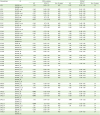
Finally, we utilized datasets available in the Kaplan-Meier plotter database to evaluate OS based on chemokine signatures in TP53WT and TP53m serous OC. Tables 1 and 2 show HRs for OS based on chemokine and chemokine receptor signatures in TP53WT and TP53m serous OC, respectively. High expression levels of CXCL12 (HR=1.84; 95% CI=1.05–3.22) and CCL21 (HR=1.76; 95% CI=1.01–3.06) were associated with poor OS in TP53WT serous OC (Fig. 5A and B). In a similar study, EOC patients with highly expressed CXCL12 had poor survival compared to patients with lower levels (13). High expression of CXCR2 (HR=1.34; 95% CI=1.06–1.69) was associated with poor OS in TP53m serous OC, while high expressions of CXCL9 (HR=0.78; 95% CI=0.62–0.98), CCL5 (HR=0.77; 95% CI=0.61–0.98), CXCR4 (HR=0.75; 95% CI=0.59–0.94), CXCL11 (HR=0.72; 95% CI=0.57–0.91), and CXCL13 (HR=0.64; 95% CI=0.51–0.81) were associated with better OS (Fig. 5A and C). CXCR2 was correlated with poor OS and recurrence-free survival (RFS) in non-metastatic patients with non-clear cell renal carcinoma patients (38). In addition, analysis of 12 studies with a total of 2,461 cancer patients including laryngeal squamous cell carcinoma, lung cancer and gastric cancer indicated that high expression level of CXCR2 was associated with poor OS and RFS (39). Overexpression of CXCR2 was associated with poor OS and disease-free survival of patients with HGSOC (40). These studies suggest poorer OS in TP53m serous OC with high levels of CXCR2 (Table 2, Fig. 5A and C). CXCL9 and CCL5 released by IL18-stimulated natural killer cells aid in the recruitment of effector T cells by promoting type I response against cancer (414243). Consistently, high expression levels of CXCL9 and CCL5 in TP53m have better OS (Table 1, Fig. 5A and C). EOC patients with high expression of CXCL12 and low expression of CXCR4 showed worse survival (13). Accordingly, high expression of CXCL12 in TP53WT serous OC has worse OS, while high levels of CXCR4 in TP53m serous OC has better OS (Tables 1 and 2, Fig. 5).
Table 1

HRs for OS based on expression levels of chemokines in serous OC

in-18-e29-i001
Chemokines ID TP53 mutation TP53WT
HR 95% CI No. of cases HR 95% CI No. of cases
XCL1 206365_at 1.01 0.80–1.27 493 0.87 0.50–1.53 91
206366_x_at
XCL2 214567_s_at 0.86 0.69–1.09 493 0.71 0.40–1.25 91
CCL1 207533_at 1.09 0.86–1.37 493 1.14 0.66–1.98 91
CCL2 216598_s_at 0.87 0.69–1.10 493 0.71 0.40–1.26 91
CCL3 205114_s_at 0.94 0.75–1.19 493 0.90 0.52–1.57 91
CCL4 204103_at 0.90 0.71–1.13 493 1.17 0.67–2.03 91
CCL5 204655_at 0.77 0.61–0.98 493 1.24 0.71–2.16 91
1405_i_at
CCL7 208075_s_at 0.89 0.71–1.12 493 0.75 0.43–1.32 91
CCL8 214038_at 0.79 0.63–1.00 493 0.76 0.43–1.36 91
CCL11 210133_at 0.97 0.77–1.22 493 0.92 0.53–1.62 91
206407_s_at
CCL13 216714_at 0.92 0.73–1.16 493 0.86 0.49–1.50 91
CCL14 205392_s_at 0.89 0.70–1.12 493 1.38 0.79–2.41 91
CCL15 210390_s_at 1.11 0.88–1.40 493 0.93 0.53–1.62 91
CCL16 207354_at 1.22 0.97–1.53 493 0.82 0.47–1.43 91
CCL17 207900_at 1.24 0.98–1.56 493 0.78 0.45–1.35 91
209924_at
CCL18 32128_at 0.87 0.69–1.10 493 0.81 0.47–1.42 91
CCL19 210072_at 0.93 0.74–1.17 493 1.42 0.81–2.49 91
CCL20 205476_at 0.86 0.68–1.08 493 1.14 0.65–1.98 91
CCL21 204606_at 0.99 0.79–1.25 493 1.76 1.01–3.06 91
CCL22 207861_at 1.08 0.86–1.37 493 0.95 0.54–1.65 91
210548_at
CCL23 210549_s_at 1.08 0.86–1.36 493 1.28 0.74–2.23 91
CCL24 221463_at 1.00 0.79–1.25 493 1.56 0.89–2.75 91
CCL25 206988_at 1.12 0.89–1.41 493 1.23 0.71–2.13 91
CCL26 223710_at 0.98 0.66–1.47 111 16
CCL27 207955_at 0.96 0.76–1.21 493 0.72 0.41–1.27 91
CCL28 238750_at 1.04 0.70–1.56 111 16
224240_s_at
CXCL1 204470_at 0.83 0.66–1.05 493 0.97 0.56–1.68 91
CXCL2 209774_x_at 0.86 0.68–1.08 493 1.20 0.69–2.08 91
CXCL3 207850_at 0.95 0.76–1.20 493 1.32 0.76–2.31 91
CXCL4 206390_x_at 1.02 0.81–1.28 493 1.57 0.89–2.74 91
CXCL5 214974_x_at 0.97 0.77–1.23 493 1.29 0.73–2.27 91
207852_at
215101_s_at
CXCL6 206336_at 0.85 0.67–1.07 493 1.12 0.64–1.97 91
CXCL7 214146_s_at 1.10 0.87–1.38 493 1.32 0.76–2.30 91
CXCL8 202859_x_at 0.98 0.78–1.24 493 0.85 0.49–1.48 91
211506_s_at
CXCL9 203915_at 0.78 0.62–0.98 493 1.12 0.64–1.98 91
CXCL10 204533_at 0.80 0.63–1.01 493 0.66 0.36–1.20 91
CXCL11 211122_s_at 0.72 0.57–0.91 493 0.69 0.39–1.21 91
210163_at
CXCL12 203666_at 1.05 0.84–1.33 493 1.84 1.05–3.22 91
209687_at
CXCL13 205242_at 0.64 0.51–0.81 493 0.94 0.53–1.67 91
CXCL14 218002_s_at 1.16 0.92–1.46 493 1.14 0.66–1.98 91
CXCL16 223454_at 0.82 0.55–1.22 111 16
CXCL17 226960_at 0.99 0.66–1.47 111 16
CX3CL1 823_at 1.13 0.90–1.43 493 0.94 0.54–1.63 91
203687_at
Bold HR: p<0.05 increase or decrease.
Table 2

HRs for OS based on expression levels chemokine receptors in serous OC

in-18-e29-i002
Chemokine receptor ID TP53 mutation TP53WT
HR 95% CI No. of cases HR 95% CI No. of cases
XCR1 221468_at 0.99 0.78–1.25 493 1.05 0.60–1.83 91
CCR1 205099_s_at 0.92 0.73–1.16 493 1.03 0.59–1.80 91
205098_at
CCR2 206978_at 0.94 0.75–1.19 493 1.56 0.90–2.72 91
207794_at
CCR3 208304_at 1.14 0.90–1.43 493 0.83 0.47–1.45 91
CCR4 208376_at 1.15 0.91–1.45 493 0.84 0.48–1.48 91
217970_s_at
220671_at
CCR5 206991_s_at 0.98 0.78–1.23 493 0.92 0.53–1.60 91
CCR6 206983_at 1.25 0.99–1.58 493 1.13 0.65–1.96 91
CCR7 206337_at 1.00 0.79–1.25 493 1.05 0.61–1.83 91
CCR8 208059_at 1.03 0.82–1.30 493 0.94 0.54–1.63 91
CCR9 206887_at 1.13 0.90–1.43 493 1.04 0.58–1.87 91
207445_s_at
CCR10 220565_at 1.07 0.85–1.35 493 1.17 0.68–2.04 91
CXCR1 207094_at 0.92 0.73–1.16 493 1.23 0.70–2.14 91
CXCR2 207008_at 1.34 1.06–1.69 493 0.82 0.47–1.44 91
CXCR3 207681_at 1.20 0.95–1.51 493 0.80 0.46–1.40 91
217119_s_at
CXCR4 209201_x_at 0.75 0.59–0.94 493 0.92 0.53–1.62 91
211919_s_at
217028_at
CXCR5 206126_at 1.00 0.80–1.26 493 0.96 0.55–1.68 91
216734_s_at
CXCR6 206974_at 0.89 0.71–1.12 493 1.09 0.63–1.90 91
211469_s_at
CXCR7 212977_at 0.98 0.78–1.23 493 0.88 0.50–1.53 91
CX3CR1 205898_at 1.05 0.83–1.32 493 1.66 0.95–2.90 91
Bold HR: p<0.05 increase or decrease.
Figure 5

Chemokine and chemokine receptor signatures associated with OS in TP53WT and TP53m serous OC. (A) HR of chemokines and chemokine receptors that were either positively or negatively related with OS in TP53WT and TP53m serous OC using GEO and TCGA datasets available in the Kaplan-Meier plotter database (http://kmplot.com/analysis/index.php?p=service&cancer=ovar). (B) Kaplan-Meier OS for CXCL12 and CCL21 in TP53WT serous OC (n=91). (C) Kaplan-Meier OS for CXCR2, CXCL9, CCL5, CXCR4, CXCL11 and CXCL13 in TP53m serous OC (n=493). Blue and red letters indicate low and high expression of chemokines and chemokine receptors, respectively.

in-18-e29-g005
The present study has several limitations. Less common OC subtypes such as clear cell, mucinous and endometrioid OC had limited data available. Also, we could not perform OS analysis for TP53WT and TP53m endometrioid OC because of limited data number. Even OS analysis for serous OC, a representative OC, has unbalanced number between TP53WT (n=91) and TP53m (n=493) serous OC because of more frequent mutation of OC compared to other cancer types (Fig. 1). We cannot exclude the possibility that these TP53WT serous OC most likely represent LGSOC (24), since published TCGA data encompassing only HGSOC show that above 95% of tumors are TP53m (9). Therefore, differences in chemokine signatures between, and survival of patients with TP53WT and TP53m serous OC may be a reflection of different subtype. Further studies and additional data will help to overcome these limitations.
In conclusion, a distinct chemokine signature may be seen in TP53WT and TP53m OC and be associated with better or worse OS, suggesting possible utility as a biomarker for OC prognosis. In particular, CXCR2 has worse OS in TP53m serous OC, the most representative OC, and may be a potential target for OC therapy.

Abbreviations

CI

confidence interval

EOC

epithelial ovarian cancer

GEO

Gene Expression Omnibus

HGSOC

high-grade serous ovarian cancer

HR

hazard ratio

LGSOC

low-grade serous ovarian cancer

NCBI

National Center for Biotechnology Information

OC

ovarian cancer

OS

overall survival

PFS

progression-free survival

RFS

recurrence-free survival

TCGA

The Cancer Genome Atlas

TP53

tumor suppressor protein p53

TP53m

tumor suppressor protein p53 mutant

TP53WT

tumor suppressor protein p53 wild-type

ACKNOWLEDGEMENTS

This research was supported, in whole or in part, by National Institutes of Health as the following grants: R01ES024756 (E.L.), NIAID SC1AI089073 (D.S.), NCI SC1CA200519 (D.S.) and U54CA163069 (D.S., A.B., M.M.W.). Its contents are solely the responsibility of the authors and do not necessarily represent the official views of NIH. In addition, the authors gratefully acknowledge the members and supporters of the Vanderbilt Ovarian Cancer Alliance (VOCAL).

Notes

Conflict of Interest The authors declare no potential conflicts of interest.

Author Contributions

  • Conceptualization: Son DS, Lee ES, Beeghly-Fadiel A, Whalen MM.

  • Data curation: Son DS, Ignacio RMC, Wilson AJ, Beeghly-Fadiel A.

  • Formal analysis: Son DS, Ignacio RMC, Wilson AJ, Beeghly-Fadiel A.

  • Funding acquisition: Son DS, Lee ES, Wilson AJ, Beeghly-Fadiel A, Whalen MM.

  • Investigation: Son DS, Ignacio RMC, Lee ES, Wilson AJ, Beeghly-Fadiel A.

  • Methodology: Son DS, Ignacio RMC, Beeghly-Fadiel A.

  • Supervision: Son DS.

  • Validation: Son DS, Beeghly-Fadiel A.

  • Visualization: Son DS.

  • Writing - original draft: Son DS, Ignacio RMC.

  • Writing - review & editing: Son DS, Ignacio RMC, Lee ES, Wilson AJ, Beeghly-Fadiel A, Whalen MM.

References

1. Siegel RL, Miller KD, Jemal A. Cancer statistics, 2018. CA Cancer J Clin. 2018; 68:7–30.
crossref
2. Doubeni CA, Doubeni AR, Myers AE. Diagnosis and management of ovarian cancer. Am Fam Physician. 2016; 93:937–944.
3. National Cancer Institute (US). Surveillance, Epidemiology, and End Results program. Statistical summaries: cancer stat fact sheets (ovary) and cancer statistics review (CSR), 1975??013 [Internet]. accessed on 11 June 2016. Available at http://seer.cancer.gov/statistics/summaries.html.
4. National Cancer Institute (US). Surveillance, Epidemiology, and End Results (SEER) program. SEER*Stat Database: NAACCR Incidence Data – CiNA Analytic File -, for Expanded Races, Custom File With County, ACS Facts and Figures projection Project (which includes data from CDC's National Program of Cancer Registries (NPCR), CCCR's Provincial and Territorial Registries, and the NCI's Surveillance, Epidemiology and End Results (SEER) Registries), certified by the North American Association of Central Cancer Registries (NAACCR) as meeting high-quality incidence data standards for the specified time periods, submitted December 2016. Bethesda, MD: National Cancer Institute, Division of Cancer Control and Population Sciences, Surveillance Research Program, Surveillance Systems Branch;2016.
5. Rojas V, Hirshfield KM, Ganesan S, Rodriguez-Rodriguez L. Molecular characterization of epithelial ovarian cancer: implications for diagnosis and treatment. Int J Mol Sci. 2016; 17:E2113.
crossref
6. Barbieri F, Bajetto A, Florio T. Role of chemokine network in the development and progression of ovarian cancer: a potential novel pharmacological target. J Oncol. 2010; 2010:426956.
crossref
7. Kurman RJ, Shih IM. The dualistic model of ovarian carcinogenesis: revisited, revised, and expanded. Am J Pathol. 2016; 186:733–747.
8. Hall J, Paul J, Brown R. Critical evaluation of p53 as a prognostic marker in ovarian cancer. Expert Rev Mol Med. 2004; 6:1–20.
crossref
9. Cancer Genome Atlas Research Network. Integrated genomic analyses of ovarian carcinoma. Nature. 2011; 474:609–615.
10. Shih IM, Kurman RJ. Ovarian tumorigenesis: a proposed model based on morphological and molecular genetic analysis. Am J Pathol. 2004; 164:1511–1518.
11. Chan WY, Cheung KK, Schorge JO, Huang LW, Welch WR, Bell DA, Berkowitz RS, Mok SC. Bcl-2 and p53 protein expression, apoptosis, and p53 mutation in human epithelial ovarian cancers. Am J Pathol. 2000; 156:409–417.
crossref
12. Pils D, Pinter A, Reibenwein J, Alfanz A, Horak P, Schmid BC, Hefler L, Horvat R, Reinthaller A, Zeillinger R, et al. In ovarian cancer the prognostic influence of HER2/neu is not dependent on the CXCR4/SDF-1 signalling pathway. Br J Cancer. 2007; 96:485–491.
crossref
13. Popple A, Durrant LG, Spendlove I, Rolland P, Scott IV, Deen S, Ramage JM. The chemokine, CXCL12, is an independent predictor of poor survival in ovarian cancer. Br J Cancer. 2012; 106:1306–1313.
crossref
14. Haghnegahdar H, Du J, Wang D, Strieter RM, Burdick MD, Nanney LB, Cardwell N, Luan J, Shattuck-Brandt R, Richmond A. The tumorigenic and angiogenic effects of MGSA/GRO proteins in melanoma. J Leukoc Biol. 2000; 67:53–62.
crossref
15. Rubie C, Frick VO, Wagner M, Schuld J, Gräber S, Brittner B, Bohle RM, Schilling MK. ELR+ CXC chemokine expression in benign and malignant colorectal conditions. BMC Cancer. 2008; 8:178.
crossref
16. Palacios-Arreola MI, Nava-Castro KE, Castro JI, García-Zepeda E, Carrero JC, Morales-Montor J. The role of chemokines in breast cancer pathology and its possible use as therapeutic targets. J Immunol Res. 2014; 2014:849720.
crossref
17. Di Minin G, Bellazzo A, Dal Ferro M, Chiaruttini G, Nuzzo S, Bicciato S, Piazza S, Rami D, Bulla R, Sommaggio R, et al. Mutant p53 reprograms TNF signaling in cancer cells through interaction with the tumor suppressor DAB2IP. Mol Cell. 2014; 56:617–629.
crossref
18. Son DS, Kabir SM, Dong YL, Lee E, Adunyah SE. Inhibitory effect of tumor suppressor p53 on proinflammatory chemokine expression in ovarian cancer cells by reducing proteasomal degradation of IκB. PLoS One. 2012; 7:e51116.
crossref
19. Hacke K, Rincon-Orozco B, Buchwalter G, Siehler SY, Wasylyk B, Wiesmüller L, Rösl F. Regulation of MCP-1 chemokine transcription by p53. Mol Cancer. 2010; 9:82.
crossref
20. Perez-Llamas C, Lopez-Bigas N. Gitools: analysis and visualisation of genomic data using interactive heat-maps. PLoS One. 2011; 6:e19541.
crossref
21. Gyorffy B, Lánczky A, Szállási Z. Implementing an online tool for genome-wide validation of survival-associated biomarkers in ovarian-cancer using microarray data from 1287 patients. Endocr Relat Cancer. 2012; 19:197–208.
crossref
22. Cerami E, Gao J, Dogrusoz U, Gross BE, Sumer SO, Aksoy BA, Jacobsen A, Byrne CJ, Heuer ML, Larsson E, et al. The cBio cancer genomics portal: an open platform for exploring multidimensional cancer genomics data. Cancer Discov. 2012; 2:401–404.
crossref
23. Gao J, Aksoy BA, Dogrusoz U, Dresdner G, Gross B, Sumer SO, Sun Y, Jacobsen A, Sinha R, Larsson E, et al. Integrative analysis of complex cancer genomics and clinical profiles using the cBioPortal. Sci Signal. 2013; 6:pl1.
crossref
24. Vang R, Levine DA, Soslow RA, Zaloudek C, Shih IM, Kurman RJ. Molecular alterations of TP53 are a defining feature of ovarian high-grade serous carcinoma: a rereview of cases lacking TP53 mutations in The Cancer Genome Atlas ovarian study. Int J Gynecol Pathol. 2016; 35:48–55.
crossref
25. Ness RB, Cottreau C. Possible role of ovarian epithelial inflammation in ovarian cancer. J Natl Cancer Inst. 1999; 91:1459–1467.
crossref
26. Ignacio RM, Kabir SM, Lee ES, Adunyah SE, Son DS. NF-κB-mediated CCL20 reigns dominantly in CXCR2-driven ovarian cancer progression. PLoS One. 2016; 11:e0164189.
crossref
27. Yuan L, Wan J, Huang C, Liang J, Liu M, Yue C, Li L. Evaluation of serum CCL18 as a potential biomarker for ovarian cancer. Cancer Biomark. 2017; 21:97–104.
crossref
28. Gu XL, Ou ZL, Lin FJ, Yang XL, Luo JM, Shen ZZ, Shao ZM. Expression of CXCL14 and its anticancer role in breast cancer. Breast Cancer Res Treat. 2012; 135:725–735.
crossref
29. Tessema M, Klinge DM, Yingling CM, Do K, Van Neste L, Belinsky SA. Re-expression of CXCL14, a common target for epigenetic silencing in lung cancer, induces tumor necrosis. Oncogene. 2010; 29:5159–5170.
crossref
30. Shellenberger TD, Wang M, Gujrati M, Jayakumar A, Strieter RM, Burdick MD, Ioannides CG, Efferson CL, El-Naggar AK, Roberts D, et al. BRAK/CXCL14 is a potent inhibitor of angiogenesis and a chemotactic factor for immature dendritic cells. Cancer Res. 2004; 64:8262–8270.
crossref
31. Hromas R, Broxmeyer HE, Kim C, Nakshatri H, Christopherson K 2nd, Azam M, Hou YH. Cloning of BRAK, a novel divergent CXC chemokine preferentially expressed in normal versus malignant cells. Biochem Biophys Res Commun. 1999; 255:703–706.
crossref
32. Sjöberg E, Augsten M, Bergh J, Jirström K, Östman A. Expression of the chemokine CXCL14 in the tumour stroma is an independent marker of survival in breast cancer. Br J Cancer. 2016; 114:1117–1124.
crossref
33. Belperio JA, Keane MP, Arenberg DA, Addison CL, Ehlert JE, Burdick MD, Strieter RM. CXC chemokines in angiogenesis. J Leukoc Biol. 2000; 68:1–8.
34. Liu M, Guo S, Stiles JK. The emerging role of CXCL10 in cancer (Review). Oncol Lett. 2011; 2:583–589.
crossref
35. Monteagudo C, Martin JM, Jorda E, Llombart-Bosch A. CXCR3 chemokine receptor immunoreactivity in primary cutaneous malignant melanoma: correlation with clinicopathological prognostic factors. J Clin Pathol. 2007; 60:596–599.
crossref
36. Furuya M, Suyama T, Usui H, Kasuya Y, Nishiyama M, Tanaka N, Ishiwata I, Nagai Y, Shozu M, Kimura S. Up-regulation of CXC chemokines and their receptors: implications for proinflammatory microenvironments of ovarian carcinomas and endometriosis. Hum Pathol. 2007; 38:1676–1687.
crossref
37. Yeudall WA, Vaughan CA, Miyazaki H, Ramamoorthy M, Choi MY, Chapman CG, Wang H, Black E, Bulysheva AA, Deb SP, et al. Gain-of-function mutant p53 upregulates CXC chemokines and enhances cell migration. Carcinogenesis. 2012; 33:442–451.
crossref
38. An H, Xu L, Chang Y, Zhu Y, Yang Y, Chen L, Lin Z, Xu J. CXC chemokine receptor 2 is associated with postoperative recurrence and survival of patients with non-metastatic clear-cell renal cell carcinoma. Eur J Cancer. 2015; 51:1953–1961.
crossref
39. Qiao B, Luo W, Liu Y, Wang J, Liu C, Liu Z, Chen S, Gu J, Qi X, Wu T. The prognostic value of CXC chemokine receptor 2 (CXCR2) in cancers: a meta-analysis. Oncotarget. 2017; 9:15068–15076.
crossref
40. Yang G, Rosen DG, Liu G, Yang F, Guo X, Xiao X, Xue F, Mercado-Uribe I, Huang J, Lin SH, et al. CXCR2 promotes ovarian cancer growth through dysregulated cell cycle, diminished apoptosis, and enhanced angiogenesis. Clin Cancer Res. 2010; 16:3875–3886.
crossref
41. Nesbeth Y, Scarlett U, Cubillos-Ruiz J, Martinez D, Engle X, Turk MJ, Conejo-Garcia JR. CCL5-mediated endogenous antitumor immunity elicited by adoptively transferred lymphocytes and dendritic cell depletion. Cancer Res. 2009; 69:6331–6338.
crossref
42. Wong JL, Berk E, Edwards RP, Kalinski P. IL-18-primed helper NK cells collaborate with dendritic cells to promote recruitment of effector CD8+ T cells to the tumor microenvironment. Cancer Res. 2013; 73:4653–4662.
crossref
43. Muralidhar GG, Barbolina MV. Chemokine receptors in epithelial ovarian cancer. Int J Mol Sci. 2013; 15:361–376.
crossref
TOOLS
Similar articles