

 PDF
PDF Citation
 Print
Print


Abbreviations
AIDS
acquired immunodeficiency syndrome
BAPTA/AM
1,2-bis(2-aminophenoxy)ethane-N,N,N′,N′-tetraacetic acid acetoxymethyl ester
Con A
concanavalin A
DC
dendritic cell
DG
diacylglycerol
ER
endoplasmic reticulum
IFN-γ
Interferon-gamma
IP3
inositol 1,4,5-triphosphate
LPS
lipopolysaccharide
MLR
mixed lymphocyte reaction
PKC
protein kinase C
PMA
phorbol myristate acetate
TGN
thapsigargin
INTRODUCTION
Intracellular Ca2+ plays a vital second messenger in the activation of lymphocytes (1). For instance, stimulation of T cell receptors induces hydrolysis of phosphatidylinositol 4,5-biphosphate to produce the second messengers, inositol 1,4,5-triphosphate (IP3) and diacylglycerol (DG). IP3 induces an increase in intracellular Ca2+ concentration. IP3 binds to its receptor on the surface of the endoplasmic reticulum (ER), and triggers the opening of the Ca2+ channel, resulting in the release of Ca2+ into the cytoplasm (2). DG induces activation of protein kinase C (PKC). The combined action of IP3 and DG leads to the activation and proliferation of T cells. Ca2+ signaling is essential not only for T cell activation, but also for immune tolerance to self-antigens (2). Pharmacological agents such as A23187, which increase intracellular Ca2+ concentrations, are mitogenic for lymphocytes (3) and synergize with direct PKC activators, such as phorbol esters, in inducing activation and proliferation of lymphocytes (4). The combined use of ionomycin and phorbol myristate acetate (PMA) is an effective method to produce a variety of cytokines (5).
Thapsigargin (TGN), a sesquiterpene lactone isolated from a plant Thapsia garganica, increases intracellular Ca2+ concentration without the involvement of IP3 (67). TGN elevates the cytosolic Ca2+ concentration from intracellular stores by inhibiting ER Ca2+-ATPase. Much like ionomycin, TGN induces the activation of several intracellular signaling pathways, resulting in the production of a variety of cytokines. TGN elevates the production of IL-6 from mouse peritoneal macrophages (8), interferon-gamma (IFN-γ), IL-1β, and IL-6 from lipopolysaccharide (LPS)-stimulated rat peritoneal macrophages (9), tumor necrosis factor-α and IL-6 from LPS-activated macrophages (10), IFN-γ from murine macrophages and human peripheral blood mononuclear cells (11), and IL-6 from bone marrow-derived dendritic cells (DCs) (12). TGN has also been reported to inhibit the production of nitric oxide, IL-10, and RANTES in macrophages stimulated with LPS, or LPS plus IFN-γ (13).
The effects of TGN on the activation of normal T cells are not clearly defined yet. Here, we examine the effects of TGN on T cells. Our results show that TGN at low nanomolar concentrations is a significant inducer of IL-2 production, but is inhibitory to IL-2 production and T cell proliferation in micromolar concentrations. Our results also show that the IL-2 production-inducing activity of TGN is much more prominent when T cells are primed with concanavalin A (Con A) or anti-CD3 mAb.
MATERIALS AND METHODS
Reagents
1,2-bis(2-aminophenoxy)ethane-N,N,N′,N′-tetraacetic acid acetoxymethyl ester (BAPTA/AM), phorbol 12-myristate 13-acetate, and TGN were purchased from Sigma-Aldrich (St. Louis, MO, USA). Anti-mouse CD3ε mAb was purchased from BD Biosciences (San Diego, CA, USA).
Generation of DCs from bone marrow cells
DCs were generated as described previously (14). Briefly, the bone marrow cells obtained from femurs of C57BL/6 mice were cultured in 6-well plates (5×10⁶ cells/well) in a culture medium with 40 ng/ml granulocyte macrophage-colony stimulating factor and 40 ng/ml IL-4 (both from Creagene, Seongnam, Korea). After 3 days, the non-adherent cells were removed by gently shaking the plate and then replacing the medium. On day 4, the non-adherent cells were again removed by the same method. On day 6, half the culture medium was replaced with fresh medium. The DCs were harvested by gentle pipetting on day 7.
Preparation of T cells
Total T cells were purified from the spleens of BALB/c mice by adding the spleen cells to a nylon wool column and incubating for 1 h to remove adherent cells. CD4+ T cells were isolated from the adherent cell-depleted spleen cell population using the CD4+ isolation kit (Militeny Biotec, Bergisch Gladbach, Germany).
Mixed lymphocyte reaction (MLR)
To assay the effects of TGN on lymphocyte proliferation in the MLR, CD4+ T cells isolated from BALB/c mice were seeded in 96-well round bottomed plates (2×10⁵ cells/well), and were stimulated with DCs (1×10⁵ cells/well) generated from bone marrow cells of C57BL/6 mice for 72 h in the presence of various concentrations of TGN. DNA synthesis was measured by incorporating [3H]-thymidine, which was added before the final 18 h of culture.
XTT assay
The DOBW cells were cultured for 48 h in the presence of TGN. To measure cell viability, 50 μl of a solution containing 1 mg/ml XTT [2,3-bis(2-methoxy-4-nitro-5-sulfophenyl)-2H-tetrazolium-5-carboxanilide] (Sigma-Aldrich) and 0.383 mg/ml phenazine methosulfate (PMS, Fluka, Buchs, Switzerland) diluted in PBS were added to each well and incubated for 4 h at 37°C. Absorbance was measured at 460 nm.
RESULTS AND DISCUSSION
The T cell proliferation-inducing activity of TNG was examined in an allogeneic MLR. In this experiment, the highest concentration of TGN was 3 μM, and the lower TGN concentrations were the ones diluted in 3-fold. As shown in Fig. 1A, TGN produced a ‘bell-shaped’ dose-responsive curve, with full response at approximately 100 nM (Fig. 1A). High concentrations of TGN (3 μM), however, significantly reduced alloantigen-primed T cell proliferation. TGN also increased T cell proliferation in mouse splenocyte cultures that were primed with Con A (Fig. 1B). The maximum level of T cell proliferation occurred at approximately 100 nM. Con A-primed T cell proliferation was also significantly reduced by TGN when its concentration was 3 μM. The cytotoxic activity of TGN was examined using DOBW cells, which are cells from an ovalbumin-specific CD4 T cell hybridoma (15), using an XTT assay. TGN was cytotoxic to DOBW cells at 1 μM or above (Fig. 1C).
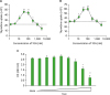 | Figure 1Effects of TGN on T cell proliferation. (A) Indicated amounts of TGN was added to allogeneic MLRs, which is composed of DCs (1×10⁵ cells/well) generated from bone marrow cells of C57BL/6 mice and CD4+ T cells (2×10⁵ cells/well) isolated from spleens of BALB/c mice. DNA synthesis was measured by [3H]-thymidine incorporation for the final 18 h of 72 h culture period. The highest concentration of TGN was 3 μM, and the lower TGN concentrations were the ones diluted in 3-fold. The dotted lines indicate the amounts of [3H]-thymidine incorporation in the absence of TGN. (B) Indicated amounts of TGN were added to cultures of splenocytes isolated from C57BL/6 mouse spleens along with Con A (1 μg/ml). After 48 h, DNA synthesis was measured by [3H]-thymidine incorporation for the final 18 h of the 48 h culture period. (C) The same concentrations of TGN as Fig. 1A were added to cultures of CD4+ DOBW cells. After 48 h, cell viability was measured using an XTT assay. Each data point represents the mean±standard deviation of values obtained from 3 individual experiments.OD, optical density.
*p<0.05, **p<0.01 compared with untreated control.
|
The T cell proliferation-inducing activity of TGN is due to the increased production of IL-2 from TGN-stimulated T cells (Fig. 2). The effects of TGN on IL-2 production was first examined in mouse splenocyte cultures primed with or without 1 μg/ml Con A (Fig. 2A). TGN alone increased IL-2 production significantly, with maximum response at 50 nM. The amounts of IL-2 produced from 25 nM TGN-stimulated lymphocytes were almost the same as that of 1 μg/ml Con A-stimulated lymphocytes. It is noteworthy that the IL-2 production-inducing activity of TGN reaches its maximum level at 50 nM, and diminishes at higher concentration of TGN (200 nM). This phenomenon may explain why TGN exerts a bell-shaped dose-response curve in Fig. 1A and B. This phenomenon may also explain the reason for the earlier observation which showed that TGN alone did not induce IL-2 production from lymphocytes (16). In that study, TGN was used at 2 μM, which is cytotoxic and inhibitory to IL-2 production according to our present study. TGN profoundly increased IL-2 production when used simultaneously with Con A (Fig. 2A). In fact, the IL-2 production-inducing activity of low nanomolar concentrations of TGN was previously noted in lymphocytes isolated from acquired immunodeficiency syndrome (AIDS) patients (17).
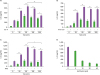 | Figure 2Effects of TGN on IL-2 production from T cells. (A) Indicated amounts of TGN and 1 μg/ml Con A were added simultaneously to cultures of splenocytes (5×10⁵ cells/well) isolated from C57BL/6 mouse spleens. After 48 h, the culture supernatants were harvested and measured for IL-2 content. (B) CD4+ T cells (2×10⁵ cells/well) isolated from spleens of C57BL/6 mice were cultured in the presence or absence of TGN in 96-well culture plates that were pre-coated with anti-CD3 mAb (1 μg/ml) overnight. After 48 h, the culture supernatants were harvested and measured for IL-2 content. (C) Indicated amounts of TGN and 20 ng/ml PMA were added simultaneously to cultures of CD4+ T cells (2×10⁵ cells/well) isolated from spleens of C57BL/6 mice. After 48 h, the culture supernatants were harvested and measured for IL-2 content. (D) Indicated amounts of BAPTA/AM and 50 nM TGN were added to CD4+ T cell cultures which were performed in 96-well plates that were pre-coated with anti-CD3 mAb (0.1 μg/well) overnight. After 48 h, the culture supernatants were harvested and measured for IL-2 content. Each data point represents the mean±standard deviation of values obtained from 3 individual experiments.
*p<0.05, **p<0.01 compared with untreated control; #p<0.05, ##p<0.01 compared with matched group.
|
The IL-2 production-inducing activity of TGN was also examined using T cells purified from mouse spleens. TGN alone increased IL-2 production from purified T cells significantly, with maximum response at 50 nM (Fig. 2B). Furthermore, TGN increased IL-2 production profoundly when used along with anti-CD3 mAb (Fig. 2B). We then examined the combination effects of various concentrations of TGN with 20 ng/ml PMA, which mimics the action of DG in PKC activation. TGN together with PMA induced IL-2 production potently in T cells purified from mouse spleens (Fig. 2C).
We added various concentrations of BAPTA/AM, an intracellular Ca2+ chelator, to cultures of T cells stimulated with PMA (20 ng/ml) and TGN (25 nM), to confirm that the increase of intracellular Ca2+ is the main mechanism of TGN's activity on IL-2 production. BAPTA/AM blocked IL-2 production dose-dependently, with complete nullification at approximately 20 μM (Fig. 2D), confirming that TGN's activity on IL-2 production is mostly due to the increase of intracellular Ca2+.
Subsequently, we examined whether TGN could also increase any cytokine production other than IL-2 in antigen-primed T cells. In this experiment, the total T cells purified from mouse spleens were cultured in plates pre-coated with anti-CD3 mAb (1 μg/ml) in the presence or absence of TGN. TGN (25 nM) alone or in combination with anti-CD3 mAb did not induce IFN-γ or IL-4 production significantly (Fig. 3).
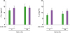 | Figure 3Effects of TGN on IFN-γ and IL-4 production from T cells. CD4+ T cells isolated from C57BL/6 mouse spleens were cultured in the presence or absence of TGN in 96-well culture plates that were pre-coated with anti-CD3 mAb (0.1 μg/well) overnight. After 48 h, the culture supernatants were harvested and measured for IL-2 content. Each data point represents the mean±standard deviation of values obtained from 3 individual experiments.
|
TGN does not activate PKC; instead it discharges intracellular Ca2+ stores by specific inhibition of the ER Ca2+-ATPase resulting in rapid increase of intracellular Ca2+ concentration (7). Due to this activity, TGN induces acute responses in a large number of cell types (1819). Thus, TGN did not receive much attention as a pharmacological agent that modulates immune responses. TGN, however, came to attention in recent years since the development of TGN analogue-based prodrug for the treatment of solid tumors (20). TGN has been known to increase cytokine production in macrophages and DCs (910111221). The effects of TGN on cytokine production by T cells are not clearly defined yet. TGN has been reported to have no effect on IL-2 production in Jurkat T cells (16), while there are reports showing that TGN increases IL-2 production from lymphocytes isolated from AIDS patients (17). This discrepancy might be due to the difference in the concentration of TGN used, as well as difference in the cell type used. The present study shows that TGN exerts a ‘bell-shaped’ dose-response curve in inducing T cell proliferation in allogeneic MLR and Con A-stimulated lymphocyte cultures. It also shows that TGN alone is a significant inducer of IL-2 production only in low nanomolar concentrations with full activity at approximately 50 nM. In addition, this study shows that the IL-2 production-inducing activity of TGN is much more potent when T cells are primed with Con A or anti-CD3. Thus, the present study sheds a new light on the potentiation of antigen-primed T cells using low nanomolar concentrations of TGN.
 XML Download