Journal List > Immune Netw > v.18(3) > 1108109

Bai, Aerts, Verma, Ordway, and Chan: Epidemiologic Evidence of and Potential Mechanisms by Which Second-Hand Smoke Causes Predisposition to Latent and Active Tuberculosis

Abstract

Many studies have linked cigarette smoke (CS) exposure and tuberculosis (TB) infection and disease although much fewer have studied second-hand smoke (SHS) exposure. Our goal is to review the epidemiologic link between SHS and TB as well as to summarize the effects SHS and direct CS on various immune cells relevant for TB. PubMed searches were performed using the key words “tuberculosis” with “cigarette,” “tobacco,” or “second-hand smoke.” The bibliography of relevant papers were examined for additional relevant publications. Relatively few studies associate SHS exposure with TB infection and active disease. Both SHS and direct CS can alter various components of host immunity resulting in increased vulnerability to TB. While the epidemiologic link of these 2 health maladies is robust, more definitive, mechanistic studies are required to prove that SHS and direct CS actually cause increased susceptibility to TB.

Abbreviations

AMP

antimicrobial peptide

BCG

Bacillus Calmette-Guérin

CS

cigarette smoke

CTLA

cytotoxic T-lymphocyte-associated antigen

IFN

interferon

LTBI

latent tuberculosis infection

MAIT

mucosal associated invariant T

mprF

multiple peptide resistance factor

MTB

Mycobacterium tuberculosis

PD-1

programmed cell death protein 1

S. aureus

Staphylococcus aureus

SHS

second-hand smoke

TB

tuberculosis

TGF

transforming growth factor

TNF

tumor necrosis factor

Treg

T regulatory cell

TST

tuberculin skin test

INTRODUCTION

Cigarette smoking is increasing worldwide and by reasonable inference, second-hand smoke (SHS) exposure is likely increasing in parallel (1). SHS, also called environmental tobacco smoke, involuntary smoke, and passive smoke, is comprised mainly, by up to 85%, of “sidestream” smoke (the smoke given off by a burning cigarette or another tobacco product) and to a lesser extent (up to 15%) of “mainstream” smoke (the smoke inhaled and exhaled by a smoker). Not surprisingly, sidestream smoke and hence SHS contains >7,000 distinct chemical compounds including nicotine. Indeed, nicotine has been found in the exhaled breath, oral fluids, and blood of individuals exposed to SHS and second-hand vapors from electronic cigarettes (23). SHS exposure has been shown to cause many of the same diseases caused by direct smoking (4). In fact, sidestream smoke — the major component of SHS — may be more harmful, in the context of toxicity and tumorogenicity, than mainstream smoke (5).
Tuberculosis (TB) is an ongoing global public health crisis (6). The geographic overlap of the prevalence of cigarette smoking and the number of latent and active TB cases is striking (7). Mathematical models project that cigarette smoke (CS) exposure contributes to millions of TB cases and deaths worldwide and is a significant driver of the TB pandemic (89). SHS exposure is likely more common in TB endemic countries than in the U.S. because of greater smoking prevalences and less stringent public smoking bans in those countries. There appears to be no safe levels of SHS exposure (10). Indeed, small airway epithelial cells exposed in vivo to the lowest detectable level of CS — as that seen with SHS — displayed abnormal gene expression (11). Our goals are to review the epidemiological studies that examined the relationship between SHS exposure and TB as well as summarize the effects SHS/direct CS exposure have on immune cells that play a role in host defense and/or the pathogenesis of TB.

METHODS

We reviewed the literature — using PubMed searches — on SHS exposure and its association with various clinical forms of TB including latent tuberculosis infection (LTBI) and active TB (12). We also searched for the effects of SHS/direct CS on the functions of macrophages, neutrophils, T effector cells, CD8+ mucosal associated invariant T (MAIT) cells, B cells, and T regulatory cells (Tregs).

EPIDEMIOLOGIC STUDIES LINKING SHS EXPOSURE WITH TB INFECTION AND DISEASE

TB infection

As part of a larger analysis, we previously reported 5 studies that examined the association between LTBI and SHS exposure (1314151617). In a cross-sectional study of nearly 8,000 high school students, those with larger tuberculin skin test (TST) reactivity (mode up to 16 mm induration) were twice as likely to have 2 smoking parents than to have one or no smoking parents (16). In 2 separate studies from South Africa of up to 1,500 children, those who were contacts of smokers with active TB or who lived with 2 or more smokers with active TB were more likely to have a positive TST than contacts of non-smokers with active TB (1314). In over 7,000 Spaniards, those in contact with smokers diagnosed with active TB were more likely to have a positive TST than subjects in contact with non-smokers with TB (15). In 95 children from the Indian subcontinent who were contacts of adults with active TB, the determining risk factors for transmission of infection were younger age, severe malnutrition, absence of Bacillus Calmette-Guérin (BCG) vaccination, contact with a sputum-positive adult, and exposure to SHS (17). More recently, 2 studies reported meta-analyses on the association of SHS exposure and LTBI or active TB (1819). Patra and colleagues analyzed 18 studies and found a significant association between SHS and LTBI with a pooled relative risk (RR) of 1.64 (95% confidence interval [CI], 1.00–2.83) but observed great heterogeneity in the studies (18). Dogar and co-workers (19) performed a meta-analysis on 12 studies and found that SHS exposure was associated with LTBI although this did not reach statistical significance (RR, 1.19; 95% CI, 0.90–1.57); marked variability in the results were also found, which was attributed to differences in the diagnostic criteria used among the studies analyzed.

Primary progressive TB

In infants and young children, active TB cases are almost always due to progression of primary infection, since they are unlikely old enough to have LTBI (20). Four studies in children found a significant association between SHS and primary progressive TB (21222324). Altet et al. (21) also showed a greater association between primary progressive TB in children and the amount smoked in the household. In 1 study from Thailand and 2 from India, children with active TB were more likely to have had SHS exposure (222324). Patra et al. (18) performed a meta-analysis of the studies that examined the association between SHS and active TB in children and found a 3-fold greater association than those without SHS exposure.

Active TB in adults

We previously summarized the numerous studies that investigated the association between cigarette smoking and active TB with the vast majority noting a significant link (12). That report included four case-control studies which examined both direct and SHS exposure; all found that active TB patients were more likely to be smokers (present or past) and/or have SHS exposure (25262728). In the one study that only examined SHS and active TB in adults, over 15,000 elderly, never-smoking, married Chinese women were followed prospectively; 117 cases of active TB were identified (29). The women with TB were more likely to have had SHS exposure than those without TB (29). While no studies have examined the frequency and amount of SHS exposure and its association with active TB, it appears that the intensity of CS exposure — as measured by number of cigarettes smoked per day, number of years smoked, or number of pack-years smoked — is significantly associated with active TB (2526273031323334353637). In 2 recent meta-analysis investigations, SHS was also significantly associated with active TB (1819) although the association with adults was not as great as that seen with children (18).

Limitations of epidemiologic studies linking SHS/direct CS exposure and TB

Epidemiologic studies can at best show an association between SHS/direct CS exposure and LTBI or active TB but cannot show any direct causal link. A potential confounder that may perhaps lead to increased TB transmission among those exposed to SHS and/or direct CS is congregation among smokers — leading to potential increase in TB transmission if there was a person with active TB in the group (12). Since cigarette smoking is generally associated with a lower socioeconomic status, with secondary link to malnutrition and crowded living conditions, these are additional confounders for acquiring TB (12). In studies of SHS exposure and TB contact investigation, it is not always clear whether SHS refers to the index TB case or the contacts. In either case, the active TB patient would be exposed to direct CS, SHS, or both and thus the increased association between SHS exposure and active TB/LTBI may be due to exacerbation of cough in the active TB subject from the SHS and/or direct CS exposure, resulting in greater transmission of Mycobacterium tuberculosis (MTB). Similarly, if indeed direct CS exposure is a risk factor for TB, those with SHS exposure may simply be at greater risk for TB because, by definition of having SHS exposure, they may be exposed more frequently to individuals (smokers) with active TB.
In summary, epidemiological studies show SHS exposure is associated with active TB and LTBI but such links are compromised by the great variability in the results (121819). Moreover, these studies do not show a cause-and-effect relationship and there is the issue of potential confounders such as malnutrition, crowded living conditions, and HIV co-infection. Even if there is a causal link between SHS/direct CS exposure and TB, epidemiologic studies cannot elucidate the mechanism(s). Thus, in the next 2 sections, we are going to: 1) tabulate the murine studies which investigated the effects of CS exposure (direct CS + SHS) on MTB infection and 2) summarize the main functions of key immune cells in control of MTB and the effects of SHS/direct CS on their functions.

MURINE STUDIES ON SHS/DIRECT CS AND TB

To the best of our knowledge, there have been three in vivo murine studies examining the effects of CS on control of MTB infection (383940). Each used CS exposure levels — based on total suspended particulate — that were comprised of both SHS and direct CS exposure. Each revealed that CS exposure impaired the ability of mice to control MTB infection (383940). We summarized the key findings of these mouse studies by immune cell types (Table 1).
Table 1

Effects of CS exposure on murine models of MTB infection

in-18-e22-i001
Murine cell type Findings in CS-exposed mice compared to air-exposed mice
Macrophages Reduced number of IL-12 and TNFα-positive splenic and lung macrophages; increased number of IL-10-positive splenic macrophages (40).
Dendritic cells Reduced number of IL-12 and TNFα-positive splenic and lung dendritic cells; increased number of IL-10-positive splenic dendritic cells (40).
T effector cells Reduced number of TH1 cells with reduced IFNγ production per cell (383940); also reduced influx of IFNγ and TNFα-positive CD4+ and CD8+ T cells into the lungs and spleens (3840); increased influx of IL-4-positive TH2 cells in the lungs (39); CS also impaired (DNA) vaccinated mice to MTB-specific antigen by reducing the number of antigen-specific TH1 cells (38).

THE EFFECTS OF SHS/DIRECT CS EXPOSURE ON IMMUNE CELLS RELEVANT FOR TB

CS significantly increases the number of innate and adaptive immune cells into the lungs (41) and yet epidemiologic evidence and experimental models indicate CS leads to a predisposition to TB (383940424344). This paradox supports the notion that the host-protective anti-MTB cell types are compromised and/or there is activation of immunosuppressive cells by SHS/direct CS. While CS extract impairs human macrophages in controlling MTB infection, the mechanisms remain unknown (4045). Hence, we highlight below some of the key immune cells relevant in host immune response to MTB and how SHS/direct CS may affect their functions.

Macrophage function against TB and potential effects of SHS/direct CS

M1 macrophage phenotype is characterized by production of pro-inflammatory cytokines (tumor necrosis factor [TNF] α, IL-12), chemokines IL-8 and monocyte chemoattractant protein-1, and nitric oxide as well as biasing T cells toward the TH1 phenotype and interferon (IFN) γ-producing CD8+ T cells (464748). M1 effector functions against MTB include phagosome-lysosome fusion, autophagy, apoptosis, efferocytosis, and granuloma formation (4950). Generally, M2 macrophages, the alternatively-activated or “deactivated” macrophage phenotype (in the context of host-defense against pathogens), are characterized by increased activity of arginase, matrix metalloproteinase (MMP), transforming growth factor (TGF) β, IL-10, and prostaglandin E2, and would not be expected to be host-protective during active MTB replication/infection (5152). Efferocytosis of MTB-infected neutrophils leads to macrophage activation through the actions of TNFα and heat shock proteins (53). Efferocytosis may assist in clearing MTB infection, enhance antigen presentation to T cells, limit potentially injurious inflammation, and increase phagosome-lysosome fusion through uptake of neutrophil azurophilic granules (5455). Table 2 lists the potential effects of SHS, direct CS, or nicotine on anti-TB macrophage function; whether murine cells (M), human cells (H), or both (M/H) were studied are noted for each of the studies cited.
Table 2

Potential effects of SHS/direct CS/direct or secondary nicotine on macrophage function against TB

in-18-e22-i002
Macrophage phenotype and function Effects of SHS/direct CS/direct or secondary nicotine
M1 phenotype Skewed macrophages to the M2 phenotype (52)(H). Induced PD-L1/2, resulting in a deactivated phenotype (565758)(M/H,H,H).
Cell surface markers Smoker AM had less expression of CD11a, CD71, and CD54; smoker AM have greater density of CD11c (but not percentage of AM positive for CD11c) (59)(H).
Phagocytosis Effect of SHS/direct CS on phagocytosis is conflicting, from no effect to inhibition of phagocytosis (40456061)(M,H,H,H).
Efferocytosis Inhibited efferocytosis (556263)(M/H,H,H)
Phagosome-lysosome fusion Inhibited phagosome-lysosome fusion (646566)(R,H,M/H)
Autophagy Inhibited autophagy by CS (5167)(M/H,H) and nicotine (68)(M/H).
Apoptosis Inhibited apoptosis (6970)(H,M/H).
M, murine; H, human; M/H, murine and human; AM, alveolar macrophage.

Neutrophil function against TB and potential effects of SHS/direct CS

Lung neutrophils are one of the most commonly MTB-infected cells in TB patients and smokers have 3- to 4-fold more lung neutrophils than non-smokers (7172). Thus, whatever impact CS may have on neutrophils is likely relevant with MTB infection. Some studies show neutrophils restrict the growth or kill MTB whereas others show neutrophils contribute to excessive lung inflammation and may enhance mycobacterial dissemination (737475). Such discrepancies may be due to phenotypic differences in neutrophils as the CD16hiCD62Llo neutrophil subtype is considered immunosuppressive (76). Table 3 lists the potential effects of SHS or direct CS on anti-TB neutrophil function.
Table 3

Potential effects of SHS/direct CS on neutrophil function against TB

in-18-e22-i003
Neutrophil phenotype and function Effects of SHS/direct CS
Migration of neutrophils Smokers have three- to four-fold more neutrophils in the lungs than non-smokers, likely due to CS induction of IL-8 and IL-17, the latter through CS differentiation of TH17 cells (7177)(H,M).
Neutrophil activation PD-L1 is increased on neutrophils of active TB patients (78)(H) and CS, nicotine, and lipopolysaccharide (a common contaminant of cigarettes) are known to induce PD-L1/2 expression (5758)(M,H), theoretically deactivating neutrophils, leading to formation of non-protective, necrotic granulomas (75)(M/H).
Oxidative burst CS inhibited endogenous oxidative burst (79)(H).
Induction of (N2) neutrophils Lipopolysaccharide found in cigarettes induced the expression of immunosuppressive N2 (CD16hiCD62LloCD11bhiCD54hi) neutrophils, which inhibit T cell proliferation (76)(H).
NETs CS inhibited endogenous oxidative burst — which is required for NETs formation; thus, CS could inhibit formation of NETs (7980)(H,M/H)
M, murine; H, human; M/H, murine and human; NET, neutrophil extracellular trap.

T effector cell and B lymphocyte function against TB and potential effects of SHS/direct CS

Increasing evidence indicates that a TH1/TH2/TH17 balance is vital to establish control of active TB disease (81828384). MTB-specific TH1 cells (IL-12+, IFNγ+, TNFα+) stimulate inflammation, help skew macrophages to the M1 phenotype, and initiate antibacterial effector functions in infected macrophages (8586). Early expansion of TH2 cells (IL-4+, IL-13+) antagonizes protective cellular immunity, resulting in a partial or complete loss of TB control (468788). TH17 cells (IL-17+, IL-22+) recruit neutrophils through the chemoattractant actions of IL-17, resulting in improved early granuloma formation and increased cooperation between neutrophils and macrophages in killing MTB (89). However, sustained exposure of neutrophils to IL-17 can polarize their phenotype to one that is less effective in controlling MTB, causing more immunopathology (89). While increased programmed cell death protein 1 (PD-1) expression on T cells have been shown to impair human host immunity to MTB (90919293), several groups have shown that PD-1 knockout mice were paradoxically more susceptible to MTB, perhaps related to excessive inflammation and tissue injury (including neutrophil-mediated necrosis) in the complete absence of PD-1 (90949596).
CD8+ T cells are also required for optimal host defense against human MTB disease (4697). CD8+ T cells recognize protein and lipid antigens that are either class I MHC or CD1 restricted, respectively (9899). CD8+ T cells have the capability to recognize and kill MTB-infected macrophages (100101). Humans infected with MTB have expansion of cytotoxic CD8+ T cells capable of producing various cytokines (97). CD8+ T cells have also been identified in granulomas and pleural fluid of TB patients (102103104) and cloning these cells demonstrated TB antigen specificity (105). Additional reports demonstrated MTB-specific CD8+ T cells in BCG vaccinated individuals, and both active and latent TB patients (106107).
MAIT cells are a subset of CD8+ T cells that recognize bacteria-derived metabolites of riboflavin (vitamin B2) biosynthesis pathway that are presented on major histocompatibility complex-related protein-1 (MR1) located on the cell surface of antigen-presenting cells; interestingly, cell surface expression of MR1 is dependent on the presence of an exogenous, bacterial metabolite ligand (108). In contrast to conventional T cells, MAIT cells are functional prior to exposure to antigens although exposure to MR1-restricted microbial antigens induce MAIT cell expansion. The semi-invariant T cell receptors (TRAV1-2/TRAJ33) of MAIT cells that recognized MR1-restricted antigens are comprised of α and β subunits. When activated, MAIT cells are host-protective through the expression of IFNγ and TNFα, and may induce lysis of target cells through granulysin and perforin (109). In healthy humans, MAIT cells are already enriched in the respiratory tract but with active TB, the MAIT cell population is further increased in the lungs but decreased in the peripheral blood (108110111). But the MAIT cells in patients with active TB may be compromised as they exhibit increased expression of PD-1 and blockade of PD-1 signaling resulted in a significantly higher number of IFNγ-producing MAIT cells upon ex vivo stimulation with BCG (93).
There is increasingly convincing evidence that B cells — through production of antibodies but also by biasing specific CD4+ T cell phenotypic activation through cytokine production and antigen presentation to T cells — are an important component in an effective host immune response against MTB (112113114115116). While MTB is traditionally considered an intracellular pathogen, extracellular bacilli are a significant component of the total bacterial population. In this regard, there are at least 2 circumstances in which binding of specific antibodies to mycobacterial antigens optimizes antigen presentation; such optimization is perhaps required since many MTB cell wall components contain lipids and carbohydrates that are not typically recognized by MHC molecules. In 1 instance, mycobacterial antigens bind to cell surface antibodies on B cells, the immune complexes are internalized, and the antigens are processed and presented on class II MHC on B cells to T cells. Alternatively and/or concomitantly, free mycobacterial antigen-antibody complexes are able to bind to FcγR on the surfaces of professional antigen presenting cells (dendritic cells and macrophages) and be internalized. The antigens are then processed and presented to T cells. In turn, T cells are able to reciprocate and help B cells produce antibodies (117). In addition to enhancing antigen presentation, IgG can increase phagocytosis of MTB through increased binding of IgG-opsonized bacteria to FcγR or complement receptors present on macrophage cell surface, the latter occurring when C3 or C4 are bound directly to the mycobacteria or to IgG (114). Another potential antibody-mediated effector mechanism is opsonization of infected macrophages that express mycobacterial antigens on their cell surfaces with subsequent Fc binding to FcγRIII on cytolytic lymphocytes (114). In contrast to the active roles of B cells in anti-MTB immunity upon binding to stimulatory FcγR, binding of immune complexes to inhibitory Fc receptors such as FcγRIIB inhibits dendritic cell maturation, resulting in impaired T cell activation. Thus, antibodies produced by B cells may have both stimulatory and inhibitory effect on host immunity against MTB.
B and T cells are also able to reciprocally skew the differentiation of each other into specific phenotypes through the production of cytokines (112118). Remarkably, the specific subsets of B and T cells involved in this interaction are similar; i.e., B effector-1 cells (Be-1) produce IFNγ, IL-12, TNFα, IL-10, and IL-6 to skew naïve CD4+ T cells to the TH1 IFNγ+ phenotype and, in a reciprocal fashion, TH1 cells induce Be-1 differentiation. Similarly, Be-2 cells produce IL-2, lymphotoxin, IL-4, IL-13, IL-10, and IL-6 to induce TH2 IL-4+ differentiation and vice versa (112). Furthermore, B cell-derived TNFα serves to not only induce antibody production but also promote expansion of CD4+ T cell subsets, depending on the prevailing cytokine millieu.
Mice deficient in B cells or the common γ chain of FcγR are compromised in controlling an MTB infection, which may be due, in part, to increased IL-10 production (119120). Conversely, mice with genetic disruption of the inhibitory FcγRIIB have greater capacity to control MTB and have increased TH1 response, in part through increased IL-12 expression (120). In addition to the aforementioned host-protective mechanisms of B cells against MTB, they also contribute to the formation of acute and chronic granulomas in response to MTB (112121122123). In MTB infection of non-human primates, depletion of B cells with rituximab resulted in altered local T cell responses in granulomas (increased number of IL-2+, IL-10+, and IL-17+ T cells), decreased inflammation, and increased bacterial burden in individual granulomas although there was significant heterogeneity seen between the granulomas even in the same animal (124).
While B cells appear to play a lesser role than T cells in a respiratory mucosal vaccine against MTB (125), the humoral response also attenuates the potentially tissue damaging neutrophilic response, in part, by reducing the neutrophil chemokine IL-17 and the TH17 response (126). Indeed, B cell deficient mice have increased neutrophilia at the site of experimental immunization, with reduced dendritic cell migration to regional lymph nodes and decreased vaccine-induced TH1 response (126). As a promising therapeutic, monoclonal antibodies directed against various MTB cell wall components protected mice against MTB, as evinced by reduced bacterial load, containment from disseminated disease, and attenuated inflammation (127). Table 4 lists the potential effects of SHS or direct CS on anti-TB effector lymphocyte function.
Table 4

Potential effects of SHS/direct CS on effector lymphocyte function against TB

in-18-e22-i004
Lymphocyte subtype Effects of SHS/direct CS
TH1 cells Reduced number of TH1 cells and decreased production of IFNγ per cell (128129)(H,H). Potential mechanisms include CS-derived reactive oxygen species-induced necrosis of T cells (130)(H), T cell exhaustion as CS increases PD-L1 on antigen presenting cells, leading to engagement of programmed death (PD-1) receptors on T cells (589192131)(H,H,H,M/H), downregulation of CD28, and upregulation of CTLA-4 (132)(M). In adolescents exposed to SHS, the percentages of blood memory CD4+ and CD3+ T cells (CD45RO+) were inversely related to the degree of SHS exposure although there was a direct relationship between the percentages of naïve CD3+ and CD4+ T cells (CD45RA+) and degree of SHS exposure (133)(H). In contrast, T cells from both CS-exposed mice and human smokers fluxed calcium, proliferated and produced IFNγ at levels comparable to T cells from control mice and non-smokers (134)(M/H).
TH2 cells Increased influx of IL-4-positive TH2 cells in the lungs (39)(M).
TH17 cells In mice, CS increased TH17 cells but only after relatively long-term exposure (135)(M). The percentage of TH17 cells is increased in the blood of healthy cigarette smokers (136)(H) and CS exposure of human lung explants increased IL-17 expression (137)(H).
CD8+ cells Smokers have increased CD8+ cells in their airways than non-smokers (138)(H). CS increased the proliferation and inhibited the apoptosis of CD8+ T effector cells but increased both the proliferation and apoptosis of CD8+ Tregs (139)(H). CS exposure of mice increased CD8+ (and CD4+) T cells (140)(M).
MAIT cells Human subjects exposed to CS had reduced circulating levels of CD26hiCD161hi MAIT cells (141)(H).
B cells Chronic smoking is known to cause a polyclonal B cell lymphocytosis (142)(H). In adolescents exposed to SHS, there was no difference in the percentages of blood B lymphocytes and the degree of SHS exposure (133)(H). B cells from CS-exposed mice and human smokers fluxed calcium, proliferated and produced immunoglobulins at levels comparable to control mice and non-smokers (134)(M/H). In contrast, others have shown that CS-exposed or nicotine-exposed rodents have reduced number of B cells as well as reduced calcium flux and decreased proliferative capacity in response to antigenic stimulation although the amount of experimental CS exposure in these studies were greater than that typically seen in human smokers (143144)(M,M).
M, murine; H, human; M/H, murine and human.

Treg function against TB and potential effects of SHS/direct CS

Tregs secrete immunosuppressive cytokines such as IL-9, IL-10, IL-35, and TGFβ, increase consumption of IL-2, suppress Foxp3-negative TH1, TH2, and TH17 cells, and quell antigen-presenting cells (145146). Early in TB infection, excess Treg activity is associated with production anti-inflammatory of mediators (IL-10+, TGFβ+) that may promote MTB growth and persistence. In experimental animals, the influx of Tregs to the lungs with MTB infection worsens disease (147). Numerous studies have demonstrated that CD25+ and Foxp3+ Tregs are elevated in the circulation and at the site of infection in patients with active pulmonary TB (148149) and suppress the proliferation of and IFNγ production by CD4+CD25 T cells in an in vitro co-culture system (150). Conversely, in vitro depletion of CD25hi Tregs from peripheral blood mononuclear cells obtained from active TB patients resulted in further increase in IFNγ levels induced by specific antigens (151152). Other studies have confirmed these findings and determined that CD4+CD25+ Tregs in active TB co-expressed Foxp3, cytotoxic T-lymphocyte-associated antigen (CTLA)-4, glucocorticoid-induced tumor necrosis factor receptor (GITR), PD-1 and CD39 at the protein level (150153154) and also higher levels of CD45RO and human leukocyte antigen-antigen D related (HLA-DR) together with lower levels of CD127 (155) and CD45RA compared to CD4+CD25 T cells (154156157158). Patients with active TB have increased Tregs in their peripheral blood and bronchoalveolar lavage compared to subjects with latent TB infection or healthy controls (159). As would be predicted, co-culture of these isolated Tregs with T effector cells and MTB-infected monocyte-derived macrophages or alveolar macrophages significantly decreased mycobacterial killing (159). However, Treg function is also essential to control tissue damaging inflammation. Tregs express Foxp3+ CTLA-4 that have been linked to reduce TH17 cytokines such as IL-17 and IL-23 in subjects with LTBI (160161). Table 5 lists the potential effects of SHS, direct CS, or nicotine on Treg function.
Table 5

Potential effects of SHS/direct CS/direct or secondary nicotine on Treg function

in-18-e22-i005
Tregs Effects of SHS/direct CS/direct or secondary nicotine
Tregs In man, CS exposure enhanced the function of Tregs in the lungs (162163164)(H,H,M). A possible mechanism by which CS enhances Treg function is the ability of CS to induce PD-L1/2 on antigen-presenting cells, resulting in increased engagement to PD-1 on Tregs, enhancing the immunosuppressive function of Tregs (5658165)(M/H,H,H).
In mice, Tregs express the α7 nicotinic acetylcholine receptor and upon binding to nicotine, there is an upregulation of both the transcription factor Foxp3 and the cell surface molecule CTLA-4, proteins that characterize Tregs and dampen Foxp3-negative T effector cell activity, respectively (164)(M). By producing TGFβ and IL-10, CS-induced Tregs can further suppress M1 macrophage activation against MTB as well as induce macrophages to differentiate to the M2/deactivated phenotype (51)(M/H). Nicotine induced murine Tregs to produce TGFβ and nicotine-exposed Tregs impair macrophage activity against MTB in murine macrophages (68)(M/H).
M, murine; H, human; M/H, murine and human.

CAN CS ENHANCE MTB VIRULENCE?

To the best of our knowledge, there has been no investigations to determine if CS influences the virulence of MTB. However, MTB is exposed to endogenous and exogenous reactive oxygen species — abundantly present in CS — and it is known that oxidative as well as reductive stressors may affect several physiologic functions of MTB, including intracellular signaling, synthesis of nucleic acids and proteins, and drug resistance (166). As in other host-pathogen interactions, MTB and host cells are likely altering its own redox balance to gain advantage over the other. McEachern and co-workers (167) showed in Staphylococcus aureus (S. aureus) that CS extract induced the expression of the multiple peptide resistance factor (mprF) protein, which adds lysine residues to acidic phospholipids (which have relative negative charge prior to lysine adducts) on the bacterial cell surface, converting them to basic phospholipids (relatively positive charge after addition of lysine residues), creating an unfavorable charge environment for the cationic antimicrobial peptide (AMP) LL-37 to bind bacteria, thereby generating a more virulent S. aureus (Fig. 1, left-side). Thus, it is highly relevant that MTB possess the lysX gene — a fusion gene comprised of both an mprF-like gene and the lysU gene — that encodes a protein with both lysyl transferase and lysyl-tRNA synthetase activities (168). A catalytic product of this 2-domain protein is lysinylated phosphatidylglycerol, a basic and positively-charged phospholipid, which imparts MTB resistance to cationic AMPs. Hence, it would be very interesting to determine whether CS could induce lysX gene expression, provoking greater MTB virulence through addition of lysine-mediated positive charge on MTB cell surface (Fig. 1, right-side).
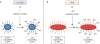
Figure 1

A hypothesized mechanism by which CS may affect virulence of MTB. (A) With S. aureus, CS induces the expression of mprF, which adds positively charged lysine residues to the bacterial membrane phospholipids. With greater positive charge on its cell membrane, S. aureus is better able to repel cationic AMP+. (B) Similar to the mprF molecule in S. aureus, MTB possess the lysX gene, which encodes a 2-domain protein with lysyl transferase and lysyl-tRNA synthetase activities that also adds positively charged lysine residues to phosphatidylglycerol, converting the acidic, negatively-charged phospholipid to a basic, positively-charged phospholipid. While it is not known if CS can induce lysX gene, this may be another potential mechanism by which CS increases susceptibility to TB by inducing a more virulent MTB strain.

in-18-e22-g001

SUMMARY

Both SHS and direct CS exposure are associated with increased risk for LTBI and active TB. This susceptibility may be due to increased exposure to the tubercle bacilli from the increased cough seen in smokers and those exposed to SHS, suppressed anti-TB immunity, enhanced activity of immunosuppressive N2 neutrophils or Tregs, or a combination of any of these factors. These generalized statements are likely an oversimplification of what is actually occurring since each cell type is not always salutorious or deleterious to the host; i.e., the temporal and coordinated influx (or absence) of each cell type may be necessary not only during the initial phase of the host-protective inflammatory response but also the necessary resolution of inflammation once the infection is under control. In addition, the potential effects of CS in inducing greater virulence in MTB remains to be determined but a plausibility given such findings in other bacteria. Finding stronger evidence of a causal link between SHS/direct CS exposure may provide greater impetus to implement public health policies to further reduce SHS/direct CS exposure in TB-endemic countries, potentially providing another important measure to help eradicate TB.

Abbreviations

AMP

antimicrobial peptide

BCG

Bacillus Calmette-Guérin

CS

cigarette smoke

CTLA

cytotoxic T-lymphocyte-associated antigen

IFN

interferon

LTBI

latent tuberculosis infection

MAIT

mucosal associated invariant T

mprF

multiple peptide resistance factor

MTB

Mycobacterium tuberculosis

PD-1

programmed cell death protein 1

S. aureus

Staphylococcus aureus

SHS

second-hand smoke

TB

tuberculosis

TGF

transforming growth factor

TNF

tumor necrosis factor

Treg

T regulatory cell

TST

tuberculin skin test

ACKNOWLEDGEMENTS

We are grateful to the Potts Memorial Foundation, the Department of Veterans Affairs, Veterans Health Administration, Office of Research and Development, 1 I01 CX001452-01A1 (EDC), and the National Institutes of Health, R21 AI121605-01 for their generosity in helping to fund this work.

Notes

Conflict of Interest The authors declare no potential conflicts of interest.

Author Contributions

  • Data curation: Bai X, Aerts SL, Chan ED.

  • Investigation: Bai X, Aerts SL, Chan ED.

  • Writing - original draft: Bai X, Aerts SL, Verma D, Ordway DJ, Chan ED.

References

1. Slama K. Global perspective on tobacco control. Part I. The global state of the tobacco epidemic. Int J Tuberc Lung Dis. 2008; 12:3–7.
2. Gallart-Mateu D, Elbal L, Armenta S, de la Guardia M. Passive exposure to nicotine from e-cigarettes. Talanta. 2016; 152:329–334.
crossref
3. Jarvis MJ, Feyerabend C, Bryant A, Hedges B, Primatesta P. Passive smoking in the home: plasma cotinine concentrations in non-smokers with smoking partners. Tob Control. 2001; 10:368–374.
crossref
4. Office on Smoking and Health (US). The Health Consequences of Involuntary Exposure to Tobacco Smoke: a Report of the Surgeon General. Atlanta, GA: Centers for Disease Control and Prevention;2006.
5. Schick S, Glantz S. Philip Morris toxicological experiments with fresh sidestream smoke: more toxic than mainstream smoke. Tob Control. 2005; 14:396–404.
crossref
6. World Health Organization (CH). Global Tuberculosis Report 2014. Geneva: World Health Organization;2014.
7. Pai M, Mohan A, Dheda K, Leung CC, Yew WW, Christopher DJ, Sharma SK. Lethal interaction: the colliding epidemics of tobacco and tuberculosis. Expert Rev Anti Infect Ther. 2007; 5:385–391.
crossref
8. Basu S, Stuckler D, Bitton A, Glantz SA. Projected effects of tobacco smoking on worldwide tuberculosis control: mathematical modelling analysis. BMJ. 2011; 343:d5506.
crossref
9. van Zyl-Smit R, Dheda K. Partners in crime: the deadly synergy of tuberculosis and tobacco smoke? Mycobact Dis. 2012; 2:e111.
10. U.S. Department of Health and Human Services. The Health Consequences of Involuntary Smoking: a Report of the Surgeon General. Washington, D.C.: U.S. Department of Health and Human Services;1986. p. 1–363.
11. Strulovici-Barel Y, Omberg L, O'Mahony M, Gordon C, Hollmann C, Tilley AE, Salit J, Mezey J, Harvey BG, Crystal RG. Threshold of biologic responses of the small airway epithelium to low levels of tobacco smoke. Am J Respir Crit Care Med. 2010; 182:1524–1532.
crossref
12. Bishwakarma R, Kinney WH, Honda JR, Mya J, Strand MJ, Gangavelli A, Bai X, Ordway DJ, Iseman MD, Chan ED. Epidemiologic link between tuberculosis and cigarette/biomass smoke exposure: limitations despite the vast literature. Respirology. 2015; 20:556–568.
crossref
13. den Boon S, Verver S, Marais BJ, Enarson DA, Lombard CJ, Bateman ED, Irusen E, Jithoo A, Gie RP, Borgdorff MW, et al. Association between passive smoking and infection with Mycobacterium tuberculosis in children. Pediatrics. 2007; 119:734–739.
crossref
14. du Preez K, Mandalakas AM, Kirchner HL, Grewal HM, Schaaf HS, van Wyk SS, Hesseling AC. Environmental tobacco smoke exposure increases Mycobacterium tuberculosis infection risk in children. Int J Tuberc Lung Dis. 2011; 15:1490–1496. i
crossref
15. Godoy P, Caylà JA, Carmona G, Camps N, Álvarez J, Alsedà M, Minguell S, Rodés A, Altet N, Pina JM, et al. Smoking in tuberculosis patients increases the risk of infection in their contacts. Int J Tuberc Lung Dis. 2013; 17:771–776.
crossref
16. Kuemmerer JM, Comstock GW. Sociologic concomitants of tuberculin sensitivity. Am Rev Respir Dis. 1967; 96:885–892.
17. Singh M, Mynak ML, Kumar L, Mathew JL, Jindal SK. Prevalence and risk factors for transmission of infection among children in household contact with adults having pulmonary tuberculosis. Arch Dis Child. 2005; 90:624–628.
crossref
18. Patra J, Bhatia M, Suraweera W, Morris SK, Patra C, Gupta PC, Jha P. Exposure to second-hand smoke and the risk of tuberculosis in children and adults: a systematic review and meta-analysis of 18 observational studies. PLoS Med. 2015; 12:e1001835.
crossref
19. Dogar OF, Pillai N, Safdar N, Shah SK, Zahid R, Siddiqi K. Second-hand smoke and the risk of tuberculosis: a systematic review and a meta-analysis. Epidemiol Infect. 2015; 143:3158–3172.
crossref
20. Iseman MD. A Clinician's Guide to Tuberculosis. Philadelphia, PA: Lippincott Williams & Wilkins;2000. p. 253–269.
21. Altet MN, Alcaide J, Plans P, Taberner JL, Saltó E, Folguera LI, Salleras L. Passive smoking and risk of pulmonary tuberculosis in children immediately following infection. A case-control study. Tuber Lung Dis. 1996; 77:537–544.
crossref
22. Patra S, Sharma S, Behera D. Passive smoking, indoor air pollution and childhood tuberculosis: a case control study. Indian J Tuberc. 2012; 59:151–155.
23. Ramachandran R, Indu PS, Anish TS, Nair S, Lawrence T, Rajasi RS. Determinants of childhood tuberculosis--a case control study among children registered under revised National Tuberculosis Control Programme in a district of South India. Indian J Tuberc. 2011; 58:204–207.
24. Tipayamongkholgul M, Podhipak A, Chearskul S, Sunakorn P. Factors associated with the development of tuberculosis in BCG immunized children. Southeast Asian J Trop Med Public Health. 2005; 36:145–150.
25. Alcaide J, Altet MN, Plans P, Parrón I, Folguera L, Saltó E, Domínguez A, Pardell H, Salleras L. Cigarette smoking as a risk factor for tuberculosis in young adults: a case-control study. Tuber Lung Dis. 1996; 77:112–116.
crossref
26. Ariyothai N, Podhipak A, Akarasewi P, Tornee S, Smithtikarn S, Thongprathum P. Cigarette smoking and its relation to pulmonary tuberculosis in adults. Southeast Asian J Trop Med Public Health. 2004; 35:219–227.
27. Pokhrel AK, Bates MN, Verma SC, Joshi HS, Sreeramareddy CT, Smith KR. Tuberculosis and indoor biomass and kerosene use in Nepal: a case-control study. Environ Health Perspect. 2010; 118:558–564.
crossref
28. Woldesemayat EM, Datiko DG, Lindtjørn B. Use of biomass fuel in households is not a risk factor for pulmonary tuberculosis in South Ethiopia. Int J Tuberc Lung Dis. 2014; 18:67–72.
crossref
29. Leung CC, Lam TH, Ho KS, Yew WW, Tam CM, Chan WM, Law WS, Chan CK, Chang KC, Au KF. Passive smoking and tuberculosis. Arch Intern Med. 2010; 170:287–292.
crossref
30. Crampin AC, Glynn JR, Floyd S, Malema SS, Mwinuka VK, Ngwira BM, Mwaungulu FD, Warndorff DK, Fine PE. Tuberculosis and gender: exploring the patterns in a case control study in Malawi. Int J Tuberc Lung Dis. 2004; 8:194–203.
31. Gajalakshmi V, Peto R, Kanaka TS, Jha P. Smoking and mortality from tuberculosis and other diseases in India: retrospective study of 43000 adult male deaths and 35000 controls. Lancet. 2003; 362:507–515.
crossref
32. Kolappan C, Gopi PG. Tobacco smoking and pulmonary tuberculosis. Thorax. 2002; 57:964–966.
crossref
33. Leung CC, Li T, Lam TH, Yew WW, Law WS, Tam CM, Chan WM, Chan CK, Ho KS, Chang KC. Smoking and tuberculosis among the elderly in Hong Kong. Am J Respir Crit Care Med. 2004; 170:1027–1033.
crossref
34. Leung CC, Yew WW, Law WS, Tam CM, Leung M, Chung YW, Cheung KW, Chan KW, Fu F. Smoking and tuberculosis among silicotic patients. Eur Respir J. 2007; 29:745–750.
crossref
35. Lin HH, Ezzati M, Chang HY, Murray M. Association between tobacco smoking and active tuberculosis in Taiwan: prospective cohort study. Am J Respir Crit Care Med. 2009; 180:475–480.
crossref
36. Singh PN, Yel D, Kheam T, Hurd G, Job JS. Cigarette smoking and tuberculosis in Cambodia: findings from a national sample. Tob Induc Dis. 2013; 11:8.
crossref
37. Yen YF, Yen MY, Lin YS, Lin YP, Shih HC, Li LH, Chou P, Deng CY. Smoking increases risk of recurrence after successful anti-tuberculosis treatment: a population-based study. Int J Tuberc Lung Dis. 2014; 18:492–498.
crossref
38. Feng Y, Kong Y, Barnes PF, Huang FF, Klucar P, Wang X, Samten B, Sengupta M, Machona B, Donis R, et al. Exposure to cigarette smoke inhibits the pulmonary T-cell response to influenza virus and Mycobacterium tuberculosis . Infect Immun. 2011; 79:229–237.
crossref
39. Shaler CR, Horvath CN, McCormick S, Jeyanathan M, Khera A, Zganiacz A, Kasinska J, Stampfli MR, Xing Z. Continuous and discontinuous cigarette smoke exposure differentially affects protective Th1 immunity against pulmonary tuberculosis. PLoS One. 2013; 8:e59185.
crossref
40. Shang S, Ordway D, Henao-Tamayo M, Bai X, Oberley-Deegan R, Shanley C, Orme IM, Case S, Minor M, Ackart D, et al. Cigarette smoke increases susceptibility to tuberculosis--evidence from in vivo and in vitro models. J Infect Dis. 2011; 203:1240–1248.
crossref
41. Kuschner WG, D'Alessandro A, Wong H, Blanc PD. Dose-dependent cigarette smoking-related inflammatory responses in healthy adults. Eur Respir J. 1996; 9:1989–1994.
crossref
42. Bates MN, Khalakdina A, Pai M, Chang L, Lessa F, Smith KR. Risk of tuberculosis from exposure to tobacco smoke: a systematic review and meta-analysis. Arch Intern Med. 2007; 167:335–342.
crossref
43. Lin HH, Ezzati M, Murray M. Tobacco smoke, indoor air pollution and tuberculosis: a systematic review and meta-analysis. PLoS Med. 2007; 4:e20.
crossref
44. Slama K, Chiang CY, Enarson DA, Hassmiller K, Fanning A, Gupta P, Ray C. Tobacco and tuberculosis: a qualitative systematic review and meta-analysis. Int J Tuberc Lung Dis. 2007; 11:1049–1061.
45. van Zyl-Smit RN, Binder A, Meldau R, Semple PL, Evans A, Smith P, Bateman ED, Dheda K. Cigarette smoke impairs cytokine responses and BCG containment in alveolar macrophages. Thorax. 2014; 69:363–370.
crossref
46. Ottenhoff TH, Lewinsohn DA, Lewinsohn DM. Human CD4 and CD8 T cell responses to Mycobacterium tuberculosis: antigen specificity, function, implications and applications. In : Kaufmann SH, Britton WJ, editors. Handbook of Tuberculosis: Immunology and Cell Biology. Weinheim: Wiley-Blackwell;2008. p. 119–155.
47. Lee DH, Ghiasi H. Roles of M1 and M2 macrophages in herpes simplex virus 1 infectivity. J Virol. 2017; 91:e00578–e00517.
crossref
48. Mills CD, Ley K. M1 and M2 macrophages: the chicken and the egg of immunity. J Innate Immun. 2014; 6:716–726.
crossref
49. Bai X, Feldman NE, Chmura K, Ovrutsky AR, Su WL, Griffin L, Pyeon D, McGibney MT, Strand MJ, Numata M, et al. Inhibition of nuclear factor-kappa B activation decreases survival of Mycobacterium tuberculosis in human macrophages. PLoS One. 2013; 8:e61925.
50. Schlesinger LS, Azad AK, Torrelles JB, Roberts E, Vergne I, Deretic V. Determinants of phagocytosis, phagosome biogenesis and autophagy for Mycobacterium tuberculosis. In : Kaufmann SH, Britton WJ, editors. Handbook of Tuberculosis: Immunology and Cell Biology. Weinheim: Wiley-Blackwell;2008. p. 1–22.
51. Martinez FO, Helming L, Gordon S. Alternative activation of macrophages: an immunologic functional perspective. Annu Rev Immunol. 2009; 27:451–483.
crossref
52. Shaykhiev R, Krause A, Salit J, Strulovici-Barel Y, Harvey BG, O'Connor TP, Crystal RG. Smoking-dependent reprogramming of alveolar macrophage polarization: implication for pathogenesis of chronic obstructive pulmonary disease. J Immunol. 2009; 183:2867–2883.
crossref
53. Persson YA, Blomgran-Julinder R, Rahman S, Zheng L, Stendahl O. Mycobacterium tuberculosis-induced apoptotic neutrophils trigger a pro-inflammatory response in macrophages through release of heat shock protein 72, acting in synergy with the bacteria. Microbes Infect. 2008; 10:233–240.
crossref
54. Jena P, Mohanty S, Mohanty T, Kallert S, Morgelin M, Lindstrøm T, Borregaard N, Stenger S, Sonawane A, Sørensen OE. Azurophil granule proteins constitute the major mycobactericidal proteins in human neutrophils and enhance the killing of mycobacteria in macrophages. PLoS One. 2012; 7:e50345.
crossref
55. Minematsu N, Blumental-Perry A, Shapiro SD. Cigarette smoke inhibits engulfment of apoptotic cells by macrophages through inhibition of actin rearrangement. Am J Respir Cell Mol Biol. 2011; 44:474–482.
crossref
56. Francisco LM, Sage PT, Sharpe AH. The PD-1 pathway in tolerance and autoimmunity. Immunol Rev. 2010; 236:219–242.
crossref
57. Robays LJ, Lanckacker EA, Moerloose KB, Maes T, Bracke KR, Brusselle GG, Joos GF, Vermaelen KY. Concomitant inhalation of cigarette smoke and aerosolized protein activates airway dendritic cells and induces allergic airway inflammation in a TLR-independent way. J Immunol. 2009; 183:2758–2766.
crossref
58. Yanagita M, Kobayashi R, Kojima Y, Mori K, Murakami S. Nicotine modulates the immunological function of dendritic cells through peroxisome proliferator-activated receptor-γ upregulation. Cell Immunol. 2012; 274:26–33.
crossref
59. Sköld CM, Lundahl J, Halldén G, Hallgren M, Eklund A. Chronic smoke exposure alters the phenotype pattern and the metabolic response in human alveolar macrophages. Clin Exp Immunol. 1996; 106:108–113.
crossref
60. Ando M, Sugimoto M, Nishi R, Suga M, Horio S, Kohrogi H, Shimazu K, Araki S. Surface morphology and function of human pulmonary alveolar macrophages from smokers and non-smokers. Thorax. 1984; 39:850–856.
crossref
61. Berenson CS, Garlipp MA, Grove LJ, Maloney J, Sethi S. Impaired phagocytosis of nontypeable Haemophilus influenzae by human alveolar macrophages in chronic obstructive pulmonary disease. J Infect Dis. 2006; 194:1375–1384.
crossref
62. Hodge S, Hodge G, Ahern J, Jersmann H, Holmes M, Reynolds PN. Smoking alters alveolar macrophage recognition and phagocytic ability: implications in chronic obstructive pulmonary disease. Am J Respir Cell Mol Biol. 2007; 37:748–755.
crossref
63. Subramaniam R, Mukherjee S, Chen H, Keshava S, Neuenschwander P, Shams H. Restoring cigarette smoke-induced impairment of efferocytosis in alveolar macrophages. Mucosal Immunol. 2016; 9:873–883.
crossref
64. Harris JO, Gonzalez-Rothi RJ. Abnormal phagolysosome fusion in pulmonary alveolar macrophages of rats exposed chronically to cigarette smoke. Am Rev Respir Dis. 1984; 130:467–471.
65. Oberley-Deegan RE, Lee YM, Morey GE, Cook DM, Chan ED, Crapo JD. The antioxidant mimetic, MnTE-2-PyP, reduces intracellular growth of Mycobacterium abscessus . Am J Respir Cell Mol Biol. 2009; 41:170–178.
crossref
66. Oberley-Deegan RE, Rebits BW, Weaver MR, Tollefson AK, Bai X, McGibney M, Ovrutsky AR, Chan ED, Crapo JD. An oxidative environment promotes growth of Mycobacterium abscessus . Free Radic Biol Med. 2010; 49:1666–1673.
67. Monick MM, Powers LS, Walters K, Lovan N, Zhang M, Gerke A, Hansdottir S, Hunninghake GW. Identification of an autophagy defect in smokers' alveolar macrophages. J Immunol. 2010; 185:5425–5435.
crossref
68. Bai X, Stitzel JA, Bai A, Zambrano CA, Phillips M, Marrack P, Chan ED. Nicotine impairs macrophage control of Mycobacterium tuberculosis. Am J Respir Cell Mol Biol. 2017; 57:324–333.
crossref
69. Kojima J, Araya J, Hara H, Ito S, Takasaka N, Kobayashi K, Fujii S, Tsurushige C, Numata T, Ishikawa T, et al. Apoptosis inhibitor of macrophage (AIM) expression in alveolar macrophages in COPD. Respir Res. 2013; 14:30.
crossref
70. Sopori M. Effects of cigarette smoke on the immune system. Nat Rev Immunol. 2002; 2:372–377.
crossref
71. Blidberg K, Palmberg L, Dahlén B, Lantz AS, Larsson K. Increased neutrophil migration in smokers with or without chronic obstructive pulmonary disease. Respirology. 2012; 17:854–860.
crossref
72. Eum SY, Kong JH, Hong MS, Lee YJ, Kim JH, Hwang SH, Cho SN, Via LE, Barry CE 3rd. Neutrophils are the predominant infected phagocytic cells in the airways of patients with active pulmonary TB. Chest. 2010; 137:122–128.
crossref
73. Corleis B, Korbel D, Wilson R, Bylund J, Chee R, Schaible UE. Escape of Mycobacterium tuberculosis from oxidative killing by neutrophils. Cell Microbiol. 2012; 14:1109–1121.
crossref
74. Eruslanov EB, Lyadova IV, Kondratieva TK, Majorov KB, Scheglov IV, Orlova MO, Apt AS. Neutrophil responses to Mycobacterium tuberculosis infection in genetically susceptible and resistant mice. Infect Immun. 2005; 73:1744–1753.
crossref
75. Lowe DM, Redford PS, Wilkinson RJ, O'Garra A, Martineau AR. Neutrophils in tuberculosis: friend or foe? Trends Immunol. 2012; 33:14–25.
crossref
76. Pillay J, Kamp VM, van Hoffen E, Visser T, Tak T, Lammers JW, Ulfman LH, Leenen LP, Pickkers P, Koenderman L. A subset of neutrophils in human systemic inflammation inhibits T cell responses through Mac-1. J Clin Invest. 2012; 122:327–336.
crossref
77. Harrison OJ, Foley J, Bolognese BJ, Long E 3rd, Podolin PL, Walsh PT. Airway infiltration of CD4+ CCR6+ Th17 type cells associated with chronic cigarette smoke induced airspace enlargement. Immunol Lett. 2008; 121:13–21.
crossref
78. McNab FW, Berry MP, Graham CM, Bloch SA, Oni T, Wilkinson KA, Wilkinson RJ, Kon OM, Banchereau J, Chaussabel D, et al. Programmed death ligand 1 is over-expressed by neutrophils in the blood of patients with active tuberculosis. Eur J Immunol. 2011; 41:1941–1947.
crossref
79. Dunn JS, Freed BM, Gustafson DL, Stringer KA. Inhibition of human neutrophil reactive oxygen species production and p67phox translocation by cigarette smoke extract. Atherosclerosis. 2005; 179:261–267.
crossref
80. Papayannopoulos V, Zychlinsky A. NETs: a new strategy for using old weapons. Trends Immunol. 2009; 30:513–521.
crossref
81. Herrera MT, Torres M, Nevels D, Perez-Redondo CN, Ellner JJ, Sada E, Schwander SK. Compartmentalized bronchoalveolar IFN-γ and IL-12 response in human pulmonary tuberculosis. Tuberculosis (Edinb). 2009; 89:38–47.
crossref
82. Gerosa F, Nisii C, Righetti S, Micciolo R, Marchesini M, Cazzadori A, Trinchieri G. CD4+ T cell clones producing both interferon-gamma and interleukin-10 predominate in bronchoalveolar lavages of active pulmonary tuberculosis patients. Clin Immunol. 1999; 92:224–234.
crossref
83. Lienhardt C, Azzurri A, Amedei A, Fielding K, Sillah J, Sow OY, Bah B, Benagiano M, Diallo A, Manetti R, et al. Active tuberculosis in Africa is associated with reduced Th1 and increased Th2 activity in vivo . Eur J Immunol. 2002; 32:1605–1613.
crossref
84. Sharma SK, Mitra DK, Balamurugan A, Pandey RM, Mehra NK. Cytokine polarization in miliary and pleural tuberculosis. J Clin Immunol. 2002; 22:345–352.
85. Kaufmann SH. Protection against tuberculosis: cytokines, T cells, and macrophages. Ann Rheum Dis. 2002; 61:Suppl 2. ii54–ii58.
crossref
86. Munk ME, Emoto M. Functions of T-cell subsets and cytokines in mycobacterial infections. Eur Respir J Suppl. 1995; 20:668s–675s.
87. Newcomb DC, Zhou W, Moore ML, Goleniewska K, Hershey GK, Kolls JK, Peebles RS Jr. A functional IL-13 receptor is expressed on polarized murine CD4+ Th17 cells and IL-13 signaling attenuates Th17 cytokine production. J Immunol. 2009; 182:5317–5321.
crossref
88. Rook GA, Hernandez-Pando R, Dheda K, Teng Seah G. IL-4 in tuberculosis: implications for vaccine design. Trends Immunol. 2004; 25:483–488.
crossref
89. Torrado E, Cooper AM. IL-17 and Th17 cells in tuberculosis. Cytokine Growth Factor Rev. 2010; 21:455–462.
crossref
90. Sable SB. Programmed death 1 lives up to its reputation in active tuberculosis. J Infect Dis. 2013; 208:541–543.
crossref
91. Singh A, Dey AB, Mohan A, Sharma PK, Mitra DK. Foxp3+ regulatory T cells among tuberculosis patients: impact on prognosis and restoration of antigen specific IFN-γ producing T cells. PLoS One. 2012; 7:e44728.
92. Singh A, Mohan A, Dey AB, Mitra DK. Inhibiting the programmed death 1 pathway rescues Mycobacterium tuberculosis-specific interferon γ-producing T cells from apoptosis in patients with pulmonary tuberculosis. J Infect Dis. 2013; 208:603–615.
crossref
93. Jiang J, Wang X, An H, Yang B, Cao Z, Liu Y, Su J, Zhai F, Wang R, Zhang G, et al. Mucosal-associated invariant T-cell function is modulated by programmed death-1 signaling in patients with active tuberculosis. Am J Respir Crit Care Med. 2014; 190:329–339.
94. Barber DL, Mayer-Barber KD, Feng CG, Sharpe AH, Sher A. CD4 T cells promote rather than control tuberculosis in the absence of PD-1-mediated inhibition. J Immunol. 2011; 186:1598–1607.
crossref
95. Lázár-Molnár E, Chen B, Sweeney KA, Wang EJ, Liu W, Lin J, Porcelli SA, Almo SC, Nathenson SG, Jacobs WR Jr. Programmed death-1 (PD-1)-deficient mice are extraordinarily sensitive to tuberculosis. Proc Natl Acad Sci U S A. 2010; 107:13402–13407.
crossref
96. Tousif S, Singh Y, Prasad DV, Sharma P, Van Kaer L, Das G. T cells from programmed death-1 deficient mice respond poorly to Mycobacterium tuberculosis infection. PLoS One. 2011; 6:e19864.
97. Lin PL, Flynn JL. CD8 T cells and Mycobacterium tuberculosis infection. Semin Immunopathol. 2015; 37:239–249.
crossref
98. Stenger S, Mazzaccaro RJ, Uyemura K, Cho S, Barnes PF, Rosat JP, Sette A, Brenner MB, Porcelli SA, Bloom BR, et al. Differential effects of cytolytic T cell subsets on intracellular infection. Science. 1997; 276:1684–1687.
crossref
99. Lamb JR, Rees AD, Bal V, Ikeda H, Wilkinson D, De Vries RR, Rothbard JB. Prediction and identification of an HLA-DR-restricted T cell determinant in the 19-kDa protein of Mycobacterium tuberculosis . Eur J Immunol. 1988; 18:973–976.
crossref
100. Woodworth JS, Behar SM. Mycobacterium tuberculosis-specific CD8+ T cells and their role in immunity. Crit Rev Immunol. 2006; 26:317–352.
crossref
101. Woodworth JS, Wu Y, Behar SM. Mycobacterium tuberculosis-specific CD8+ T cells require perforin to kill target cells and provide protection in vivo . J Immunol. 2008; 181:8595–8603.
crossref
102. Randhawa PS. Lymphocyte subsets in granulomas of human tuberculosis: an in situ immunofluorescence study using monoclonal antibodies. Pathology. 1990; 22:153–155.
crossref
103. Guzman J, Bross KJ, Würtemberger G, Freudenberg N, Costabel U. Tuberculous pleural effusions: lymphocyte phenotypes in comparison with other lymphocyte-rich effusions. Diagn Cytopathol. 1989; 5:139–144.
crossref
104. Manca F, Rossi G, Valle MT, Lantero S, Li Pira G, Fenoglio D, De Bruin J, Costantini M, Damiani G, Balbi B, et al. Limited clonal heterogeneity of antigen-specific T cells localizing in the pleural space during mycobacterial infection. Infect Immun. 1991; 59:503–513.
crossref
105. Rees A, Scoging A, Mehlert A, Young DB, Ivanyi J. Specificity of proliferative response of human CD8 clones to mycobacterial antigens. Eur J Immunol. 1988; 18:1881–1887.
crossref
106. Mohagheghpour N, Gammon D, Kawamura LM, van Vollenhoven A, Benike CJ, Engleman EG. CTL response to Mycobacterium tuberculosis: identification of an immunogenic epitope in the 19-kDa lipoprotein. J Immunol. 1998; 161:2400–2406.
107. Lalvani A, Brookes R, Wilkinson RJ, Malin AS, Pathan AA, Andersen P, Dockrell H, Pasvol G, Hill AV. Human cytolytic and interferon γ-secreting CD8+ T lymphocytes specific for Mycobacterium tuberculosis . Proc Natl Acad Sci U S A. 1998; 95:270–275.
crossref
108. Gold MC, Napier RJ, Lewinsohn DM. MR1-restricted mucosal associated invariant T (MAIT) cells in the immune response to Mycobacterium tuberculosis . Immunol Rev. 2015; 264:154–166.
crossref
109. Le Bourhis L, Dusseaux M, Bohineust A, Bessoles S, Martin E, Premel V, Coré M, Sleurs D, Serriari NE, Treiner E, et al. MAIT cells detect and efficiently lyse bacterially-infected epithelial cells. PLoS Pathog. 2013; 9:e1003681.
crossref
110. Le Bourhis L, Martin E, Péguillet I, Guihot A, Froux N, Coré M, Lévy E, Dusseaux M, Meyssonnier V, Premel V, et al. Antimicrobial activity of mucosal-associated invariant T cells. Nat Immunol. 2010; 11:701–708.
crossref
111. Gold MC, Cerri S, Smyk-Pearson S, Cansler ME, Vogt TM, Delepine J, Winata E, Swarbrick GM, Chua WJ, Yu YY, et al. Human mucosal associated invariant T cells detect bacterially infected cells. PLoS Biol. 2010; 8:e1000407.
crossref
112. Chan J, Mehta S, Bharrhan S, Chen Y, Achkar JM, Casadevall A, Flynn J. The role of B cells and humoral immunity in Mycobacterium tuberculosis infection. Semin Immunol. 2014; 26:588–600.
crossref
113. Maglione PJ, Chan J. How B cells shape the immune response against Mycobacterium tuberculosis. Eur J Immunol. 2009; 39:676–686.
crossref
114. Rao M, Valentini D, Poiret T, Dodoo E, Parida S, Zumla A, Brighenti S, Maeurer M. B in TB: B Cells as mediators of clinically relevant immune responses in tuberculosis. Clin Infect Dis. 2015; 61:Suppl 3. S225–S234.
crossref
115. Achkar JM, Chan J, Casadevall A. B cells and antibodies in the defense against Mycobacterium tuberculosis infection. Immunol Rev. 2015; 264:167–181.
crossref
116. du Plessis WJ, Walzl G, Loxton AG. B cells as multi-functional players during Mycobacterium tuberculosis infection and disease. Tuberculosis (Edinb). 2016; 97:118–125.
crossref
117. Crotty S. A brief history of T cell help to B cells. Nat Rev Immunol. 2015; 15:185–189.
crossref
118. du Plessis WJ, Kleynhans L, du Plessis N, Stanley K, Malherbe ST, Maasdorp E, Ronacher K, Chegou NN, Walzl G, Loxton AG. The functional response of B cells to antigenic stimulation: a preliminary report of latent tuberculosis. PLoS One. 2016; 11:e0152710.
crossref
119. Maglione PJ, Xu J, Chan J. B cells moderate inflammatory progression and enhance bacterial containment upon pulmonary challenge with Mycobacterium tuberculosis. J Immunol. 2007; 178:7222–7234.
crossref
120. Maglione PJ, Xu J, Casadevall A, Chan J. Fc gamma receptors regulate immune activation and susceptibility during Mycobacterium tuberculosis infection. J Immunol. 2008; 180:3329–3338.
crossref
121. Choi HS, Rai PR, Chu HW, Cool C, Chan ED. Analysis of nitric oxide synthase and nitrotyrosine expression in human pulmonary tuberculosis. Am J Respir Crit Care Med. 2002; 166:178–186.
crossref
122. Tsai MC, Chakravarty S, Zhu G, Xu J, Tanaka K, Koch C, Tufariello J, Flynn J, Chan J. Characterization of the tuberculous granuloma in murine and human lungs: cellular composition and relative tissue oxygen tension. Cell Microbiol. 2006; 8:218–232.
crossref
123. Kozakiewicz L, Phuah J, Flynn J, Chan J. The role of B cells and humoral immunity in Mycobacterium tuberculosis infection. Adv Exp Med Biol. 2013; 783:225–250.
124. Phuah J, Wong EA, Gideon HP, Maiello P, Coleman MT, Hendricks MR, Ruden R, Cirrincione LR, Chan J, Lin PL, et al. Effects of B cell depletion on early Mycobacterium tuberculosis infection in cynomolgus macaques. Infect Immun. 2016; 84:1301–1311.
crossref
125. Khera AK, Afkhami S, Lai R, Jeyanathan M, Zganiacz A, Mandur T, Hammill J, Damjanovic D, Xing Z. Role of B cells in mucosal vaccine-induced protective CD8+ T cell immunity against pulmonary tuberculosis. J Immunol. 2015; 195:2900–2907.
crossref
126. Kozakiewicz L, Chen Y, Xu J, Wang Y, Dunussi-Joannopoulos K, Ou Q, Flynn JL, Porcelli SA, Jacobs WR Jr, Chan J. B cells regulate neutrophilia during Mycobacterium tuberculosis infection and BCG vaccination by modulating the interleukin-17 response. PLoS Pathog. 2013; 9:e1003472.
127. Glatman-Freedman A. The role of antibody-mediated immunity in defense against Mycobacterium tuberculosis: advances toward a novel vaccine strategy. Tuberculosis (Edinb). 2006; 86:191–197.
crossref
128. Hagiwara E, Takahashi KI, Okubo T, Ohno S, Ueda A, Aoki A, Odagiri S, Ishigatsubo Y. Cigarette smoking depletes cells spontaneously secreting Th1 cytokines in the human airway. Cytokine. 2001; 14:121–126.
crossref
129. Meuronen A, Majuri ML, Alenius H, Mäntylä T, Wolff H, Piirilä P, Laitinen A. Decreased cytokine and chemokine mRNA expression in bronchoalveolar lavage in asymptomatic smoking subjects. Respiration. 2008; 75:450–458.
crossref
130. Wickenden JA, Clarke MC, Rossi AG, Rahman I, Faux SP, Donaldson K, MacNee W. Cigarette smoke prevents apoptosis through inhibition of caspase activation and induces necrosis. Am J Respir Cell Mol Biol. 2003; 29:562–570.
crossref
131. Keir ME, Butte MJ, Freeman GJ, Sharpe AH. PD-1 and its ligands in tolerance and immunity. Annu Rev Immunol. 2008; 26:677–704.
crossref
132. Zhang S, Petro TM. The effect of nicotine on murine CD4 T cell responses. Int J Immunopharmacol. 1996; 18:467–478.
crossref
133. Vardavas CI, Plada M, Tzatzarakis M, Marcos A, Warnberg J, Gomez-Martinez S, Breidenassel C, Gonzalez-Gross M, Tsatsakis AM, Saris WH, et al. Passive smoking alters circulating naïve/memory lymphocyte T-cell subpopulations in children. Pediatr Allergy Immunol. 2010; 21:1171–1178.
crossref
134. Zavitz CC, Gaschler GJ, Robbins CS, Botelho FM, Cox PG, Stampfli MR. Impact of cigarette smoke on T and B cell responsiveness. Cell Immunol. 2008; 253:38–44.
crossref
135. Wang H, Peng W, Weng Y, Ying H, Li H, Xia D, Yu W. Imbalance of Th17/Treg cells in mice with chronic cigarette smoke exposure. Int Immunopharmacol. 2012; 14:504–512.
crossref
136. Vargas-Rojas MI, Ramírez-Venegas A, Limón-Camacho L, Ochoa L, Hernández-Zenteno R, Sansores RH. Increase of Th17 cells in peripheral blood of patients with chronic obstructive pulmonary disease. Respir Med. 2011; 105:1648–1654.
crossref
137. Chang Y, Al-Alwan L, Alshakfa S, Audusseau S, Mogas AK, Chouiali F, Nair P, Baglole CJ, Hamid Q, Eidelman DH. Upregulation of IL-17A/F from human lung tissue explants with cigarette smoke exposure: implications for COPD. Respir Res. 2014; 15:145.
crossref
138. Mikko M, Forsslund H, Cui L, Grunewald J, Wheelock AM, Wahlström J, Sköld CM. Increased intraepithelial (CD103+) CD8+ T cells in the airways of smokers with and without chronic obstructive pulmonary disease. Immunobiology. 2013; 218:225–231.
crossref
139. Chen G, Zhou M, Chen L, Meng ZJ, Xiong XZ, Liu HJ, Xin JB, Zhang JC. Cigarette smoke disturbs the survival of CD8+ Tc/Tregs partially through muscarinic receptors-dependent mechanisms in chronic obstructive pulmonary disease. PLoS One. 2016; 11:e0147232.
140. Moerloose KB, Pauwels RA, Joos GF. Short-term cigarette smoke exposure enhances allergic airway inflammation in mice. Am J Respir Crit Care Med. 2005; 172:168–172.
crossref
141. Ammitzbøll C, Börnsen L, Romme Christensen J, Ratzer R, Romme Nielsen B, Søndergaard HB, von Essen MR, Sellebjerg F. Smoking reduces circulating CD26hiCD161hi MAIT cells in healthy individuals and patients with multiple sclerosis. J Leukoc Biol. 2017; 101:1211–1220.
crossref
142. Reimer P, Weissinger F, Tony HP, Koniczek KH, Wilhelm M. Persistent polyclonal B-cell lymphocytosis--an important differential diagnosis of B-cell chronic lymphocytic leukemia. Ann Hematol. 2000; 79:327–331.
crossref
143. Kalra R, Singh SP, Savage SM, Finch GL, Sopori ML. Effects of cigarette smoke on immune response: chronic exposure to cigarette smoke impairs antigen-mediated signaling in T cells and depletes IP3-sensitive Ca2+ stores. J Pharmacol Exp Ther. 2000; 293:166–171.
144. Savage SM, Donaldson LA, Cherian S, Chilukuri R, White VA, Sopori ML. Effects of cigarette smoke on the immune response. II. Chronic exposure to cigarette smoke inhibits surface immunoglobulin-mediated responses in B cells. Toxicol Appl Pharmacol. 1991; 111:523–529.
crossref
145. Schmidt A, Oberle N, Krammer PH. Molecular mechanisms of Treg-mediated T cell suppression. Front Immunol. 2012; 3:51.
crossref
146. Shevach EM. Mechanisms of Foxp3+ T regulatory cell-mediated suppression. Immunity. 2009; 30:636–645.
crossref
147. Shang S, Harton M, Tamayo MH, Shanley C, Palanisamy GS, Caraway M, Chan ED, Basaraba RJ, Orme IM, Ordway DJ. Increased Foxp3 expression in guinea pigs infected with W-Beijing strains of M. tuberculosis . Tuberculosis (Edinb). 2011; 91:378–385.
crossref
148. Ribeiro-Rodrigues R, Resende Co T, Rojas R, Toossi Z, Dietze R, Boom WH, Maciel E, Hirsch CS. A role for CD4+CD25+ T cells in regulation of the immune response during human tuberculosis. Clin Exp Immunol. 2006; 144:25–34.
crossref
149. Guyot-Revol V, Innes JA, Hackforth S, Hinks T, Lalvani A. Regulatory T cells are expanded in blood and disease sites in patients with tuberculosis. Am J Respir Crit Care Med. 2006; 173:803–810.
crossref
150. Chen X, Zhou B, Li M, Deng Q, Wu X, Le X, Wu C, Larmonier N, Zhang W, Zhang H, et al. CD4+CD25+Foxp3+ regulatory T cells suppress Mycobacterium tuberculosis immunity in patients with active disease. Clin Immunol. 2007; 123:50–59.
crossref
151. Hougardy JM, Place S, Hildebrand M, Drowart A, Debrie AS, Locht C, Mascart F. Regulatory T cells depress immune responses to protective antigens in active tuberculosis. Am J Respir Crit Care Med. 2007; 176:409–416.
crossref
152. Fletcher HA, Pathan AA, Berthoud TK, Dunachie SJ, Whelan KT, Alder NC, Sander CR, Hill AV, McShane H. Boosting BCG vaccination with MVA85A down-regulates the immunoregulatory cytokine TGF-β1. Vaccine. 2008; 26:5269–5275.
crossref
153. Chiacchio T, Casetti R, Butera O, Vanini V, Carrara S, Girardi E, Di Mitri D, Battistini L, Martini F, Borsellino G, et al. Characterization of regulatory T cells identified as CD4+CD25highCD39+ in patients with active tuberculosis. Clin Exp Immunol. 2009; 156:463–470.
crossref
154. Li L, Lao SH, Wu CY. Increased frequency of CD4+CD25high Treg cells inhibit BCG-specific induction of IFN-γ by CD4+ T cells from TB patients. Tuberculosis (Edinb). 2007; 87:526–534.
crossref
155. Hougardy JM, Verscheure V, Locht C, Mascart F. In vitro expansion of CD4+CD25high Foxp3+CD127low/− regulatory T cells from peripheral blood lymphocytes of healthy Mycobacterium tuberculosis-infected humans. Microbes Infect. 2007; 9:1325–1332.
crossref
156. He XY, Xiao L, Chen HB, Hao J, Li J, Wang YJ, He K, Gao Y, Shi BY. T regulatory cells and Th1/Th2 cytokines in peripheral blood from tuberculosis patients. Eur J Clin Microbiol Infect Dis. 2010; 29:643–650.
crossref
157. Mahan CS, Thomas JJ, Boom WH, Rojas RE. CD4+ CD25high Foxp3+ regulatory T cells downregulate human Vδ2+ T-lymphocyte function triggered by anti-CD3 or phosphoantigen. Immunology. 2009; 127:398–407.
crossref
158. Dieli F, Troye-Blomberg M, Ivanyi J, Fournié JJ, Krensky AM, Bonneville M, Peyrat MA, Caccamo N, Sireci G, Salerno A. Granulysin-dependent killing of intracellular and extracellular Mycobacterium tuberculosis by Vγ9/Vδ2 T lymphocytes. J Infect Dis. 2001; 184:1082–1085.
crossref
159. Semple PL, Binder AB, Davids M, Maredza A, van Zyl-Smit RN, Dheda K. Regulatory T cells attenuate mycobacterial stasis in alveolar and blood-derived macrophages from patients with tuberculosis. Am J Respir Crit Care Med. 2013; 187:1249–1258.
crossref
160. Babu S, Bhat SQ, Kumar NP, Kumaraswami V, Nutman TB. Regulatory T cells modulate Th17 responses in patients with positive tuberculin skin test results. J Infect Dis. 2010; 201:20–31.
crossref
161. Ibrahim L, Salah M, Abd El Rahman A, Zeidan A, Ragb M. Crucial role of CD4+CD25+Foxp3+ T regulatory cell, interferon-γ and interleukin-16 in malignant and tuberculous pleural effusions. Immunol Invest. 2013; 42:122–136.
crossref
162. Barceló B, Pons J, Ferrer JM, Sauleda J, Fuster A, Agustí AG. Phenotypic characterisation of T-lymphocytes in COPD: abnormal CD4+CD25+ regulatory T-lymphocyte response to tobacco smoking. Eur Respir J. 2008; 31:555–562.
crossref
163. Ménard L, Rola-Pleszczynski M. Nicotine induces T-suppressor cells: modulation by the nicotinic antagonist D-tubocurarine and myasthenic serum. Clin Immunol Immunopathol. 1987; 44:107–113.
crossref
164. Wang DW, Zhou RB, Yao YM, Zhu XM, Yin YM, Zhao GJ, Dong N, Sheng ZY. Stimulation of α7 nicotinic acetylcholine receptor by nicotine increases suppressive capacity of naturally occurring CD4+CD25+ regulatory T cells in mice in vitro . J Pharmacol Exp Ther. 2010; 335:553–561.
crossref
165. Saresella M, Marventano I, Longhi R, Lissoni F, Trabattoni D, Mendozzi L, Caputo D, Clerici M. CD4+CD25+Foxp3+PD1- regulatory T cells in acute and stable relapsing-remitting multiple sclerosis and their modulation by therapy. FASEB J. 2008; 22:3500–3508.
crossref
166. Kumar A, Farhana A, Guidry L, Saini V, Hondalus M, Steyn AJ. Redox homeostasis in mycobacteria: the key to tuberculosis control? Expert Rev Mol Med. 2011; 13:e39.
crossref
167. McEachern EK, Hwang JH, Sladewski KM, Nicatia S, Dewitz C, Mathew DP, Nizet V, Crotty Alexander LE. Analysis of the effects of cigarette smoke on staphylococcal virulence phenotypes. Infect Immun. 2015; 83:2443–2452.
crossref
168. Maloney E, Stankowska D, Zhang J, Fol M, Cheng QJ, Lun S, Bishai WR, Rajagopalan M, Chatterjee D, Madiraju MV. The two-domain LysX protein of Mycobacterium tuberculosis is required for production of lysinylated phosphatidylglycerol and resistance to cationic antimicrobial peptides. PLoS Pathog. 2009; 5:e1000534.
TOOLS
Similar articles