Journal List > Immune Netw > v.17(5) > 1108082

Krause, Azouz, Shin, and Kumar: Understanding the Pathogenesis of Zika Virus Infection Using Animal Models

Abstract

Zika virus (ZIKV) is a member of Flaviviridae family that has emerged as a pathogen of significant public health importance. The rapid expansion of ZIKV in the South and Central America has recently gained medical attention emphasizing the capacity of ZIKV to spread to non-endemic regions. ZIKV infection during pregnancy has been demonstrated to cause microcephaly and other fetal developmental abnormalities. An increased incidence of Guillain-Barre syndrome, an immune mediated neuropathy of the peripheral nervous system, has also been reported in ZIKV-infected patients in French Polynesia and Brazil. No effective therapies currently exist for treating patients infected with ZIKV. Despite the relatively short time interval, an intensive effort by the global scientific community has resulted in development of animal models to study multiple aspects of ZIKV biology. Several animal models have been established to investigate pathogenesis of ZIKV in adults, pregnant mothers, and developing fetuses. Here we review the remarkable progress of newly developed small and large animal models for understanding ZIKV pathogenesis.

Abbreviations

CNS

central nervous system

IFN

interferon

IRF

interferon regulatory factor

NHP

non-human primate

WT

wild-type

ZIKV

Zika virus

INTRODUCTION

Zika virus (ZIKV) is an emerging mosquito-borne pathogen that is part of the Spondweni serocomplex of the genus Flavivirus, family Flaviviridae. ZIKV is closely related to other pathogens of public health importance including yellow fever virus (YFV), dengue virus (DENV), Japanese encephalitis virus (JEV), and West Nile virus (WNV). The ZIKV genome is comprised of a single-stranded, positive-sense 11-kb RNA that contains three structural genes (capsid [C], precursor of membrane [M], and envelope [E]) and seven nonstructural genes (NS1, NS2A, NS2B, NS3, NS4A, NS4B, and NS5) (1).
Little is known about ZIKV pathogenesis, however it is thought that after an infected mosquito bite, viral replication occurs in local dendritic cells with subsequent spread to lymph nodes and the bloodstream. Viremia is generally seen within 3 to 4 days after onset of symptoms, and approximately 80% of individuals infected with ZIKV have no symptoms. Patients with symptomatic ZIKV infection usually present with a mild febrile illness characterized by fever, rash, arthralgia, myalgia, headache, and conjunctivitis (12). During the first week after onset of symptoms, ZIKV infection can often be diagnosed by performing quantitative RT-PCR (qRT-PCR) on serum specimens. ZIKV-specific IgM and neutralizing antibodies typically develop toward the end of the first week of illness (1).
ZIKV was isolated in 1947 from the blood of a sentinel rhesus monkey in the Zika forest of Uganda. ZIKV caused only sporadic cases of infection in Africa and Southeast Asia until 2007, when the first large outbreak occurred on the island of Yap in the Federated States of Micronesia. Another outbreak in French Polynesia in 2013 was notable for being associated with an increase in cases of Guillain-Barré syndrome (12). In 2015, the virus was first reported in Brazil and since then has spread through several additional countries in South and Central America and the Caribbean. ZIKV outbreaks have also been recorded in the United States (34567). Simultaneously, several of these countries have seen a dramatic increase in the incidence of infants born with microcephaly (128910).
During the recent epidemic in Latin America, ZIKV infection has been linked to the development of severe fetal abnormalities that include spontaneous abortion, stillbirth, hydranencephaly, microcephaly, and placental insufficiency that may cause intrauterine growth restriction (1811). Unlike most other flaviviruses, ZIKV has the potential for significant human-to-human transmission through sexual and vertical routes (91012). Phylogenetic analysis of ZIKV genomes reveals African and Asian strains of ZIKV as two distinct lineages (113). The African lineage viruses have caused sporadic human infections in the last century, resulting in mild, febrile disease symptoms (114151617). The Asian lineage has however emerged at a larger scale displaying vector-borne as well as human-to-human transmission, causing fetal abnormalities and neuronal disease in humans (111181920). Comparison of isolates from Brazil and French Polynesia show 87% to 90% sequence similarity to the original MR 766 strain from Uganda (13).
No effective therapies currently exist for treating patients infected with ZIKV. Recently several drugs and therapeutic candidates have been evaluated in cell culture and animal models. These anti-ZIKV drugs include drugs targeting virus entry into the cells and helicase protein, inhibitors of NS3 protein, methyltransferase inhibitors, and interferons (IFNs). The viral polymerase inhibitor 7-Deaza-2'-C-methyladenosine (7DMA) has been demonstrated to efficiently inhibit ZIKV replication, reduce viremia, and delay ZIKV-associated morbidity and mortality in a mouse model (21). Sofosbuvir, an RdRp inhibitor approved by the US Food and Drug Administration for the treatment of hepatitis C virus (HCV) infection, efficiently inhibits infection and replication of several ZIKV strains in human cells and isolated neuronal stem cells (222324). Similarly, sofosbuvir treatment protects mice against ZIKV-associated morbidity and mortality. The small molecule drug candidate BCX4430, a broad-spectrum antiviral, has also been demonstrated to be protective against ZIKV-associated mortality in a mouse model (25). In addition, several reports demonstrated anti-ZIKV activities of T-705 (2126272829). T-705 (favipiravir) is a novel antiviral compound that selectively and potently inhibits the RdRp common to several RNA viruses, including influenza virus (303132). Currently, there are no licensed vaccines for ZIKV prevention. World Health Organization has announced that ZIKV vaccine development is a top priority, and various approaches are being tried including inactivated virus, live attenuated virus, recombinant E protein, virus like particle, DNA vaccine, mRNA based vaccine and peptides (33). Purified inactivated ZIKV vaccine has been shown to be effective against ZIKV challenge in both mice and rhesus monkeys (3435). Griffin et al. (36) has reported that a DNA vaccine coding ZIKV prM-E protects mice against ZIKV-associated tissue damage.

ANIMAL MODELS OF ZIKV INFECTION

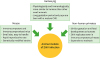
The factors that contributed to the emergence, global spread of ZIKV and change in the disease phenotype are not well understood. Animal models of ZIKV infection and disease are critical to study ZIKV pathogenesis, including pregnancy outcomes and evaluation of vaccines and therapeutics. Recently, ZIKV infection and disease have been evaluated in non-pregnant and pregnant animals, as well as a large panel of immunodeficient transgenic mice. Here we summarize the animal models that have been used to study ZIKV pathogenesis and to develop vaccine and therapeutic strategies (Fig. 1).
Figure 1
Characteristics of animals models of ZIKV infection.
in-17-287-g001

SMALL ANIMAL MODELS OF ZIKV INFECTION

Immunocompetent mouse models

Prior to the recent ZIKV epidemics, few animal models of ZIKV infection existed. From the first isolation of ZIKV in 1947 until 2015, there were only three studies which tested the virus' pathogenic potential in animal models (373839). In the first publication on ZIKV infection in a mouse model, inoculation of ZIKV via intracranial route caused neurological disease in suckling or adult mice (37). However, infection of adult immunocompetent mice with ZIKV via intraperitoneal route did not cause disease, indicating that intracranial route of inoculation is necessary to establish any successful infection. This study used the prototype MR 766 strain of ZIKV, which had undergone extensive passage in suckling mouse brains (40). Recently, ZIKV infection and disease have been evaluated in non-pregnant and pregnant immunocompetent mice using contemporary ZIKV strains. After peripheral (subcutaneous, intra-peritoneal, and intravenous) ZIKV inoculation, no clinical disease and little or no virus was detected in wild-type (WT) C57BL/6, Swiss Webster, BALB/c, and CD-1 mice (344142). Many different strains of ZIKV have been examined in similar mouse studies and regardless of the strain of ZIKV, similar results have been reported (43). Similarly, no fetal defects were observed after peripheral inoculation of ZIKV into pregnant WT C57BL/6 mice (44). Intravenous inoculation of pregnant WT SJL mice with a Brazilian ZIKV strain caused intrauterine growth restriction of developing fetuses, cortical malformations, and ocular defects (45). However, SJL mice are not fully immunocompetent and express lower levels of IFN-stimulated genes than C57BL/6, and this difference may account for the varied susceptibility to the ZIKV (43). It has been demonstrated that the ZIKV NS5 protein inhibits type I IFN response in a species-specific fashion, which might explain why adult WT mice are more resistant to the infection (4647). ZIKV infection has also been evaluated in immunocompetent neonatal mice (414849). ZIKV inoculation in one-day old immunocompetent C57BL/6 or Swiss mice via subcutaneous or intracranial route resulted in neurological disease characterized by tremors, ataxia, and seizures with evidence of ZIKV infection in the brain (4849). Since ZIKV is not naturally adapted to replicate in immunocompetent mice, their use in ZIKV research is limited. Nevertheless, immunocompetent mice have been used to assess the immunogenicity of vaccine candidates as well as their protective efficacy against viremia (3443).

Immunocompromised mouse models

In order to develop a mouse model that can support ZIKV replication and disease, several groups have evaluated the ZIKV infection in immunocompromised adult mice. Several of these models have altered IFN responses, which are an important component of antiviral defense. Type I IFNs, which include multiple forms of IFN-α and one IFN-β, signal through the same receptor, termed IFNAR1, and have been commonly associated with innate immune responses to viruses (50). Both the IFN-α/β receptor and downstream signaling molecules, including STAT-1, are critical for protection from viral infection as mice deficient in these factors do not mount effective anti-viral responses (5051). IFN regulatory factor (IRF), in particular IRF3 and IRF7, are essential for regulating the type I IFN response following viral infections (51). Type II IFN, or IFN-γ, which signals through a distinct receptor, IFNGR1, is the canonical cytokine of adaptive Th1 immunity and is essential for immune responses to intracellular pathogens (52). Animals lacking the receptor for type I IFN including A129 mice (129S2 Ifnar1tm1Agt) and Ifnar1−/− C57BL/6 mice or mice deficient in transcription factors IRF-3/5/7−/− are highly susceptible to both African and Asian-lineage ZIKV and sustain infection with high viral loads in the brain (4142535455). These animals developed severe ZIKV disease including hind-limb weakness, paralysis and death after peripheral (subcutaneous, intra-peritoneal, and intravenous) inoculation of ZIKV. Severity of ZIKV infection in these immunocompromised mice is age dependent, as older mice (11 week-old) are less susceptible to infection than younger mice (3-5 week-old) (4142). Mice lacking both the type I and type II IFN receptors (AG129, 129/Sv Ifnar1tm1Agt Ifngr1tm1Agt) demonstrated greater susceptibility and more severe disease following ZIKV infection than A129 mice (21254256). Analysis of the tissues from ZIKV-infected A129 and AG129 mice demonstrated that the highest viral loads were in testes and brain. The presence of virus in the testes is consistent with the reports of sexual transmission of ZIKV. In addition, Stat2−/− mice are also highly susceptible to ZIKV infection. After peripheral ZIKV inoculation, Stat2−/− mice display neurological symptoms and virus was detected in the central nervous system (CNS), gonads and other visceral organs (57). Stat2−/− mice lack both type I and type III IFN signaling. Subcutaneous inoculation of pregnant Ifnar1−/− C57BL/6 mice at gestation days 6.5 and 7.5 with an Asian ZIKV strain resulted in fetal death and reabsorption in most of the fetuses while those that survived the infection had intrauterine growth restriction and growth impairment (44). For these experiments, Ifnar1−/− female mice were mated with WT sires resulting in fetuses that were heterozygous for IFNAR1. Thus, despite the fetuses having the ability to respond to type I IFN, severe outcomes still were observed, suggesting that a type I IFN response in the fetus is not sufficient to protect from ZIKV-induced injury (44). Together, these immunocompromised mouse models have been used to demonstrate ZIKV ability to cause fetal abnormalities, deterioration of gonadal tissue and infection through sexual route (434458596061). Therefore, these susceptible mouse models also have been used extensively to evaluate candidate therapies and vaccines for efficacy against ZIKV replication (244362636465). While these immunodeficient mice are useful, they are inherently biased toward producing disease based on their genetic background. Furthermore, lethality is the main endpoint without assessing actual clinical disease.
In an attempt to produce infection models that do not rely upon knockout mice, several groups have explored the temporal blockade of type 1 IFN in immune intact mice using polyclonal and monoclonal antibodies targeting either type 1 IFNs directly or the IFNAR1 receptor. The major advantage of this approach is that it allows immune responses to be elicited in immunologically competent mice with type 1 IFN blockade only induced at the time of infection. It has been demonstrated that adult immunocompetent C57BL/6 mice treated with anti-IFNAR1 antibodies (that suppress expression of type 1 IFN) before infection are highly susceptible to mouse-adapted African ZIKV-Dakar strain (5866). These mice develop severe ZIKV-mediated disease accompanied by significant neuroinflammation and mortality. Similarly, fetuses from mice with prior exposure to a blocking antibody against anti-IFNAR1 before ZIKV infection also resulted in intrauterine growth restriction (44).

Guinea pig model

Initial experiments conducted in 1950s showed that guinea pigs inoculated via intracranial route with the African ZIKV strain MR 766 developed no signs of infection (37). These studies used the prototype MR 766 strain of ZIKV, which had undergone extensive passage in suckling mouse brains. Recently, it has been demonstrated that guinea pigs are susceptible to infection by a contemporary Asian strain of ZIKV (67). Upon subcutaneous inoculation with PRVABC59 strain of ZIKV, guinea pigs demonstrated clinical signs of infection characterized by fever, lethargy, hunched back, ruffled fur, and decrease in mobility. ZIKV was detected in the serum using qRT-PCR and plaque assay. ZIKV infection resulted in a dramatic increase in protein levels of multiple cytokines, chemokines and growth factors in the serum. ZIKV RNA was detected in the spleen and brain, with the highest viral load in the brain (67). The guinea pig is more physiologically and immunologically similar to humans than other small animals. Specifically, the guinea pig's reproductive physiology and estrous cycle are similar to humans. Also, placentation in the guinea pig occurs in a manner similar to that of humans, and both guinea pig and human placentas are classified as hemomonochorial (68). Guinea pigs have a long gestation period and pups are born with a mature CNS, which makes this species a promising subject for studies of in utero transfer of ZIKV and neurological manifestations in infants (69).

LARGE ANIMAL MODELS OF ZIKV INFECTION

Non-human primate (NHP) models

NHPs also are being used to study ZIKV pathogenesis. In contrast to mice, immunocompetent macaque monkeys are ideal to study ZIKV because of the similarity in gestation and fetal development as compared to humans. Rhesus macaques are susceptible to infection by both African and Asian-lineage ZIKV (4370717273). ZIKV-infected rhesus macaques developed viremia that peaked 2 to 6 days after infection and became undetectable by day 10. ZIKV was also detected in various organs, urine, saliva, and cerebrospinal fluid of some animals (7071). Similar to rhesus macaques, ZIKV infection was detected in several tissues of cynomolgus macaques including the male reproductive tract, intestines, and the brain and spinal cord (7475). Pregnant rhesus macaques infected with ZIKV developed viremia lasting 30 to 55 days (7072). Subcutaneous inoculation of a pregnant pigtail macaque with an Asian-lineage ZIKV resulted in reduced growth of the fetal brain (7677). Autopsy analysis from fetal brain demonstrated ZIKV infection and substantial pathology to the CNS. ZIKV was detected in the placenta, fetal brain and liver, and maternal brain, eyes, spleen, and liver (77). ZIKV infection induced T-cell responses and protected NHPs from ZIKV re-infection and from heterologous ZIKV infection (7074). Therefore, NHP models have been utilized for testing protective efficacy of novel vaccines and therapeutics against ZIKV (357378). The NHPs reproductive cycle is similar to the human reproductive cycle, which has its clear benefits in the study of disease outcomes in pregnant mothers infected with ZIKV, but this comes with the caveat that the NHP models will produce data at a much slower rate. NHP models are very expensive to maintain and require a great amount of space and time when compared to other animal models.

CONCLUSION

Both small and large animal models have been established to investigate ZIKV pathogenesis and to develop vaccine and therapeutic strategies. ZIKV does not cause infection and clinical disease in weaned immunocompetent mice. Immunocompromised mice including IFN dysregulated mice have successfully reproduced clinical disease or demonstrated lethality after ZIKV infection. Immunocompetent guinea pigs are susceptible to infection by a contemporary strain of ZIKV. Similarly, NHPs have been demonstrated to be susceptible to infection by ZIKV. Together, these animal models of ZIKV pathogenesis can be utilized to evaluate candidate therapies and vaccines against ZIKV infection. However, each of these models has limitations that must be considered in the experimental design and interpretation of results.

Abbreviations

CNS

central nervous system

IFN

interferon

IRF

interferon regulatory factor

NHP

non-human primate

WT

wild-type

ZIKV

Zika virus

ACKNOWLEDGEMENTS

This work was supported by a grant (P30GM114737) from the Centers of Biomedical Research Excellence, National Institute of General Medical Sciences, grant (R21NS099838) from National Institute of Neurological Disorders and Stroke, and grant (R21OD024896) from the Office of the Director, National Institutes of Health.

Notes

Conflict of Interest The authors declared no potential conflicts of interest.

Author Contributions

  • Conceptualization: Kumar M.

  • Funding acquisition: Kumar M.

  • Visualization: Shin OS, Kumar M.

  • Writing - original draft: Krause KK, Azouz F, Kumar M.

  • Writing - review & editing: Krause KK, Azouz F, Shin OS, Kumar M.

References

1. Musso D, Gubler DJ. Zika virus. Clin Microbiol Rev. 2016; 29:487–524.
crossref
2. Klase ZA, Khakhina S, Schneider Ade B, Callahan MV, Glasspool-Malone J, Malone R. Zika fetal neuropathogenesis: etiology of a viral syndrome. PLoS Negl Trop Dis. 2016; 10:e0004877.
crossref
3. Culjat M, Darling SE, Nerurkar VR, Ching N, Kumar M, Min SK, Wong R, Grant L, Melish ME. Clinical and imaging findings in an infant with Zika embryopathy. Clin Infect Dis. 2016; 63:805–811.
crossref
4. Kumar M, Ching L, Astern J, Lim E, Stokes AJ, Melish M, Nerurkar VR. Prevalence of antibodies to Zika virus in mothers from Hawaii who delivered babies with and without microcephaly between 2009–2012. PLoS Negl Trop Dis. 2016; 10:e0005262.
crossref
5. Walker WL, Lindsey NP, Lehman JA, Krow-Lucal ER, Rabe IB, Hills SL, Martin SW, Fischer M, Staples JE. Zika virus disease cases - 50 states and the district of Columbia, January 1–July 31, 2016. MMWR Morb Mortal Wkly Rep. 2016; 65:983–986.
crossref
6. Likos A, Griffin I, Bingham AM, Stanek D, Fischer M, White S, Hamilton J, Eisenstein L, Atrubin D, Mulay P, et al. Local mosquito-borne transmission of Zika virus - Miami-Dade and Broward Counties, Florida, June–August 2016. MMWR Morb Mortal Wkly Rep. 2016; 65:1032–1038.
crossref
7. Adams L, Bello-Pagan M, Lozier M, Ryff KR, Espinet C, Torres J, Perez-Padilla J, Febo MF, Dirlikov E, Martinez A, et al. Update: ongoing Zika virus transmission - Puerto Rico, November 1, 2015–July 7, 2016. MMWR Morb Mortal Wkly Rep. 2016; 65:774–779.
crossref
8. Mlakar J, Korva M, Tul N, Popović M, Poljšak-Prijatelj M, Mraz J, Kolenc M, Resman Rus K, Vesnaver Vipotnik T, Fabjan Vodušek V, et al. Zika virus associated with microcephaly. N Engl J Med. 2016; 374:951–958.
crossref
9. D’Ortenzio E, Matheron S, Yazdanpanah Y, de Lamballerie X, Hubert B, Piorkowski G, Maquart M, Descamps D, Damond F, Leparc-Goffart I. Evidence of sexual transmission of Zika virus. N Engl J Med. 2016; 374:2195–2198.
crossref
10. Russell K, Hills SL, Oster AM, Porse CC, Danyluk G, Cone M, Brooks R, Scotland S, Schiffman E, Fredette C, et al. Male-to-female sexual transmission of Zika virus-United States, January–April 2016. Clin Infect Dis. 2017; 64:211–213.
crossref
11. Ferguson NM, Cucunubá ZM, Dorigatti I, Nedjati-Gilani GL, Donnelly CA, Basáñez MG, Nouvellet P, Lessler J. Epidemiology. Countering the Zika epidemic in Latin America. Science. 2016; 353:353–354.
12. Oster AM, Russell K, Stryker JE, Friedman A, Kachur RE, Petersen EE, Jamieson DJ, Cohn AC, Brooks JT. Update: interim guidance for prevention of sexual transmission of Zika virus--United States, 2016. MMWR Morb Mortal Wkly Rep. 2016; 65:323–325.
crossref
13. Faria NR, Azevedo RD, Kraemer MU, Souza R, Cunha MS, Hill SC, Thézé J, Bonsall MB, Bowden TA, Rissanen I, et al. Zika virus in the Americas: early epidemiological and genetic findings. Science. 2016; 352:345–349.
crossref
14. Herrera BB, Chang CA, Hamel DJ, Mboup S, Ndiaye D, Imade G, Okpokwu J, Agbaji O, Bei AK, Kanki PJ. Continued transmission of Zika virus in humans in West Africa, 1992–2016. J Infect Dis. 2017; 215:1546–1550.
crossref
15. Grard G, Caron M, Mombo IM, Nkoghe D, Mboui Ondo S, Jiolle D, Fontenille D, Paupy C, Leroy EM. Zika virus in Gabon (Central Africa)--2007: a new threat from Aedes albopictus? PLoS Negl Trop Dis. 2014; 8:e2681.
16. Fagbami AH. Zika virus infections in Nigeria: virological and seroepidemiological investigations in Oyo State. J Hyg (Lond). 1979; 83:213–219.
crossref
17. Saluzzo JF, Ivanoff B, Languillat G, Georges AJ. Serological survey for arbovirus antibodies in the human and simian populations of the South-East of Gabon (author’s transl). Bull Soc Pathol Exot. 1982; 75:262–266.
18. Musso D, Lanteri MC. Emergence of Zika virus: where does it come from and where is it going to? Lancet Infect Dis. 2017; 17:255.
crossref
19. Who supports Cabo Verde in managing Zika virus. Saudi Med J. 2016; 37:470–471.
20. Hennessey M, Fischer M, Staples JE. Zika virus spreads to new areas - region of the Americas, May 2015–January 2016. MMWR Morb Mortal Wkly Rep. 2016; 65:55–58.
crossref
21. Zmurko J, Marques RE, Schols D, Verbeken E, Kaptein SJ, Neyts J. The viral polymerase inhibitor 7-Deaza-2′-C-methyladenosine is a potent inhibitor of in vitro Zika virus replication and delays disease progression in a robust mouse infection model. PLoS Negl Trop Dis. 2016; 10:e0004695.
22. Sacramento CQ, de Melo GR, de Freitas CS, Rocha N, Hoelz LV, Miranda M, Fintelman-Rodrigues N, Marttorelli A, Ferreira AC, Barbosa-Lima G, et al. The clinically approved antiviral drug sofosbuvir inhibits Zika virus replication. Sci Rep. 2017; 7:40920.
crossref
23. Reznik SE, Ashby CR Jr. Sofosbuvir: an antiviral drug with potential efficacy against Zika infection. Int J Infect Dis. 2017; 55:29–30.
crossref
24. Bullard-Feibelman KM, Govero J, Zhu Z, Salazar V, Veselinovic M, Diamond MS, Geiss BJ. The FDA-approved drug sofosbuvir inhibits Zika virus infection. Antiviral Res. 2017; 137:134–140.
crossref
25. Julander JG, Siddharthan V, Evans J, Taylor R, Tolbert K, Apuli C, Stewart J, Collins P, Gebre M, Neilson S, et al. Efficacy of the broad-spectrum antiviral compound BCX4430 against Zika virus in cell culture and in a mouse model. Antiviral Res. 2017; 137:14–22.
crossref
26. Baz M, Goyette N, Griffin BD, Kobinger GP, Boivin G. In vitro susceptibility of geographically and temporally distinct Zika viruses to favipiravir and ribavirin. Antivir Ther.
27. Cai L, Sun Y, Song Y, Xu L, Bei Z, Zhang D, Dou Y, Wang H. Viral polymerase inhibitors T-705 and T-1105 are potential inhibitors of Zika virus replication. Arch Virol. 2017; 162:2847–2853.
crossref
28. Best K, Guedj J, Madelain V, de Lamballerie X, Lim SY, Osuna CE, Whitney JB, Perelson AS. Zika plasma viral dynamics in nonhuman primates provides insights into early infection and antiviral strategies. Proc Natl Acad Sci USA. 2017; 114:8847–8852.
crossref
29. Lanko K, Eggermont K, Patel A, Kaptein S, Delang L, Verfaillie CM, Neyts J. Replication of the Zika virus in different iPSC-derived neuronal cells and implications to assess efficacy of antivirals. Antiviral Res. 2017; 145:82–86.
crossref
30. Furuta Y, Gowen BB, Takahashi K, Shiraki K, Smee DF, Barnard DL. Favipiravir (T-705), a novel viral RNA polymerase inhibitor. Antiviral Res. 2013; 100:446–454.
crossref
31. Furuta Y, Takahashi K, Shiraki K, Sakamoto K, Smee DF, Barnard DL, Gowen BB, Julander JG, Morrey JD. T-705 (favipiravir) and related compounds: novel broad-spectrum inhibitors of RNA viral infections. Antiviral Res. 2009; 82:95–102.
crossref
32. Furuta Y, Takahashi K, Fukuda Y, Kuno M, Kamiyama T, Kozaki K, Nomura N, Egawa H, Minami S, Watanabe Y, et al. In vitro and in vivo activities of anti-influenza virus compound T-705. Antimicrob Agents Chemother. 2002; 46:977–981.
crossref
33. Lin HH, Yip BS, Huang LM, Wu SC. Zika virus structural biology and progress in vaccine development. Biotechnol Adv.
crossref
34. Larocca RA, Abbink P, Peron JP, Zanotto PM, Iampietro MJ, Badamchi-Zadeh A, Boyd M, Ng’ang’a D, Kirilova M, Nityanandam R, et al. Vaccine protection against Zika virus from Brazil. Nature. 2016; 536:474–478.
crossref
35. Abbink P, Larocca RA, De La Barrera RA, Bricault CA, Moseley ET, Boyd M, Kirilova M, Li Z, Ng’ang’a D, Nanayakkara O, et al. Protective efficacy of multiple vaccine platforms against Zika virus challenge in rhesus monkeys. Science. 2016; 353:1129–1132.
crossref
36. Griffin BD, Muthumani K, Warner BM, Majer A, Hagan M, Audet J, Stein DR, Ranadheera C, Racine T, De La Vega MA, et al. DNA vaccination protects mice against Zika virus-induced damage to the testes. Nat Commun. 2017; 8:15743.
crossref
37. Dick GW. Zika virus. II. Pathogenicity and physical properties. Trans R Soc Trop Med Hyg. 1952; 46:521–534.
crossref
38. Bell TM, Field EJ, Narang HK. Zika virus infection of the central nervous system of mice. Arch Gesamte Virusforsch. 1971; 35:183–193.
crossref
39. Way JH, Bowen ET, Platt GS. Comparative studies of some African arboviruses in cell culture and in mice. J Gen Virol. 1976; 30:123–130.
crossref
40. Dick GW, Kitchen SF, Haddow AJ. Zika virus. I. Isolations and serological specificity. Trans R Soc Trop Med Hyg. 1952; 46:509–520.
crossref
41. Lazear HM, Govero J, Smith AM, Platt DJ, Fernandez E, Miner JJ, Diamond MS. A mouse model of Zika virus pathogenesis. Cell Host Microbe. 2016; 19:720–730.
crossref
42. Rossi SL, Tesh RB, Azar SR, Muruato AE, Hanley KA, Auguste AJ, Langsjoen RM, Paessler S, Vasilakis N, Weaver SC. Characterization of a novel murine model to study Zika virus. Am J Trop Med Hyg. 2016; 94:1362–1369.
crossref
43. Morrison TE, Diamond MS. Animal models of Zika virus infection, pathogenesis, and immunity. J Virol. 2017; 91:e00009–e00017.
crossref
44. Miner JJ, Cao B, Govero J, Smith AM, Fernandez E, Cabrera OH, Garber C, Noll M, Klein RS, Noguchi KK, et al. Zika virus infection during pregnancy in mice causes placental damage and fetal demise. Cell. 2016; 165:1081–1091.
crossref
45. Cugola FR, Fernandes IR, Russo FB, Freitas BC, Dias JL, Guimarães KP, Benazzato C, Almeida N, Pignatari GC, Romero S, et al. The Brazilian Zika virus strain causes birth defects in experimental models. Nature. 2016; 534:267–271.
crossref
46. Grant A, Ponia SS, Tripathi S, Balasubramaniam V, Miorin L, Sourisseau M, Schwarz MC, Sánchez-Seco MP, Evans MJ, Best SM, et al. Zika virus targets human STAT2 to inhibit Type I interferon signaling. Cell Host Microbe. 2016; 19:882–890.
crossref
47. Bowen JR, Quicke KM, Maddur MS, O'Neal JT, McDonald CE, Fedorova NB, Puri V, Shabman RS, Pulendran B, Suthar MS. Zika virus antagonizes Type I interferon responses during infection of human dendritic cells. PLoS Pathog. 2017; 13:e1006164.
crossref
48. Manangeeswaran M, Ireland DD, Verthelyi D. Zika (PRVABC59) infection is associated with T cell Infiltration and neurodegeneration in CNS of immunocompetent neonatal C57Bl/6 mice. PLoS Pathog. 2016; 12:e1006004.
crossref
49. Fernandes NC, Nogueira JS, Réssio RA, Cirqueira CS, Kimura LM, Fernandes KR, Cunha MS, Souza RP, Guerra JM. Experimental Zika virus infection induces spinal cord injury and encephalitis in newborn Swiss mice. Exp Toxicol Pathol. 2017; 69:63–71.
crossref
50. Haller O, Kochs G, Weber F. The interferon response circuit: induction and suppression by pathogenic viruses. Virology. 2006; 344:119–130.
crossref
51. Suthar MS, Diamond MS, Gale M Jr. West Nile virus infection and immunity. Nat Rev Microbiol. 2013; 11:115–128.
crossref
52. Schoenborn JR, Wilson CB. Regulation of interferon-gamma during innate and adaptive immune responses. Adv Immunol. 2007; 96:41–101.
53. Dowall SD, Graham VA, Rayner E, Atkinson B, Hall G, Watson RJ, Bosworth A, Bonney LC, Kitchen S, Hewson R. A susceptible mouse model for Zika virus infection. PLoS Negl Trop Dis. 2016; 10:e0004658.
crossref
54. Li H, Saucedo-Cuevas L, Regla-Nava JA, Chai G, Sheets N, Tang W, Terskikh AV, Shresta S, Gleeson JG. Zika virus infects neural progenitors in the adult mouse brain and alters proliferation. Cell Stem Cell. 2016; 19:593–598.
crossref
55. Miner JJ, Sene A, Richner JM, Smith AM, Santeford A, Ban N, Weger-Lucarelli J, Manzella F, Rückert C, Govero J, et al. Zika virus infection in mice causes panuveitis with shedding of virus in tears. Cell Reports. 2016; 16:3208–3218.
crossref
56. Aliota MT, Caine EA, Walker EC, Larkin KE, Camacho E, Osorio JE. Characterization of lethal Zika virus infection in AG129 mice. PLoS Negl Trop Dis. 2016; 10:e0004682.
crossref
57. Tripathi S, Balasubramaniam VR, Brown JA, Mena I, Grant A, Bardina SV, Maringer K, Schwarz MC, Maestre AM, Sourisseau M, et al. A novel Zika virus mouse model reveals strain specific differences in virus pathogenesis and host inflammatory immune responses. PLoS Pathog. 2017; 13:e1006258.
crossref
58. Govero J, Esakky P, Scheaffer SM, Fernandez E, Drury A, Platt DJ, Gorman MJ, Richner JM, Caine EA, Salazar V, et al. Zika virus infection damages the testes in mice. Nature. 2016; 540:438–442.
crossref
59. Ma W, Li S, Ma S, Jia L, Zhang F, Zhang Y, Zhang J, Wong G, Zhang S, Lu X, et al. Zika virus causes testis damage and leads to male infertility in mice. Cell. 2016; 167:1511–1524.e10.
crossref
60. Yockey LJ, Varela L, Rakib T, Khoury-Hanold W, Fink SL, Stutz B, Szigeti-Buck K, Van den Pol A, Lindenbach BD, Horvath TL, et al. Vaginal exposure to Zika virus during pregnancy leads to fetal brain infection. Cell. 2016; 166:1247–1256.e4.
crossref
61. Tang WW, Young MP, Mamidi A, Regla-Nava JA, Kim K, Shresta S. A mouse model of Zika Virus sexual transmission and vaginal viral replication. Cell Reports. 2016; 17:3091–3098.
crossref
62. Deng YQ, Zhang NN, Li CF, Tian M, Hao JN, Xie XP, Shi PY, Qin CF. Adenosine analog NITD008 is a potent inhibitor of Zika virus. Open Forum Infect Dis. 2016; 3:ofw175.
crossref
63. Zhao H, Fernandez E, Dowd KA, Speer SD, Platt DJ, Gorman MJ, Govero J, Nelson CA, Pierson TC, Diamond MS, et al. Structural basis of Zika virus-specific antibody protection. Cell. 2016; 166:1016–1027.
crossref
64. Swanstrom JA, Plante JA, Plante KS, Young EF, McGowan E, Gallichotte EN, Widman DG, Heise MT, de Silva AM, Baric RS. Dengue virus envelope dimer epitope monoclonal antibodies isolated from dengue patients are protective against Zika virus. MBio. 2016; 7:e01123–e16.
crossref
65. Sapparapu G, Fernandez E, Kose N, Cao B, Fox JM, Bombardi RG, Zhao H, Nelson CA, Bryan AL, Barnes T, et al. Neutralizing human antibodies prevent Zika virus replication and fetal disease in mice. Nature. 2016; 540:443–447.
crossref
66. Smith DR, Hollidge B, Daye S, Zeng X, Blancett C, Kuszpit K, Bocan T, Koehler JW, Coyne S, Minogue T, et al. Neuropathogenesis of Zika virus in a highly susceptible immunocompetent mouse model after antibody blockade of Type I interferon. PLoS Negl Trop Dis. 2017; 11:e0005296.
crossref
67. Kumar M, Krause KK, Azouz F, Nakano E, Nerurkar VR. A Guinea pig model of Zika virus infection. Virol J. 2017; 14:75.
crossref
68. Mess A. The Guinea pig placenta: model of placental growth dynamics. Placenta. 2007; 28:812–815.
crossref
69. Padilla-Carlin DJ, McMurray DN, Hickey AJ. The Guinea pig as a model of infectious diseases. Comp Med. 2008; 58:324–340.
70. Dudley DM, Aliota MT, Mohr EL, Weiler AM, Lehrer-Brey G, Weisgrau KL, Mohns MS, Breitbach ME, Rasheed MN, Newman CM, et al. A rhesus macaque model of Asian-lineage Zika virus infection. Nat Commun. 2016; 7:12204.
crossref
71. Li XF, Dong HL, Huang XY, Qiu YF, Wang HJ, Deng YQ, Zhang NN, Ye Q, Zhao H, Liu ZY, et al. Characterization of a 2016 clinical isolate of Zika virus in non-human primates. EBioMedicine. 2016; 12:170–177.
crossref
72. Nguyen SM, Antony KM, Dudley DM, Kohn S, Simmons HA, Wolfe B, Salamat MS, Teixeira LB, Wiepz GJ, Thoong TH, et al. Highly efficient maternal-fetal Zika virus transmission in pregnant rhesus macaques. PLoS Pathog. 2017; 13:e1006378.
crossref
73. Aliota MT, Dudley DM, Newman CM, Mohr EL, Gellerup DD, Breitbach ME, Buechler CR, Rasheed MN, Mohns MS, Weiler AM, et al. Heterologous protection against Asian Zika virus challenge in rhesus macaques. PLoS Negl Trop Dis. 2016; 10:e0005168.
crossref
74. Osuna CE, Lim SY, Deleage C, Griffin BD, Stein D, Schroeder LT, Omange R, Best K, Luo M, Hraber PT, et al. Zika viral dynamics and shedding in rhesus and cynomolgus macaques. Nat Med. 2016; 22:1448–1455.
crossref
75. Koide F, Goebel S, Snyder B, Walters KB, Gast A, Hagelin K, Kalkeri R, Rayner J. Development of a Zika virus infection model in cynomolgus macaques. Front Microbiol. 2016; 7:2028.
crossref
76. Driggers RW, Ho CY, Korhonen EM, Kuivanen S, Jääskeläinen AJ, Smura T, Rosenberg A, Hill DA, DeBiasi RL, Vezina G, et al. Zika virus infection with prolonged maternal viremia and fetal brain abnormalities. N Engl J Med. 2016; 374:2142–2151.
crossref
77. Adams Waldorf KM, Stencel-Baerenwald JE, Kapur RP, Studholme C, Boldenow E, Vornhagen J, Baldessari A, Dighe MK, Thiel J, Merillat S, et al. Fetal brain lesions after subcutaneous inoculation of Zika virus in a pregnant nonhuman primate. Nat Med. 2016; 22:1256–1259.
crossref
78. Dowd KA, Ko SY, Morabito KM, Yang ES, Pelc RS, DeMaso CR, Castilho LR, Abbink P, Boyd M, Nityanandam R, et al. Rapid development of a DNA vaccine for Zika virus. Science. 2016; 354:237–240.
crossref
TOOLS
Similar articles