

 PDF
PDF ePub
ePub Citation
 Print
Print


INTRODUCTION
Bone grafting is a surgical procedure frequently performed in orthopedics and periodontics to treat patients with missing or damaged bones. Bone grafting not only helps to fill the physical gaps created by the missing or damaged bone and provides structural stability, but also stimulates the growth of bone tissues. In particular, periodontal bone grafts improve clinical outcomes in oral and maxillofacial surgery.
Although graft materials prepared from natural bones of different origin are mostly composed of hydroxyapatite (HA), because of its high biocompatibility and osteoconductivity, their performance is known to be significantly affected by their physicochemical properties [1]. For example, as autologous graft materials originate from the patient's own bone and can be used with minimal treatment, they have been reported to be the most efficient and fastest-healing bone substitute materials. In contrast, allografts are known to be not as effective as autografts, because the extensive chemical treatments required to prevent potential infectivity (e.g., from human immunodeficiency virus [HIV] and hepatitis viruses) can disrupt the hierarchical structure of bone and remove a significant amount of the growth factors necessary for efficient bone regeneration.
Physicochemical properties are even more important for xenografts [2]. Xenografts have clear advantages, as they can be mass-produced in large quantities at relatively affordable processing costs. However, as they originate from bone tissues of other species, their native osteological characteristics are quite different from those of human bone tissues. In addition, the series of chemical processes or thermal treatments performed on xenografts can further affect their physical and chemical properties. Thus, the physicochemical characteristics of a xenograft can be closely related to its overall performance for bone regeneration.
Bovine bones were the first animal bones employed for the production of xenograft materials, and several bovine xenograft products currently dominate the market share of bone graft materials. For example, Bio-Oss® (Geistlich Biomaterials, Wolhusen, Switzerland) is a well-known xenograft, composed of an osseous mineral matrix obtained from spinal bone [3]. Cerabone® (Botiss Biomaterials GmbH, Zossen, Germany) is another popular bone grafting material obtained from cancellous bone of cattle femur condyles [4].
In contrast, porcine-derived bone graft materials have only recently been developed and marketed. It has been reported that porcine bone has a similar formulation and structure to human bone [5]. The lipid content (in terms of category and structure) of porcine bone is also similar to that of human bone [6]. In addition, porcine bone has a relatively low risk of zoonosis [7]. Thus, considerable efforts are being devoted to replacing bovine bone graft materials with materials obtained from porcine bone [789]. However, studies of the physicochemical characteristics of xenografts originating from porcine bone remain very limited.
Therefore, in this work, we carried out a detailed study on the physicochemical properties of a commercially available porcine xenograft, THE Graft, and compared them with the properties of the bovine-origin xenografts, Bio-Oss® and Cerabone®. We focused on the morphology/porosity, specific surface area (SSA), 3-dimensional (3D) tomography, chemical component analysis, organic content analysis, and wettability of these materials, as these are considered to be important characteristics of graft materials. The results of our study suggest that the graft material obtained from porcine bone is comparable to the materials prepared from bovine bone in many aspects; moreover, the present material satisfies most of the key requirements for its effective application as a xenograft material.
MATERIALS AND METHODS
Materials
THE Graft (Purgo Biologics, Seongnam, Korea), Bio-Oss® (Geistlich Biomaterials), and Cerabone® (Botiss Biomaterials GmbH) were purchased and used to compare the physicochemical properties of xenografts of different origins.
Physicochemical characterization
Morphological characterization/porosity
The morphological characterization of the materials was carried out by scanning electron microscopy (SEM) using a field emission-environmental scanning electron microscope (Philips XL-30 ESEM-FEG, Philips Corp., Eindhoven, Netherlands) operating at 15.2 kV. The pore size analysis was performed by mercury intrusion porosimetry (MIP) (Autopore V 9600, Micromeritics Instrument Corp., Norcross, GA, USA). The measurements were made using 255.9 mg of THE Graft, 138.9 mg of Bio-Oss®, and 181.2 mg of Cerabone®. The total porosity per unit mass was calculated from the total intrusion volume. Mercury was applied with a pressure of 0.10–60,000.00 psia for the measurements. The contact angle of the mercury was 130°.
SSA
The SSA was measured using a nitrogen gas porosimeter (ASAP 2420, Micromeritics Instrument Corp.) after degassing 1.0 g of each sample below 0.010 mmHg at 150°C for 5 hours. The SSA was expressed as square meters per gram of mineral (m2/g).
3D tomography
The 3D tomography of graft materials was carried out by creating ultrathin (up to 10 nm) sections of frozen epoxy resin-reinforced graft samples with an ultramicrotome and stacking their corresponding 2-dimensional (2D) atomic force microscopy (AFM) images into a 3D image. The Japanese Industrial Standard for roughness (RzJIS) was measured based on the 5 highest peaks and lowest valleys over the entire sampling length. The sampling length was 50 µm [1011].
Chemical characterizations
The crystalline phase composition of the samples was determined by X-ray diffraction (XRD) using a D8 Advance diffractometer (Bruker Optics, Billerica, MA, USA) operating at 40 kV and 40 mA, with Cu Kα radiation to probe the 2θ range of 20°–80°. The ground samples (less than 0.3 g) were packed in a holder and measured with the diffractometer at scan steps of 0.02°, with a time per step of 35.4 seconds. Fourier-transform infrared (FT-IR) spectroscopy (Tensor 27, Bruker Optics) was used for the chemical analysis of the graft materials. A small amount (less than 0.3 g) of each graft sample was mixed with potassium bromide powder to produce a sample pellet. FT-IR spectroscopy was performed in transmission mode. For the elemental analysis of graft materials, inductively coupled plasma optical emission spectrometry (ICP-OES) (Optima 5300 DV, Perkin-Elmer, Waltham, MA, USA) was used. Each sample (0.1 g) was placed in a platinum crucible and dissolved at 200°C for 1 hour in nitric acid and deionized (DI) water before measurement. The ICP-OES measurements were performed based on ASTM F1581-08.
Residual protein analysis
The residual protein content in the xenograft materials was determined using both nitrogen and amino acid analysis. For the nitrogen analysis, we first quantified the nitrogen content in the sample using an elemental analyzer (Flash EA 2000 series, ThermoFisher Inc., Cambridge, UK). We multiplied the nitrogen content by the protein factor of 6.25 to obtain the amount of protein in the sample based on the Kjeldahl method [12]. For the amino acid analysis, we first completely dried 1 mg of the sample, then hydrolyzed it with HCl at 110°C for 24 hours, and derivatized the hydrolyzed amino acids with phenylisothiocyanate. After complete drying, the samples were dissolved with 200 µL of 0.05 M sodium acetate trihydrate. After centrifugation, the supernatant was analyzed with high-performance liquid chromatography using a HP 1100 Series liquid chromatography system (Agilent, Santa Clara, CA, USA) (C18 4 µm [3.9×300 mm], spectrophotometer at 254 nm). Individual amino acids were calculated using the chromatogram obtained with a standard solution (Waters Co., Milford, MA, USA).
RESULTS
Morphology and porosity of the graft materials
All examined xenografts were in granulated form. The size distribution of the granules of graft materials was determined by counting the number of granules of different sizes after sorting them with commercially available sieves of different mesh sizes. The size of the granules of each sample was uniformly distributed within the 0.15–1 mm range, with maximum counts between 0.3 and 0.6 mm (Figure 1). This suggests that THE Graft, Bio-Oss®, and Cerabone® all had a similar particle size distribution.
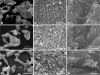
To investigate the surface morphology of THE Graft, we obtained SEM images of its granules and compared them to the corresponding images obtained for Bio-Oss® and Cerabone® samples (Figure 2). As expected, the xenograft materials were predominantly found in granular form with sizes between 0.15 and 1 mm. The majority of the examined granules contained macropores (diameter >100 µm), which are necessary to form blood vessels and induce both bone growth and reorganization around the graft material. In addition, the 3 graft materials contained a large amount of micropores (diameter <10 µm), which are required for the penetration of body fluids, ion transportation, the attachment of osteoblasts, and the precipitation of newly formed HA. Further zoomed images of both THE Graft and Bio-Oss® showed that these graft materials were composed of sub-100-nm grains with a large amount of nanoscale pores present between the grains (Figure 2C and F). In contrast, sub-100-nm grains and nanoscale pores were not observed on the surface of Cerabone® (Figure 2I).
Figure 2
Surface morphologies of the xenograft materials. (A-C) THE Graft, (D-F) Bio-Oss®, and (G-I) Cerabone®.
Next, we used MIP to compare the pore size distribution of the 3 xenograft materials (Figure 3). MIP is a simple and versatile technique used to measure the pore size distribution of a variety of porous materials. This technique, which is based on the capillary depression of mercury, can be used to measure a wide range of pore diameters, from 3 nm to 1,100 µm, as well as other parameters including porosity, average pore diameter, and bulk density. THE Graft had a large fraction of pore sizes in the 3–50 nm range and a non-negligible fraction in the 60–300 nm range. The macropores were found to be larger than 60 µm. The average pore diameter of THE Graft was 0.06 µm (Table 1). Surprisingly, Bio-Oss® showed a pore size distribution very similar to that of THE Graft, with a large fraction of pore sizes in the 3–60 nm range, some pores within the 65–280 nm range, and macropores beyond 60 µm. The average pore diameter of Bio-Oss® was also similar to that of THE Graft. In contrast, the pore size distribution of Cerabone® was significantly different from that of the other 2 materials. The Cerabone® granules had a negligible content of sub-100-nm pores: most pores were larger than 50 µm, with only a small fraction of pores between 200 nm and 1 µm. The average pore diameter of Cerabone® was 0.46 µm, which is significantly larger than that of both THE Graft and Bio-Oss®.
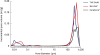
Table 1
Porosity of the graft materials

| Sample | Average pore diameter (µm) | Porosity (%) | Bulk density (g/mL) |
|---|---|---|---|
| THE Graft | 0.06 | 78.4 | 0.50 |
| Bio-Oss® | 0.06 | 70.5 | 0.54 |
| Cerabone® | 0.49 | 62.0 | 1.20 |
The porosity of THE Graft was found to be 78.4%, which is larger than that of both Bio-Oss® and Cerabone® (Table 1). Considering the comparable porosity of human trabecular bone (79.3%), this result suggests that THE Graft resembles the porous structure of human bone quite closely [13]. Since both macropores and micropores play important roles in bone formation, high porosity is crucial for the performance of graft materials [1415]. The higher porosity of THE Graft compared with both Bio-Oss® and Cerabone® suggests that THE Graft could represent an efficient xenograft material.
SSA analysis
To measure the SSA of the present materials, we carried out a Brunauer-Emmett-Teller (BET) isotherm analysis using nitrogen gas adsorption after degassing the samples below 0.010 mmHg at 150°C for 5 hours [1617]. The nitrogen gas adsorption technique can be used to measure many parameters related to the porosity of a sample, including pore volume, pore size distribution, and SSA.
The SSA of THE Graft and Bio-Oss® were similar (Table 2). In contrast, the SSA of Cerabone® was significantly smaller than the values measured for THE Graft and Bio-Oss®. Considering that both THE Graft and Bio-Oss® had a similar surface morphology and pore size distribution with a substantial amount of nanoscale pores, whereas Cerabone® did not, we believe that this difference in the SSA was closely related to the nano/microscale structure of the bone graft materials.

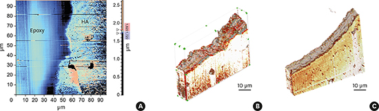
3D tomography of graft materials
Roughness is an important property of graft materials, owing to its strong influence on the adhesion, growth, proliferation, and osseointegration of osteoblast cells [18]. However, measuring the roughness of a granular graft material is a very difficult task, due to its waviness, which is associated with its shape and porosity. By creating ultrathin (up to 10 nm) sections of epoxy resin-reinforced graft samples with an ultramicrotome and stacking their corresponding 2D AFM images into a 3D image, it was possible to analyze the 3D tomography of the granules of THE Graft and Bio-Oss® and to estimate their 10-point mean roughness (RzJIS) (Figure 4). We considered it especially important to characterize these 2 samples because we found that they were very similar in terms of surface morphology, total porosity, pore size distribution, and SSA.
Figure 4
Measurement of the roughness of the grafting materials using 3D AFM. (A) The 2D AFM image of porcine graft section cut with an ultramicrotome. The 3D tomography of (B) THE Graft and (C) Bio-Oss® was obtained by stacking their 2D AFM images. The RzJIS roughness of the samples from THE Graft and Bio-Oss® was found to be 4.47 and 1.37 µm, respectively.
3D: 3-dimensional, 2D: 2-dimensional, AFM: atomic force microscopy, RzJIS: Japanese Industrial Standard for roughness, HA: hydroxyapatite.

The RzJIS of THE Graft was found to be 4.47 µm (Figure 4B). However, a parallel measurement for the Bio-Oss® sample yielded a RzJIS value of 1.37 µm, which is approximately 3 times lower than that of THE Graft. This indicates that the granules of THE Graft were rougher than those of the Bio-Oss®. Considering the significant impact of surface roughness on the performance of graft materials, this result suggests that THE Graft could represent an ideal scaffold material.
XRD analysis
XRD is frequently used to qualitatively and quantitatively characterize the chemical composition, phase identification, and crystallinity of inorganic compounds. XRD characterization has many advantages, as it can be easily carried out with a minimal sample preparation process for most powder-form samples in the air. A standard reference database is generally required to interpret data from an unknown material.
We carried out XRD analysis on THE Graft to identify its phase composition. The blue-dotted main peaks were indexed to the (002), (102), (210), (211), (112), (300), (202), (310), (222), (213), (321), and (004) planes of HA (Figure 5), respectively (JCPDS card No. 9-432), showing that THE Graft had the same crystal structure as HA (Ca10[PO4]6[OH]2) [1].
Figure 5
XRD patterns of THE Graft, Bio-Oss®, and Cerabone®. The blue dots mark the reference hydroxyapatite peaks.
XRD, X-ray diffraction.
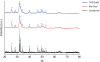
The XRD patterns of Bio-Oss® and Cerabone® showed main peaks at almost identical positions as THE Graft, suggesting that Bio-Oss® and Cerabone® were also composed of HA. Both THE Graft and Bio-Oss® had a low degree of crystallinity, whereas Cerabone® exhibited significantly stronger and narrower diffraction peaks. As HA structures with lower degree of crystallinity tend to be more degradable [1920], this finding suggests that THE Graft and Bio-Oss® will rapidly be replaced with osteoblasts.
FT-IR analysis
The chemical composition of the graft materials and their main functional groups were investigated by FT-IR analysis (Figure 6) [2122]. FT-IR is frequently used to study bone matrices, as their major components, such as HAs, carbonates, and collagen fibers, are known to absorb infrared radiation within the range of 500–4,000 cm−1. FT-IR can therefore be used to identify the chemical composition of the inorganic component of xenograft materials and to determine whether the organic component of the natural bone is present after deproteination [23].
Figure 6
FT-IR spectra of (A) THE Graft, (B) Bio-Oss®, and (C) Cerabone®.
FT-IR, fourier-transform infrared.
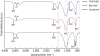
The FT-IR spectrum of THE Graft was very similar to that of Bio-Oss®, with PO43− stretching bands at 415–598 and 962–1,088 cm−1 and the O-H stretching band centered at 3,574 cm−1. We also observed H2O absorption bands at 1,636 and 3,448 cm−1 and CO32− bands at 871, 1,413, and 1,455 cm−1, which typically appear when CO32− replaces phosphate in the apatite lattice. These results confirm the presence of HA in THE Graft and Bio-Oss®. In contrast, the FT-IR spectrum of Cerabone® was slightly different from that of the other 2 graft materials, with a significantly stronger O-H stretching band at 3,574 cm−1, the O-H libration band at 632 cm−1, and a much weaker CO32− band at 871 cm−1. However, insignificant organic phases, which could be identified as amide I and amide II bands at 1,634 and 1,548 cm−1, respectively, were observed in the xenograft materials.
ICP-OES analysis
In order to investigate the Ca and P content of the graft materials, we carried out an elemental analysis of the graft materials using ICP-OES. We used ICP-OES because it is possible to analyze wide concentration ranges of multiple metallic or non-metallic elements existing in a variety of sample matrices with few issues with interference. Both the amounts of Ca and P were comparable between the inorganic graft materials (Table 3). An insignificant amount (<1% by weight) of Na, Mg, Zn, and Sr was detected in all 3 graft materials. The Ca/P molar ratio was similar for all 3 graft materials as well. This suggests that the Ca and P content of THE Graft was comparable to that of Bio-Oss® and Cerabone®, although they originated from bone tissues of different species.

Residual organic content analysis
The Kjeldahl method was the first technique used to determine the residual protein content in graft materials [1224]. Although this method may not be suitable for measuring the exact protein content in a sample, it is still a standard method used to estimate the overall protein concentration in diverse types of samples. We first measured the nitrogen content present in each sample using an elemental analyzer, and then multiplied the result by the protein factor of 6.25 to estimate the protein content in the sample. No protein was detected in the samples of THE Graft and Cerabone® (Table 4). However, small amount of protein content could be detected in the Bio-Oss® sample. To analyze the amount of amino acid content of each xenograft sample in a more accurate manner, we used the pre-column derivatization method for amino acid analysis. This method is known to be one of most reliable ways to quantify peptides and proteins in a sample, since it has detection limit lower than picomoles and can be used to identify the amount of each common amino acid [2526]. Bio-Oss® had the largest amount of amino acids among the 3 xenografts. This result is consistent with the results of the nitrogen analysis. Cerabone® had less amino acid content than Bio-Oss®, and THE Graft contained the lowest amino acid amount among the tested samples.

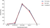
Wettability of the graft materials
Wettability is another important factor that affects the performance of a graft material. Although contact angle measurements are generally used to determine the surface energy of a flat surface, this technique is not applicable for most porous materials in granular form, such as the present graft materials. Hence, in this case we used the wicking method, based on the Washburn equation (1).
The contact angle of porous materials can be calculated by the wetting mass based on the Washburn equation (1):
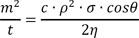
where m is the mass increase of a test liquid with viscosity η, density ρ, and surface tension of σ as a function of the flow time t, while the capillary constant of the sample, c, is determined from the size of the granules and their packing density, to estimate the surface energies of the xenograft materials.
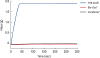
We plotted water mass wetting for THE Graft, Bio-Oss®, and Cerabone® as a function of time (Figure 7). Although Cerabone® exhibited a slightly higher wetting mass than Bio-Oss®, the wetting mass of THE Graft was considerably higher than that of the other 2 materials. Since the cosine value of the contact angle is directly related to the square of the wetting mass in equation (1), this result indicates that the wettability of THE Graft was significantly higher than that of the other 2 graft materials.
DISCUSSION
This paper summarizes comparative physicochemical characterization studies of THE Graft, Bio-Oss®, and Cerabone® that were performed to evaluate their suitability as bone void filler in dental surgery. Although both Bio-Oss® and Cerabone® originate from bovine bone, many of their properties, including the surface morphology, porosity, pore size distribution, and SSA were found to be substantially different. Bio-Oss® not only had high porosity and a large SSA, but also had nanoscale pores, which are typical characteristics of natural bone. This suggests that Bio-Oss® contains many physical characteristics of the native bovine osseous structure to a certain degree even after the deproteination process. In contrast, Cerabone® had relatively low porosity and a small SSA with negligible nanoscale pores. This indicates that a significant portion of the physical characteristics of bovine osseous structure had been altered in Cerabone®, probably due to high-temperature thermal processing procedures. Most of the physical characteristics of THE Graft, including the morphology, porosity, and SSA, were substantially equivalent to those of Bio-Oss®, despite their different origin. This suggests that THE Graft also preserves most of the physical properties of the native porcine osseous structure very well.
We believe that the different pore size distribution characterized using MIP may be closely related to the surface morphology of the graft materials composed of sub-100-nm HA grains. It is worth noting that the sub-100-nm pores observed in THE Graft and Bio-Oss® are also found in human bone, where they originate from the open gaps between HA crystals and collagen [27]. These nanoscale pores have been reported to be very important for cell adhesion, proliferation, and differentiation [192829]. It should be emphasized that osteoblasts not only show better proliferation, but also synthesize alkaline phosphatase and generate calcium-containing minerals on extracellular matrix more actively, on surfaces composed of sub-100-nm HA grains [30].
The SSA, or the total surface area of a material per unit mass, is another important parameter that determines the overall behavior of a graft material. A large surface area is a key requirement for graft materials, and not only results in a larger surface region available for the attachment of osteoblast cells, but also facilitates the exchange of nutrients and waste products and allows greater amounts of blood, proteins, and growth factors to be absorbed onto the scaffold [31].
The chemical analysis using XRD, FT-IR, and ICP-OES demonstrated that THE Graft was mainly composed of HA and showed that its chemical composition, major functional groups, and Ca/P molar ratio were very similar to those of Bio-Oss® and Cerabone®. These results indicate that chemical properties of this porcine-origin xenograft are not significantly different from those of the bovine-origin graft materials. However, it is noteworthy that the degree of crystallinity of both THE Graft and Bio-Oss® was somewhat lower than that of Cerabone®, which might facilitate the efficient replacement of graft materials with bone tissue. The wettability of THE Graft turned out to be higher than that of both Bio-Oss® and Cerabone®, which suggests that THE Graft is relatively hydrophilic and can be easily wet by body fluids after implantation. Not only protein adsorption, but also the attachment, growth, and proliferation of various types of cells, including osteoblasts, have been reported to be significantly affected by the wettability of the material surface [32]. Thus, high-energy surfaces are generally known to be highly beneficial for dental materials [33]. This high wettability of THE Graft suggests that it may have advantages in terms of protein adsorption and the resulting cell adhesion and proliferation processes after implantation. The content of the organic component of THE Graft was somewhat lower than that of both Bio-Oss® and Cerabone®. These results show that organic substances, including collagen and other organic compounds, were successfully removed from THE Graft, which is thus not affected by issues associated with the organic content.
According to the results presented in this paper, the porcine-derived THE Graft material has most of the key physicochemical characteristics required for it to be applied as a highly efficient xenograft material for bone replacement. Both Bio-Oss® and Cerabone® have been successfully used in oral surgery in the last few decades, and have gained a substantial market share. Therefore, it is expected that THE Graft, a porcine-derived xenograft, will perform as effectively as Bio-Oss® and Cerabone® as a bone void filler in oral surgical applications.
 XML Download