

 PDF
PDF ePub
ePub Citation
 Print
Print


INTRODUCTION
In recent years, restorative implant dentistry has been increasingly influenced by esthetic considerations, including the condition and color of the adjacent mucosal tissue [12]. However, the utilization of dental implants is a technique-sensitive procedure in terms of esthetics, since an error in implant positioning or soft tissue management can result in a major esthetic failure. For implant placement in esthetic sites (primarily in the anterior maxilla, teeth 14–24), when the primary goal is the establishment of esthetics and function, the submerged technique, described by Buser and von Arx [3], is the surgical technique of choice. Belser et al. [1] additionally postulated that a submucosally located implant shoulder is mandatory to avoid any visible cervical metal or margins.
The esthetic results are also dependent on marginal bone change over time. Several reports have evaluated the amount of peri-implant bone remodeling over time. These studies have reported radiographic bone remodeling ranging from 0.75 to 1 mm the first year, with minimal bone remodeling in subsequent years [45678]. The initial bone remodeling and the details of marginal bone change are paramount for predicting the stability and the location of the gingival margin, and are therefore relevant to the esthetic outcome. In recent studies, it was demonstrated that the magnitude of the initial bone remodeling in patients around 1-piece dental implants was dependent on the positioning of the rough-smooth border of the implant in the apico-coronal dimension [79].
Bone-level implants take these points into consideration, as the different length of the healing caps allows the user to perform a transgingival (1-stage) or subgingival (2-stage) procedure. Almost the whole surface of the implant is covered with a hydrophilic sandblasted and acid-etched (SLActive) surface, and only a 0.2 mm high, smooth-machined shoulder remains. This allows the implant to be placed at bone level. The described design addresses the fundamentals of optimum esthetics cited by Belser et al. [110]: optimal peri-implant soft tissue, the long-term stability of esthetic peri-implant soft tissue contours, and an emergence profile of the implant-supported restoration of the labial mucosa to match the gingival line of the adjacent teeth. The metallic tulip-shaped shoulder is eliminated to prevent the emergence of a possible grey metallic shadow through the associated soft tissue.
The purpose of this study was to assess the bone level, esthetic results, and implant success after the installation of bone-level implants in esthetic sites.
Go to : 
MATERIALS AND METHODS
In this clinical study, the patients presented a single-tooth gap in the maxilla (tooth positions 14–24), with natural or restored adjacent teeth. The implant was placed at least 8 weeks post-extraction and healed submerged for 6 weeks. Second-stage surgery was performed and a fixed provisional restoration was provided, which served as the baseline for the study. The final restoration was placed 6 months after provisional restoration. Esthetic parameters were assessed, along with bone levels at 6, 12, 24, and 36 months.
Study population and entry criteria
The study was designed as an open, single-arm, observational study, and was approved by the Institutional Review Board (IRB number: CDE-10-002) of China-Japan Friendship Hospital (Peking, China) and Seoul National University Dental Hospital (Seoul, Korea). The required sample size was calculated according to the implant stability quotient (ISQ) and its standard deviation, based on a previous pilot study, in which 10 bone-level implants were inserted into patients. The required sample size was estimated on the basis of α=0.05, power=0.80, δ=5, and σ=6.36. In the resulting calculation,
A dropout rate of 30% was assumed, increasing the number of participants required in each group to 19. The patients were recruited sequentially according to predefined inclusion and exclusion criteria and provided written informed consent. A comprehensive diagnostic assessment was performed for the thorough evaluation of each candidate (visit 1 [V1]). This included a complete oral and radiographic evaluation and the fabrication of mounted diagnostic casts. Preoperative intraoral digital images (Nikon D-100 digital camera with a 100-mm macro lens, Nikon Photo Products Inc., Tokyo, Japan) or photographs (Fujichrome Sensia 100 ASA color film, Fuji Photo Film Company Ltd., Tokyo, Japan) were made of the maxillary and mandibular ridges and dentition, and preoperative and postoperative instructions were provided orally and in written form to each patient. The inclusion criteria were as follows: the requirement for implant placement in the anterior maxilla (tooth positions 14–24) with a natural root in the adjacent tooth position; dentition opposite to the study implant; full mouth plaque index of G 25%, according to O'Leary's plaque index; substantially healed (minimum of 8 weeks) and augmented (if applicable) extraction sockets; and ridge augmentation with autogenous bone until 3 months pre-implantation in cases where bony deficiency would jeopardize the implant position. Patients were not included in the study if they met any of the following exclusion criteria: mucosal diseases (e.g., erosive lichen planus); probing pocket depth of more than 4 mm on one of the teeth immediately adjacent to the dental implant site; smoking >10 cigarettes per day, or the equivalent use of tobacco equivalents or chewing tobacco; lack of primary stability of the implant; inappropriate implant position for the prosthetic rehabilitation; the requirement for a major simultaneous augmentation procedure; and dehiscence of a vertical distance >3 mm.
Implant design and surface
The study implant was a Straumann Bone Level titanium (grade 4) implant with an SLActive surface. The endosseous diameters (3.3 and 4.1 mm) and lengths (8, 10, 12, and 14 mm) were selected according to the bone condition of the patient.
Surgical and prosthodontic procedure
Patients were prepared for aseptic surgery and local anesthesia (visit 2 [V2]). A mid-crestal incision was made in the edentulous ridge, and full-thickness mucoperiosteal flaps were elevated to expose the underlying alveolar process. Following the manufacturer's protocol, implant osteotomies were prepared by sequential cutting with externally irrigated drills (2.2-, 2.8-, and 3.5-mm twist drills), and the implants were placed with a self-tapping technique. To be included in the study, implants were required to achieve a minimum torque level of 15 Ncm during placement and to resist rotation when the same torque was applied during abutment screw tightening. Flaps were repositioned with a tension-free wound closure. During the soft tissue healing, a non-occluding removable temporary prosthesis was delivered and the sutures were removed 7 to 10 days later (visit 3 [V3]). Six weeks postoperatively, a second operation was performed to provide full-occlusion acryl resin single-tooth provisional prostheses (baseline of study; visit 4 [V4]). After 6 months of loading, patients were seen again for a final impression and insertion of the final crown (visit 5 [V5]). Follow-up appointments took place 12 months (visit 6 [V6]), 24 months (visit 7 [V7]), and 36 months (visit 8 [V8]) after baseline.
Evaluation
Wound healing and standard periodontal parameters
Wound healing at the implantation site was assessed at V3 and V4, and wounds were classified as showing normal or compromised healing. For the evaluation of soft tissue, standard soft tissue parameters such as the modified plaque index, modified sulcus bleeding index, and probing depth (PD) were recorded at 4 aspects around the implants [111213]. The plaque index was scored as follows: 0, no detection of plaque; 1, plaque only recognized by running a probe across the smooth marginal surface of the implant; 2, plaque seen by the naked eye; 3, abundance of soft matter. The sulcus bleeding index was scored as 0 if there was no bleeding when a periodontal probe was passed along the gingival margin adjacent to the implant, 1 if isolated bleeding spots were visible, 2 if blood formed a confluent red line on the margin, and 3 if there was heavy or profuse bleeding. These parameters were assessed at the final restoration visit (V5) and at 12, 24, and 36 months after baseline.
Implant stability
Magnetic resonance frequency analysis was performed using a Mentor™ device (Osstell AB, Göteborg, Sweden) and the ISQ values were documented. Measurements were made twice in each direction: the buccolingual direction from the buccal side, and the mesiodistal direction from the mesial side. The mean of the 2 measurements in each direction was recorded as the representative ISQ of that direction. The ISQ values were documented at the time of implant placement (V2), at the time of provisional abutment connection (V4), and at the final restoration (V5).
Analysis of radiographs: mesial and distal implant bone level changes
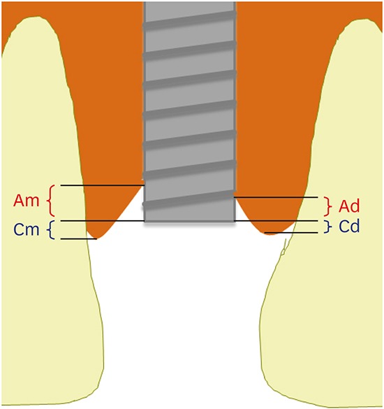
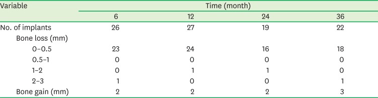
Mesial and distal implant bone level changes were evaluated on a standardized periapical radiograph immediately after implant placement (V2), at the time of fixed provisional restoration (V4), at the time of final restoration connection (V5), 12 months after the fixed provisional restoration (V6), 2 years after the fixed provisional restoration (V7), and 36 months after the fixed provisional restoration (V8). A standardized periapical radiograph was taken using an extension cone paralleling technique, with an impression bite block fabricated for each individual participant attached to the aiming device (XCP System, Dentsply Rinn Corp., Elgin, IL, USA). The radiographs were analyzed to calculate the change in bone levels, evaluated in terms of the distance between the implant shoulder and the first visible bone contact (DIB values) by a single reader using a calibrated measuring tool (Figure 1). DIB was measured at the mesial and distal aspects of each implant. Distortions were taken into account based on dimensional changes between the radiograph and the true dimensions of the implant. The primary objective in this study was to obtain a frequency distribution of bone level changes between baseline and 36 months after the connection of the abutment of a bone-level implant. For each implant, the mean value of the distal and the mesial bone level changes was calculated. The categories for the frequency distribution were as follows: bone loss: 0–0.5, 0.5–1, 1–2, and 2–3, >3 mm and bone gain. In addition to the DIB, the vertical distance from the implant shoulder to the alveolar bone crest was measured (Figure 1).
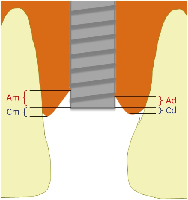 | Figure 1To calculate the change in bone levels, the distance between implant shoulder and bone contact (DIB; Am or Ad) and the distance from the implant shoulder to the alveolar bone crest (Cm or Cd) were measured in radiographs using a calibrated measuring tool.
DIB: distance between the implant shoulder and the first visible bone contact, Am: distance from the implant shoulder mesial to the bone level, Ad: distance from the implant shoulder distal to the bone level, Cm: distance from the implant shoulder mesial to the bone crest, Cd: distance from the implant shoulder distal to the bone crest.
|
Changes of interdental papilla and free gingival margin
Intraoral photographs with frontal views were taken at each visit to document the initial appearance of the soft tissue and the subsequent healing of the soft tissue after abutment connection. The mid-facial height of the implant crown and the corresponding height of the natural tooth crown were measured on these digital pictures, and any changes in the height of papilla and free gingival margin were expressed as a ratio based on the values at V4 (Figure 2).
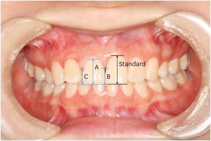 | Figure 2Changes of the interdental papilla and free gingival margin were measured using intraoral photographs. The crown length of an adjacent tooth was designated as the standard. The standard was used as the denominator in calculating the following variables: free gingival margin = length of A/length of standard, papilla mesial height = length of B/length of standard, and papilla distal height = length of C/length of standard.
|
Esthetic assessment questionnaire completed by the patient and the dentist
Intraoral pictures were analyzed according to 2 specific indices, termed the modified Pink Esthetic Score (mPES) and the modified White Esthetic Score (mWES), as described in detail by Belser et al. [14]. The mPES is composed of the following 5 variables: mesial papilla, distal papilla, curvature of the facial mucosa, level of the facial mucosa, and root convexity/soft tissue color and texture at the facial aspect of the implant site. The mWES specifically focuses on the visible part of the implant restoration (i.e., the part of the implant crown that emerges from the peri-implant mucosa) and is based on the following 5 parameters: general tooth form; outline and volume of the clinical crown; color, which includes the assessment of the hue and value in a given dimension; surface texture; and translucency and characterization.
Questionnaire analysis of esthetic satisfaction by the patient and the dentist
Patient satisfaction was investigated with the following questions, and responses were obtained using a visual analog scale (VAS) range from 0 (not satisfied at all) to 10 (fully satisfied):
Are you satisfied with your implant after installation?
Do you feel discomfort with your implant prosthesis?
Are you satisfied with the way your ceramic crown looks?
Are you satisfied with the way the gums around the implant look?
Do you feel that the shape of the gum around the implant changed (whether good or bad) as time progressed?
The satisfaction of dentists who were not directly involved in this study was also investigated in the same manner, with the following questions:
Statistical analysis
Data were analyzed using SPSS version 17.0 (SPSS Inc., Chicago, IL, USA). The Friedman test was used to compare the differences of the ISQ across visits, the marginal bone level changes, and esthetic parameters, such as the mPES, mWES, and the satisfaction questionnaire. The level of significance was set at P<0.05.
Go to :
RESULTS
Demographic characteristics of the participants
Thirty-seven subjects who received 40 implants, with 3 patients receiving 2 implants each because they had 2 non-neighboring gaps, provided written informed consent and were recruited according to the study protocol. Three of these subjects (3 implants) were withdrawn from the study by the exclusion criterion of inadequate bone volume that required major simultaneous augmentation at the time of implant placement. Thirteen subjects were lost to follow-up at 12 months (6 subjects, 8 implants) and at 36 months after provisionalization (7 subjects, 7 implants) (Figure 3). The results were based on the remaining 21 subjects (8 males and 13 females) who were treated with 22 implants. The main implant site characteristics are presented in Tables 1 and 2. All the subjects completed the study according to the protocol, and no adverse device effects were noted. All implants demonstrated successful tissue integration and clinically healthy peri-implant mucosa at the follow-up examinations. At each time point, all implants exhibited ankylotic stability, without clinical signs of peri-implant infection.
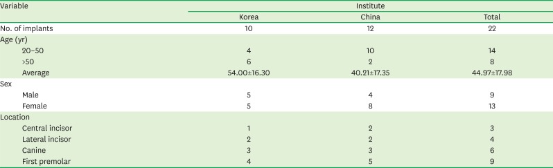
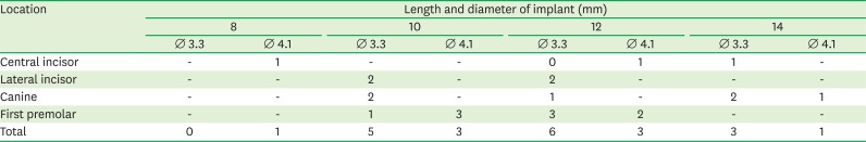
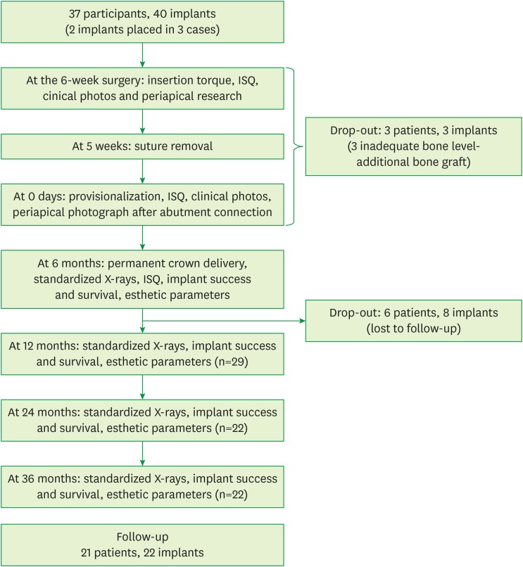
Table 1
Demographic data of the study participants (implant-based, n=22)
![]()
Table 2
The distribution of the length and diameter of the bone-level implants according to the replaced location
![]()
Wound healing and standard periodontal parameters
All implantation sites healed uneventfully and were classified as showing normal healing. The peri-implant soft tissues showed little tendency to bleed following probing, were clinically healthy from the occlusal loading, and remained stable throughout the study period. The mean PD was 1.26±1.37 mm at 12 months, 1.02±1.07 mm at 24 months, and 1.40±1.40 mm at 36 months. The differences between visits were not statistically significant (Table 3).
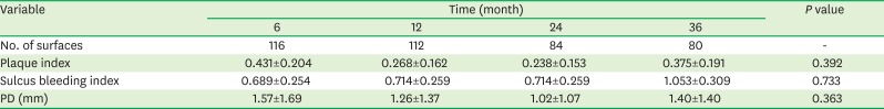
Table 3
Mucosal status at 6 (final restoration), 12, 24, and 36 months after provisional abutment connection
Data are shown as mean±standard deviation. P values were calculated using the Friedman test.
PD: probing depth.
![]()
Implant stability and marginal bone level change
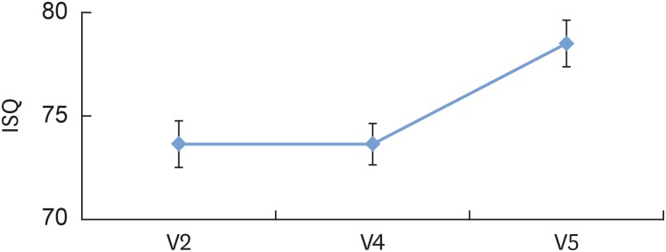
The ISQ values showed good implant stability at placement (V2), provisional abutment connection (V4), and at the final restoration (V5) (Figure 4). Statistical significance was confirmed (P=0.002) with the increasing of ISQ over time.
All implants were well integrated in the bone, and no clinical or radiographic signs of marginal radiolucency attributable to peri-implantitis or poor osseointegration were detected. None of the crowns demonstrated a radiographically detectable marginal discrepancy in either direction. Crestal bone loss occurred during the initial phase between V4 and V5, and then stabilized after V5 (final prosthesis). From implant placement to secondary surgery, the marginal bone level did not change. The mean marginal bone loss relative to baseline (fixed provisionalization) was 0.00±0.42, 0.09±0.36, 0.06±0.38, and 0.07±0.48 mm at V5, V6, V7, and V8, respectively (Figure 5). No statistically significant differences were detected between these visits or relative to baseline (P=0.931). However, the level of the crestal bone decreased significantly from 2.34±0.93 mm at V4 to 1.70±1.10 mm at V8 (Figure 6). The frequency distribution of bone level changes between baseline, 12, 24, and 36 months after the abutment connection of a bone-level implant is shown in Table 4.
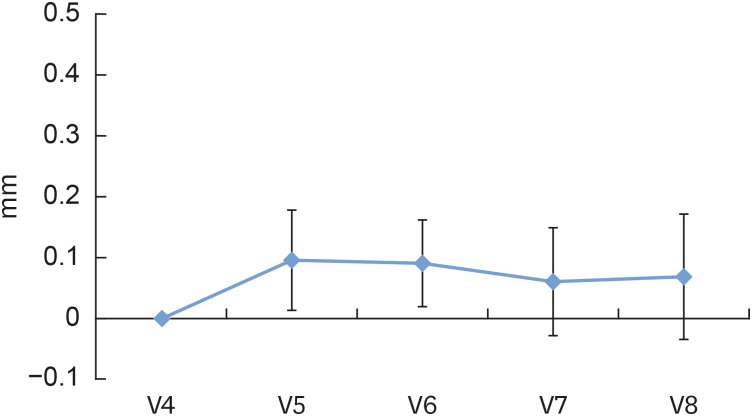 | Figure 5Marginal bone level. There was no significant difference between the visits (P=0.931; Friedman test).
V: visit, V4: provisional abutment connection, V5: final restoration, V6: 12 months, V7: 24 months, V8: 36 months.
|
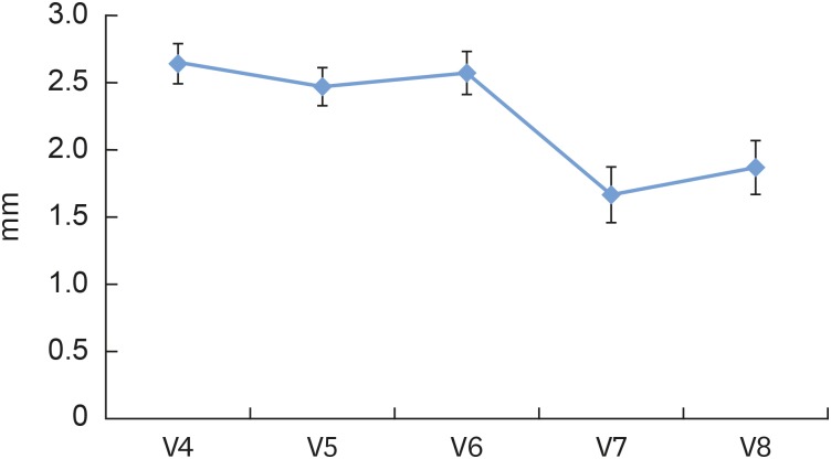 | Figure 6Mean crestal bone level. The crestal bone level significantly decreased as time passed (P=0.004; Friedman test).
V: visit, V4: provisional abutment connection, V5: final restoration, V6: 12 months, V7: 24 months, V8: 36 months.
|
Esthetic assessment of the free gingival margin and papillary height using intraoral photographic analysis
Papillary height did not show any statistically significant changes over time. From the baseline at V4, the mean change was a 2% decrease in papillary height at V5 and V6 (Figure 7) and a 3% decrease in papillary height at V7 and V8 (Figure 7). The free gingival margin values also remained stable over the study period, with a slight increase to 5% at V5, 6% at V6, 4% at V7, and 5% at V8 (also not statistically significant).
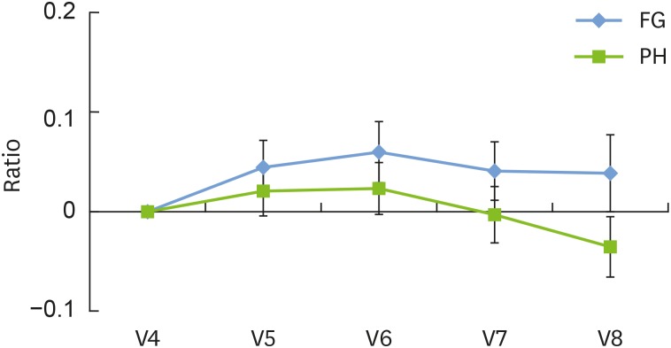 | Figure 7Changes of the FG and PH compared to that at provisional crown connection (V4). Based on the baseline at V4, FG increased by about 4%–6%, and then decreased after an increase in PH by about ±2%–3%. However, no significant difference was found between visits (P=0.213, P=0.091 for FG and PH, respectively; Friedman test).
FG: free gingival margin ratio, PH: papillary height ratio, V: visit, V5: final restoration, V6: 12 months, V7: 24 months, V8: 36 months.
|
Esthetic assessment using mPES/mWES values
The esthetic parameters at the time of provisional abutment connection, final restoration, and at 12, 24, and 36 months for all 22 implants are shown in Table 5. The esthetic outcomes were favorable; the White Esthetic Score (WES) slightly decreased at V6 compared to V5, and increased at V7 and V8. No statistically significant differences were detected between V5, V6, V7, and V8 for either the Pink Esthetic Score (PES) or WES (P=0.431, P=0.557, respectively).
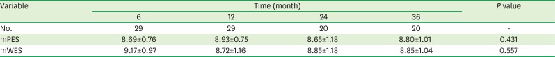
Table 5
Esthetic results at 6 (final restoration), 12, 24, and 36 months after provisional abutment connection
| Variable | Time (month) | P value | |||
|---|---|---|---|---|---|
| 6 | 12 | 24 | 36 | ||
| No. | 29 | 29 | 20 | 20 | - |
| mPES | 8.69±0.76 | 8.93±0.75 | 8.65±1.18 | 8.80±1.01 | 0.431 |
| mWES | 9.17±0.97 | 8.72±1.16 | 8.85±1.18 | 8.85±1.04 | 0.557 |
Data are shown as mean±standard deviation. P values were calculated using the Friedman test.
mPES: modified Pink Esthetic Score, mWES: modified White Esthetic Score.
![]()
Questionnaire analysis of esthetic satisfaction by patients and dentists
Patients' self-assessments and the assessments by dentists showed high esthetic satisfaction. The median VAS score of the patients was 9.30 at the time of final restoration and 9.00 at 12, 24, and 36 months. The dentists' satisfaction score was 9.00 at V5 and V6, 8.80 at V7, and 8.93 at V8 (Table 6).
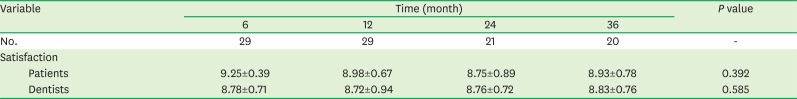
Table 6
Analysis of esthetic satisfaction by the patients and dentists, with questionnaires administered at 6 (final restoration), 12, 24, and 36 months after provisional abutment connection
| Variable | Time (month) | P value | ||||
|---|---|---|---|---|---|---|
| 6 | 12 | 24 | 36 | |||
| No. | 29 | 29 | 21 | 20 | - | |
| Satisfaction | ||||||
| Patients | 9.25±0.39 | 8.98±0.67 | 8.75±0.89 | 8.93±0.78 | 0.392 | |
| Dentists | 8.78±0.71 | 8.72±0.94 | 8.76±0.72 | 8.83±0.76 | 0.585 | |
![]()
Implant success and survival rate and treatment failure
The implant esthetic success and survival rates assessed 36 months after abutment connection were 100%. The frequency distribution of DIB changes at 36 months after the provisional prosthesis showed 1 case of bone loss over 2 mm. Therefore, the functional success rate was 95.5%. All other implant sites revealed bone resorption of less than 0.5 mm. Among these, 3 implants showed bone gain even 3 years after the abutment connection. However, no cases of esthetic failure occurred, because the facial gingival margin and papilla were stable, and did not migrate more than 20% from their planned levels, resulting in symmetry and an esthetically satisfactory gingival margin.
Go to :
DISCUSSION
Dental implant restoration of the maxillary anterior region can be functionally and esthetically challenging. In this clinical trial, maxillary anterior single-tooth gaps were restored with 4.1-mm diameter (8–14 mm in length) or 3.3-mm diameter (10–14 mm in length) bone-level implants. All surgical sites healed without complications and all implants osseointegrated. Sulcus bleeding index and PD measurements showed peri-implant gingival stability.
Peri-implant marginal bone resorption results in the loss of interdental papillae and facial gingival height, which may cause patient dissatisfaction and the functional failure of implants. At the time of second-stage surgery, 6 weeks after implant placement, all cases in our series showed surrounding bone, except for 1 where the implant was placed above the alveolar crest shoulder. Although it is generally expected for surgical trauma and bone remodeling after implant placement to cause initial bone loss [15], the mesial and distal crestal aspects of the implants and the implant rough surfaces were completely surrounded by bone at the time of provisional crown connection according to the radiographic evaluation. The initial 6-week submerging protocol, along with the inherent design and surface properties of the bone-level implants, seems to have contributed to the superb marginal bone level stability from at the time of implant placement to the provisional crown connection.
In addition, numerous studies have shown that bone resorption around the implant neck begins as the implant is uncovered and exposed to the oral cavity, because exposure of the implant to the oral cavity leads to bacterial contamination of the gap between the implant and its superstructure [1617]. At the time of implant placement (V2), the mean marginal bone level (DIB) was 0.07±0.28 mm. At the final prosthesis placement 6 months after provisional prosthetic loading (V5), the DIB increased to 0.11±0.29 mm, and changed to 0.09±0.01 mm at 12 months (V6), 0.06±0.09 mm at 24 months (V7), and 0.07±0.10 mm at 36 months (V8). Most of the patients (95.5%) showed less than 0.5 mm of bone resorption, and 3 cases (13.6%) even showed bone gain during the study. These values are smaller than have been reported in similar clinical studies of 2-piece dental implants with similar designs, in which placement of the implant-abutment interface at the level of the alveolar crest resulted in a mean marginal bone loss of 1.5–2 mm within the first year of implant placement [1819]. Peri-implant bone resorption is known to be affected by the implant surface. Chemically modified implant surfaces demonstrated a significantly greater mean percentage of bone-implant contact than surfaces with similar roughness but without chemical modification [20]. In a pilot experiment in dogs, implants with chemically modified surfaces seemed to promote bone regeneration in acute-type buccal dehiscence defects of submerged implants [21]. Therefore, less peri-implant bone loss seemed to be an effect of surface treatment.
Establishment of biologic width is also known to affect crestal bone remodeling, especially at implant sites with thin mucosa [22]. The platform-switching concept requires that the implant-abutment interface be placed away from the implant shoulder and closer toward the axis to increase the distance of the micro-gap from the bone [232425]. In our study, the implant system used for platform switching had a micro-rough surface extending to the implant shoulder, accommodating biologic width by featuring a prepared margin 1.9 mm above the shoulder. Buser et al. [26] also confirmed that a platform-switching design can reduce the marginal bone resorption at the crest level during functional loading, since 20 bone-level implants placed in the maxillary esthetic zone showed an average bone loss of only 0.44 mm at 6 years. This rate of bone loss is much less than has been observed for standard titanium implants without a platform-switching concept, where the mean DIB value was 2.18 mm [27]. Comparing the bone loss with the data of Buser et al. [28], our study showed almost the same bone remodeling at 6 months (0.10 vs. 0.09 mm) and potentially even less bone loss at 12 months (0.09 vs. 0.18 mm) after loading. Hämmerle et al. [29] also reported minimal crestal bone change in bone-level implants placed at the anterior maxilla and mandible. They reported remodeling patterns at 6, 12, 24, and 36 months, with mean values of 0.31, 0.46, 0.47, and 0.49 mm, respectively. Chemical treatment of the implant surface and an implant design providing a good seal between the implant and abutment may reduce the amount of bone remodeling during the establishment of biologic width [30].
The level of the mid-buccal peri-implant mucosal margin remained stable during the follow-up period, indicating stability of the marginal bone levels. This is consistent with the results of a study by Nisapakultorn and colleagues [31], who found that the facial marginal mucosal level, among other factors such as peri-implant biotype, implant fixture angle, and the depth of the implant platform, was affected by facial and interproximal crestal bone levels and the level of the first bone contact with the implant. In contrast, a systematic review by Cairo et al. [32] concluded that future mucosal recession around implants was not related with the peri-implant bone level. The height of the interdental papilla was stable during follow-up. Several studies have shown that the level of the interdental papilla was independent of the peri-implant marginal bone level, but associated with the marginal bone level of the adjacent teeth, which was the crestal bone level in this study [313334353637]. In our study, the crestal bone level decreased between 12 and 24 months. Similarly to the study of Cairo et al. [32], the interdental papilla seems to have been less influenced by the crestal bone level after 1 year of prosthesis connection when the mucosa became stable. Six months after the placement of final prostheses (V6), stationary papillary height and fill were observed. Although most volume change in the interdental papilla is expected to occur immediately after placement of the prosthesis, the definitive position and dimensions of the soft tissue may not settle until after 1 month. It has been found that installation of a final prosthesis on a dental implant without soft-tissue conditioning or prosthodontic treatment caused apical displacement of the mid-facial gingival margin and coronal displacement of the mesial and distal gingival margins [26]. One possible explanation for this phenomenon is the increase in pressure on the peri-implant soft tissues resulting from replacement of the healing abutment or provisional crown by a definitive crown with larger dimensions [28]. For better results, at the final prosthesis delivery, the proximal surfaces of adjacent teeth were adjusted to form surface contacts in order to prevent vertical and horizontal food impaction, and to control the distance between the crestal bone and interproximal contacts. The general contour and emergence profile of the final prosthesis were designed based on the provisional prosthesis. However, the proximal surfaces of adjacent teeth were not adjusted for the provisional placement, and this may have allowed healthier and more voluptuous interdental papilla at the final prosthesis delivery. For the health and stability of gingival tissue in contact with the prosthesis, the subgingival portion of the crown was internally stained instead of using a conventional external staining technique. Moreover, an S-shape curvature was rigidly applied to the emergence profile connecting the top of implant to the buccal gingival margin [38]. Therefore, the peri-implant gingiva at the 6-month follow-up (V6) became more esthetically pleasing than immediately after placement of the final restorations (V5).
The gingiva and peri-implant mucosa showed almost no bleeding on probing. A possible explanation might be that second-stage surgery and the delivery of the temporal prosthesis were done at the same time. Therefore, the gingiva healed according to the S-line emergence profile of the temporary prostheses without gingival recession. Moreover, it is reasonable to believe that the gingiva and peri-implant mucosa stabilized after 6 months of temporary prosthesis installation. Moreover, the emergence profile created with the temporary prosthesis was identical to that of the final prosthesis. Finally, the inner part of the crown located nearby the gingiva was coated with zirconia to increase the resistance of the gingiva to external stimuli.
The esthetic outcomes were evaluated with PES and WES, which Buser et al. [28] proposed as a comprehensive way to evaluate the pink and white dimensions of esthetics. In the present study, the mean PES slightly increased over time. However, the median did not change over the 4 visits. Lai et al. [39] also evaluated the alterations of soft tissue around single-tooth implants in the anterior maxilla with PES at 6 months post-loading of bone-level implants and concluded that the esthetic outcomes of the soft tissue improved significantly at follow-up. Meanwhile, the mean WES slightly decreased over time, although this trend was not statistically significant. Even though patients were satisfied with the final prosthesis, with better surface texture and translucency than the temporary prosthesis, they may experience a decreased level of satisfaction after 6 months due to changes in intra-oral conditions and increased esthetic expectations. In the present study, the overall results from the PES/WES were very satisfying, showing a potentially higher mean total score of 17.62 both for the PES and WES than the results reported by Buser et al. (a score of 16.75) [28].
The present prospective case series provides additional evidence that early loading for single-tooth implants in the anterior maxilla can provide successful treatment outcomes with high predictability and a low risk of complications. In this study, 3 years of follow-up demonstrated that all 22 implants achieved and maintained successful osseointegration and healthy peri-implant soft tissue according to standard clinical and radiographic parameters. Using the same parameters as previous prospective studies [1213], all 22 implants satisfied strict success criteria, resulting in a survival and functional success rate of 95.5% at the 36-month follow-up.
In conclusion, platform-switching bone-level implants placed in maxillary single-tooth gaps in the esthetic zone resulted in successful osseointegration with minimal bone remodeling, even with early functional loading 6 weeks after implant placement. All implants were osseointegrated without complications and showed minimal bone loss after 36 months of loading. The peri-implant soft tissue was esthetically pleasing and stable. In conclusion, bone-level implants appear to be a safe and reliable treatment choice for the functional and esthetic restoration of maxillary anterior single-tooth gaps.
Go to :
 XML Download