

 PDF
PDF ePub
ePub Citation
 Print
Print


INTRODUCTION
Molecular mimicry, bystander activation, and epitope spreading are representative mechanisms of autoimmunity related to microbes [1]. Epitope spreading is a phenomenon in which distinct subdominant epitopes become major targets of the immune response [23]. This induces self-tissue damage, causing the activation of autoreactive T and/or B lymphocytes as an immune response towards the persisting pathogen or resulting from direct lysis of the pathogen [4]. Antigens released from damaged tissue are taken up by antigen-presenting cells, leading to the initiation of a self-specific immune response [56]. Autoreactive cells are not found in normal subjects and do not cause clinical pathology because of their low precursor frequency and/or higher-level regulatory controls. However, due to defects in the regulatory control of autoreactive lymphocytes during tissue damage in genetically susceptible individuals, a self-specific immune response may occur [4].
The term ‘epitope spreading’ describes how the antigen-specific autoimmune response spreads to different epitopes [7]. This can occur for a single antigen, known as intramolecular epitope spreading, or between different antigens, known as intermolecular epitope spreading. The diversification and amplification of autoimmunity in individuals can be described as epitope spreading, which causes the autoimmune response to progress from the initial activation state to a chronic state [8]. Epitope spreading resulting from tissue damage is considered to play an active role in disease pathology. The process is also an important factor in the protective immune response and the mechanism that downregulates the immune response in autoimmunity. Therefore, understanding epitope spreading patterns in human autoimmune disease may facilitate the design of antigen-specific therapies [9].
Heat shock proteins (HSPs) play an important role in the association between microbial infections and autoimmunity because of their conserved amino acid sequences and strong immunogenicity [10]. Representative periodontopathic bacteria, particularly Porphyromonas gingivalis, are known to contain HSP60 family proteins. HSP60 from P. gingivalis (PgHSP60) and peptide 19 (TLVVNRLRGSLKICAVKAPG) from PgHSP60 (Pep19) are immunodominant epitopes in autoimmune disease patients [11], including those with periodontitis [12]. In addition, Pep19 has been shown to drive epitope spreading in periodontitis and periodontitis-associated autoimmune disease [13]. However, it remains unclear whether Pep19 is a dominant epitope in subjects without periodontitis or autoimmune disease.
The purpose of this study was to determine the epitope spreading pattern and verify Pep19 as an immunodominant epitope in healthy teenagers using dot immunoblot analysis. The patterns of epitope spreading in age-matched patients with type 1 diabetes mellitus (DM) and healthy 20- to 29-year old subjects were compared with those of healthy teenagers.
MATERIALS AND METHODS
Study subjects
Twenty clinically healthy subjects who were 10–19 years of age and devoid of chronic periodontitis or systemic diseases were recruited. For comparison in age-matched patients with autoimmune disease, 8 teenage patients with type 1 DM were selected. A diagnosis of type 1 DM was based on the following criteria: fasting plasma glucose level of ≥126 mg/dL or symptoms (such as polyuria, polydipsia, and unexplained weight loss) and a casual plasma glucose level of ≥200 mg/dL, or a plasma glucose level of ≥200 mg/dL 2 hours after a 75-g glucose load, or a glycated hemoglobin level of ≥6.5% [14]. To examine the effects of age in healthy subjects on immunoblot patterns against an array of target antigens, 20 clinically healthy subjects who were 20–29 years of age were also recruited. Peripheral blood was drawn from all study subjects by venipuncture to collect serum and peripheral blood mononuclear cells.
The study was approved by the Institutional Review Board of Pusan National University Hospital (approval No. PNUDH-2017-003). Written informed consent for blood collection was obtained from the study subjects. All experiments were performed under the principles of the Declaration of Helsinki.
Synthetic peptides
A total of 37 overlapping peptides (each consisting of 20 amino acids with a 5-amino acid overlapping sequence), spanning the entire protein sequence of PgHSP60 and human HSP60 (HuHSP60), were synthesized by 9-fluorenylmethoxycarbonyl solid-phase peptide synthesis (Peptron Inc., Daejeon, Korea), as previously described [11].
Dot immunoblot analysis in human serum samples
Five micrograms of synthetic peptides of PgHSP60, HuHSP60, and oxidized low-density lipoprotein (oxLDL, Kalen Biomedical, Montgomery Village, MD, USA) were spotted onto each nitrocellulose membrane. The membranes were blocked for 30 minutes with 5% skim milk. Individual serum samples from each group of subjects were added separately, and the membranes were incubated for 2 hours at room temperature. After washing the membranes with phosphate-buffered saline (PBS) with Tween® 20 (Thermo Fisher Scientific Inc., Rockford, IL, USA) for 30 minutes at room temperature, horseradish peroxidase-conjugated mouse antihuman IgG (γ-chain specific, Jackson ImmunoResearch Laboratories, West Grove, PA, USA) was added, and the membranes were incubated for 1 hour. The membranes were washed with PBS with Tween® 20 and tetramethyl benzidine was added for color development. An identical procedure was performed using serum samples from each group for PgHSP60, Mycobacterium tuberculosis HSP60 (MtHSP60), and Chlamydia pneumoniae HSP60 (CpHSP60). Each dot-blot image in each group was converted for densitometric image analysis using imaging software (Image J software version 1.45, National Institutes of Health, Bethesda, MD, USA).
Statistical analysis
The data were analyzed using SPSS software (version 22 for Windows, SPSS Inc., Chicago, IL, USA). The normality of the distribution of the data was verified by the Kolmogorov-Smirnov test. As all parameters followed a normal distribution, parametric methods were used to analyze the data. The data obtained from each group were analyzed with the independent t-test, and the differences among groups were compared using 1-way analysis of variance (ANOVA) followed by post hoc Bonferroni correction. In addition, 2-way ANOVA with post hoc Bonferroni correction was used to identify differences among groups and to explore any interactions. Statistical significance was accepted at the level of P<0.05.
RESULTS
Subject profiles
A total of 48 subjects were enrolled for this analysis of dot immunoblot patterns. The age and gender distribution of the subjects are shown in Table 1.

Dot immunoblot profiles of reactivity of serum from healthy subjects aged 10–19 years to whole HSP60 protein
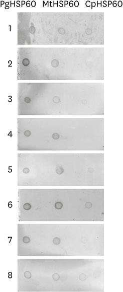
Most serum samples from healthy teenagers reacted more strongly with PgHSP60 than with the analogous targets from M. tuberculosis and C. pneumoniae (Figure 1). The highest serum immunoblot intensity was consistently observed for PgHSP60 in comparison to the HSP60 from the other 2 microorganisms. There were significant differences among the 3 groups (Table 2).
Figure 1
Dot immunoblot profiles of reactivity of serum from healthy subjects aged 10–19 years to whole PgHSP60, whole MtHSP60, and whole CpHSP60.
PgHSP60: heat shock protein 60 from P. gingivalis, MtHSP60: heat shock 60 protein from M. tuberculosis, CpHSP60: heat shock 60 protein from C. pneumoniae.
Table 2
Relative dot immunoblot intensity of reactivity of serum from H10–19 to PgHSP60, MtHSP60, and CpHSP60

| Characteristics | PgHSP60 | MtHSP60 | CpHSP60 | P valuea) |
|---|---|---|---|---|
| H10–19 | 48,097.34±12,492.67b) | 32,754.06±11,596.30c) | 4,080.48±3,917.64d) | <0.001 |
Values are presented as mean±SD.
PgHSP60: heat shock protein 60 from P. gingivalis, MtHSP60: heat shock protein 60 from M. tuberculosis, CpHSP60: heat shock protein 60 from C. pneumoniae, H10–19: healthy subjects aged 10–19 years, SD: standard deviation, ANOVA: analysis of variance.
a)P value determined by ANOVA; b,c,d)Different letters indicate statistical differences based on ANOVA with post hoc Bonferroni correction (P<0.05).
Comparison of dot immunoblot patterns among healthy subjects aged 10–19 years, age-matched patients with type 1 DM, and healthy subjects aged 20–29 years.
Most serum samples from healthy subjects and patients with type 1 DM reacted more strongly with PgHSP60 and Pep19 than with the other peptides. PgHSP60 and oxLDL consistently reacted with Pep19. Simultaneous reactivity to Pep19 with peptide 19 from HuHSP60 (Hu19) was observed, but reactivity to Hu19 without Pep19 was not observed in any subjects. Strong reactivity to peptide 9 from HuHSP60 (Hu9) and peptide 14 from HuHSP60 (Hu14) was observed only in the type 1 DM group (Figure 2).
Figure 2
Comparative dot immunoblot patterns among H10–19 (A), DM10–19 (B), and H20–29 (C). Serum samples from healthy subjects reacted exclusively to PgHSP60, Pep19, human autoantigen Hu19, and human neoantigen oxLDL. Serum reactivity to human autoantigen Hu9 is not observed in any healthy subjects but in type 1 DM. Figure 2 was reproduced and modified from Kwon et al. [13] with permission of the Journal of Periodontal Research.
H10–19: healthy subjects aged 10–19 years, DM10–19: subjects with type 1 diabetes mellitus aged 10–19 years, H20–29: healthy subjects aged 20–29 years, PgHSP60: heat shock protein 60 from P. gingivalis, Pep9, 14, 19: peptide 9, 14, and 19 from P. gingivalis heat shock protein 60, Hu9, 14, 19: peptide 9, 14, and 19 from human heat shock protein 60, oxLDL: oxidized low-density lipoprotein.

The dot immunoblot intensity between Pep19 and Hu19 was compared among groups. In all groups, the serum intensity of Pep19 was higher than that of Hu19, and there was a significant difference between Pep19 and Hu19 in the type 1 DM group. The relative intensity of the antibody reactivity to Pep19 was higher in the type 1 DM group than in the healthy groups (Table 3). In addition, the dot immunoblot intensity for Pep19 and Hu19 was compared among groups. There was a significant interaction effect between the groups of subjects and the type of peptide on the relative dot immunoblot intensity. Significant differences among all 6 groups were confirmed by the post hoc test (Table 4).
Table 3
Relative dot immunoblot intensity of reactivity of serum from H10–19, DM10–19, and H20–29 to Pep19 and Hu19

Values are presented as mean±SD.
H10–19: healthy subjects aged 10–19 years, DM10–19: subjects with type 1 diabetes mellitus aged 10–19 years, H20–29: healthy subjects aged 20–29 years, Pep19: peptide 19 from P. gingivalis heat shock protein 60, Hu19: peptide 19 from human heat shock protein 60, SD: standard deviation.
a)
P value determined by the independent t-test (P<0.05).
Table 4
Results from 2-way ANOVA with post hoc Bonferroni correction investigating the effects of the explanatory variables in Table 3 (subject groups, types of peptide, and interaction between those categories) on the relative dot immunoblot intensity

ANOVA: analysis of variance, SG: subject groups, TP: types of peptide, Pep19: peptide 19 from P. gingivalis heat shock protein 60, Hu19: peptide 19 from human heat shock protein 60, H10–19: healthy subjects aged 10–19 years, DM10–19: subjects with type 1 diabetes mellitus aged 10–19 years, H20–29: healthy subjects aged 20–29 years.
a)P<0.05.
Relatively intense antibody reactivity to Hu9 was observed in the type 1 DM group, with a significant difference from the healthy groups (Table 5).
Table 5
Relative dot immunoblot intensity of reactivity of serum from H10–19, DM10–19, and H20–29 to Hu9

| Characteristics | H10–19 | DM10–19 | H20–29 | P valuea) |
|---|---|---|---|---|
| Hu9 | 1.24±0.24b) | 40,507.32±13,112.39c) | 1.51±0.33b) | <0.001 |
Values are presented as mean±SD.
H10–19: healthy subjects aged 10–19 years, DM10–19: subjects with type 1 diabetes mellitus aged 10–19 years, H20–29: healthy subjects aged 20–29 years, Hu9: peptide 9 from human heat shock protein 60, SD: standard deviation, ANOVA: analysis of variance.
a)P value determined by ANOVA; b,c)Different letters indicate statistical differences based on ANOVA with post hoc Bonferroni correction (P<0.05).
DISCUSSION
The goal of this study was to evaluate epitope spreading using dot immunoblot pattern analysis and to ascertain the immunodominant epitope in healthy teenagers compared to age-matched patients with autoimmune disease (type 1 DM) and healthy 20- to 29-year-old subjects. In our previous research [111213], PgHSP60 and Pep19 were found to be the dominant epitopes in individuals with periodontitis and autoimmune disease. The present study showed the same results, in that the highest serum immunoblot intensity was observed with Pep19 or whole PgHSP60 in all subjects. PgHSP60 and Pep19 appeared to be the immunodominant epitopes not only in the autoimmune disease group, but also in healthy young groups, including teenagers. The presence of reactivity to Pep19 in 20- to 29-year-old subjects supports the hypothesis that Pep19 is a dominant epitope in teenagers, suggesting that its advent occurs in teenagers.
Serum samples from healthy subjects aged 10–19 years and 20–29 years showed serum reactivity to Pep19 and into Hu19, indicating intramolecular epitope spreading. Compared to the dot immunoblot intensity between Pep19 and Hu19, the serum intensity of Pep19 was higher than that of Hu19 in all groups, and there was a significant difference between Pep19 and Hu19 in the type 1 DM group.
Most serum samples showed reactivity to human neoantigen oxLDL with Pep19, suggesting intermolecular epitope spreading through neoantigen induction. This confirms the hypothesis that Pep19 oxidizes native LDL [15]. Pep19 reacted more strongly in the type 1 DM group than in the healthy groups, and showed a strong reaction in the autoimmune disease group. This corresponds to the results of our previous study, in which robust reactivity to anti-Pep19 monoclonal antibody was detected at the inflammation site (i.e., in gingival connective tissue lesions and atheromatous plaques) in contrast to normal healthy sites [13]. Based on these results, Pep19 is a key factor for HSP epitope-specific therapy for autoimmune disease control [161718]. Putative sequential intramolecular epitope spreading from Pep19 and Hu19 into human autoantigen Hu9 did not occur in healthy subjects; however, it did occur in autoimmune disease patients in the present study. This concurs with the results of previous research [1112] conducted in patients with atherosclerosis, diabetes, or rheumatoid arthritis. In our previous study, we established intramolecular epitope spreading into Hu9 in the sera of periodontitis and infection-induced autoimmune disease patients. As support for the cellular mechanisms responsible for this phenomenon, a T-cell proliferative response to Hu9 was observed [13]. Therefore, the Hu9 autoantigen is an important epitope in individuals with autoimmune disease, and Pep19 initiated the dot immunoblot reaction in both the healthy and autoimmune disease groups.
In conclusion, epitope spreading can be observed not only in autoimmune disease patients, but also in healthy young subjects, and reactivity to Pep19 was increased in autoimmune disease groups. Pep19 is an immunodominant epitope not only in autoimmune disease patients, but also in healthy young subjects. This result suggests that the Pep19-specific immune response may be an initiator that triggers autoimmune diseases.
 XML Download