

 PDF
PDF ePub
ePub Citation
 Print
Print


INTRODUCTION
A dental implant can be either simultaneously placed (in a 1-stage procedure) or placed in a second stage when the atrophic posterior maxilla is to be augmented by sinus floor elevation [12]. If primary stability of the implant is achievable, the 1-stage technique may be preferable because it would shorten the overall treatment period and allow the second operation, for placing the implant, to be skipped [34]. A randomized controlled clinical study found that the survival rate and peri-implant bone-level changes at 1 year after loading did not differ significantly between implants placed using 1- and 2-stage sinus graft procedures [5].
While 1-stage surgery has obvious advantages, it can be very difficult to achieve primary stability of the implant with this approach when there is minimal bone height (<3 mm) with poor bone quality in the posterior maxilla. In some cases, the implant can be displaced into the maxillary sinus, causing maxillary sinusitis [6]. However, improvements in surgical techniques, as well as technological advances in the characteristics of the implant surface, thread design, and surgical devices, have made it easier for clinicians to achieve initial stability of the implant [7]. Additionally, the development of bone substitute to promote bone regeneration and osseointegration has made the procedure more predictable, and has extended the applicability of the 1-stage approach [8]. Furthermore, many attempts have been made to use growth factors (e.g., bone morphogenetic protein), which are known to induce rapid bone formation in the maxillary sinus [9].
Bone morphogenetic protein requires a carrier material that serves as a scaffold for cellular growth and attachment [10]. Many reports have explored the osteopromotive effects of various carriers, with collagenated biphasic calcium phosphate (CBCP) having been used recently as a carrier material in a sinus augmentation model showing excellent osteoconductive properties [1112]. Brodie et al. [13] reported that CBCP increased the proliferation and survival rate of osteoblasts. Our previous quantitative and qualitative analyses using micro-computed tomography (CT) and histology found that CBCP could be an appropriate carrier system [14].
Several previous studies have focused on the augmented area without implant placement in a rabbit sinus model [151617], but sinus augmentation with simultaneous implant placement in a rabbit sinus model has rarely been studied [18]. We considered it important to determine whether implant osseointegration would be achieved with and without recombinant human bone morphogenetic protein-2 (rhBMP-2) loading during the early stage of healing. The aim of this study was, therefore, to determine the effect of rhBMP-2-loaded CBCP on implant placement with simultaneous sinus augmentation in rabbits.
MATERIALS AND METHODS
Animals
Five male New Zealand white rabbits weighing between 2.5 kg and 3.0 kg were selected as the experimental model. The number of animals was determined using data on newly formed bone volume from our previous study [14], and corresponded to the sample size necessary to have a 95% chance of detecting a significant difference at the 5% level. Animals were kept in separate cages under standard laboratory conditions with ad libitum access to a diet of standard laboratory pellets and water. Animal selection and care, the preparation procedures, and the surgical protocols were certified by the Institutional Animal Care and Use Committee, Yonsei Medical Center, Seoul, Korea (approval No. 2011-0262).
Experimental materials
Experimental implants
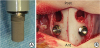
The acid-etched and sandblasted rough-surface implants used in this study (Dentium, Seoul, Korea) had a cylindrical shape with dimensions of Ø 3×6 mm (Figure 1A).
Preparation of rhBMP-2 and assessment of bioactivity
We extracted rhBMP-2 supplied by the Genoss Institute (Suwon, Korea) from inclusion bodies at room temperature, refolded it, and concentrated it. The protein was ultimately purified by heparin-affinity chromatography, filtered, and then freeze-dried. Mouse bone-marrow stromal cells and fetal bovine serum were incubated. After the supernatant was removed, rhBMP-2 was added at various concentrations to 1 mL of fresh medium. After further cultivation, rhBMP-2 activity was determined using the Alkaline Phosphatase Assay Kit (BioVision, Milpitas, CA, USA). The absorbance at 405 nm was recorded after 20 minutes of incubation at 37°C. The activity was related to the protein content in each sample using a bicinchoninic acid protein assay (Pierce, Rockford, IL, USA).
Preparation of rhBMP-2-loaded CBCP
CBCP (Osteon Collagen, Genoss Institute) with a particle size of 0.3–0.5 mm was used as the carrier of rhBMP-2 in this study. This carrier was a cylindrically shaped bone filler (Ø 6.0×5.0 mm) composed of synthetic bone (70% hydroxyapatite, 30% β-tricalcium phosphate, and a natural type I collagen). A solution of rhBMP-2 (0.1 mg/mL) was diluted in a buffer, and 0.2 mL of the resulting rhBMP-2 solution was loaded onto particles of CBCP for the experimental (BMP) group, while CBCP was soaked in saline only for the control group.
Experimental design
Two groups were allocated into both sides of the sinuses of each of the rabbits. In each animal, CBCP soaked with saline was placed on one side of the maxillary sinus to establish the control group, while CBCP loaded with rhBMP-2 was inserted on the other side, forming the BMP group. The BMP and control groups were placed at random. After bone grafting, 2 implants were placed 3 mm anterior to the holes in the left and right lateral walls, respectively. The sample size per group was 5.
Surgical procedure
All surgical procedures were performed under general anesthesia, with additional infiltration anesthesia being applied to the nasal dorsum. The protocols reported by Kim et al. [14] were used to prepare the access window and elevate the sinus membrane (SM). After the nasal dorsum of each rabbit was shaved, the surgical field was disinfected with iodine solution. The dorsal surface of the nasal bone was exposed by making a midline incision on the skin and periosteum. A trephine bur (C-reamer, Neobiotech, Seoul, Korea) was used to form 2 circular holes with diameters of 5.5 mm on both sides of the nasal bone. The SM was elevated to the hole approximately 10 mm anteriorly. While protecting the SM using a surgical curette, the implant sites were drilled to a diameter of 3 mm in front of the holes using a pilot drill followed by a final drill (2.7 mm in diameter). CBCP with rhBMP-2 or saline was packed into the hole anteriorly towards the implant site before inserting the implants. Two implants were inserted manually into the cortical bone until they were seated up to their shoulder (Figure 1B). The holes were covered with periosteum. The skin and periosteum were sutured with glyconate absorbable monofilament (4-0 Monosyn, B-Braun, Aesculap, PA, USA). At 4 weeks after surgery, the rabbits were sacrificed using an overdose of anesthesia.
Micro-CT analysis
Including the implant and surrounding tissues, all specimens were fixed in 10% formalin for 10 days, and images were then obtained using a high-resolution micro-CT system (SkyScan 1173, SkyScan, Aartselaar, Belgium) at a resolution of 35 μm (achieved using 100 kV and 100 μA). The specimens were analyzed for the remaining bone substitute material and newly formed bone, which were identified by 8-bit grayscale values from 100 to 255 and from 70 to 100, respectively. The On-Demand 3-dimensional (3D) software (Cybermed, Seoul, Korea) was utilized to obtain 3D images for making volumetric measurements of the overall augmented region, newly formed bone, implant, and remaining particles of graft materials within the region of interest.
Histological analysis
The fixed specimens were dehydrated in ethanol, embedded in methacrylate, and sectioned in the center of the augmented sinus in the sagittal plane using a diamond saw (Exakt, Apparatebau, Norderstedt, Germany). The final thickness of the reduced central section was approximately 20 μm. Each section was stained with hematoxylin and eosin. The histological slides were observed and captured digitally under a light microscope (BX50, Olympus, Tokyo, Japan).
Histomorphometric analysis
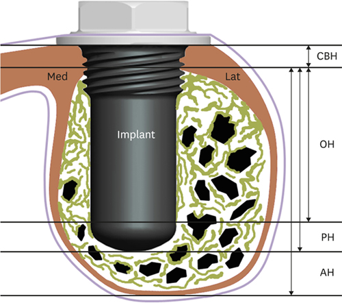
Histomorphometric measurements were made with the aid of an automated image analysis system (ImagePro Plus, Media Cybernetics, Silver Spring, MD, USA). Linear measurements, including the augmented height (AH) and protruding height into the sinus pouch of the implant, were made of each section. The cortical bone height (CBH) and the distance from the basal cortical bone to the highest point of osseointegration (OH) were measured on both the medial and lateral sides of the implant (Figure 2). Characteristics of the total augmented area, such as the areas of newly formed bone, residual materials, non-mineralized tissue, and implant, were separately and manually traced and calculated. The ratio of the total length of bone-implant contact (BIC) in the section was also obtained.
Statistical analysis
The statistical analysis was performed using the R statistical software (version 3.2.2, R Foundation for Statistical Computing, Vienna, Austria; http://www.r-project.org). A non-parametric mixed model was used for comparing radiographic and histomorphometric parameters between the 2 groups [19]. The cut-off for statistical significance was set at P<0.05. The Bonferroni correction was used for multiple comparisons.
RESULTS
Clinical observations
During the surgical procedure, rigid fixation was achieved for all implants by manual insertion into the cortical bone. Sinus augmentation surgery with simultaneous implant placement was performed without any significant problems, such as SM perforation, in any of the experimental animals. All the animals survived until the planned time and their wounds healed without any specific notable events.
Radiographic analysis: micro-CT
At 4 weeks, there were no instances of perforation in the augmented pouches in the BMP or control groups, and they were filled evenly with the remaining bone substitute and newly formed bone in both coronal and sagittal micro-CT sectional views. In both groups, the inserted part of the implant was thoroughly enveloped by newly formed bone and bone substitute in cross-sectional views (Figure 3). The amount of newly formed bone on the apex of the implant was greater in the BMP group than the control group. The remaining bone substitute was distributed more laterally than medially in sagittal views and more anteriorly than posteriorly in coronal views in both groups. The median augmented volume was significantly greater in the BMP group than in the control group (153.5 mm3 and 116.1 mm3, respectively; P=0.034), as was the median newly formed bone volume (51.6 mm3 and 46.6 mm3, respectively; P=0.019; Table 1).
Figure 3
Radiographic findings on micro-CT. False-color representation of radiographic findings in a cross-sectional view. Note that the augmented bone substitutes (green) were well maintained within the maxillary sinus, and that the implants were thoroughly enveloped by newly formed bone (purple) and augmented bone substitute. (A, B) BMP group. (C, D) Control group. (A, C) are coronal views, while (B, D) are sagittal views.
Med: medial, Lat: lateral, CT: computed tomography, Ant: anterior, Post: posterior, BMP: bone morphogenetic protein.
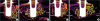
Table 1
Volumetric measurements from the micro-CT data (mm3; n=5)

Histological and histomorphometric findings
The maxillary sinus pouch was enveloped by a thin cortical layer and respiratory mucosa. The SM was intact, and there were no signs of inflammation. Osseointegration of the implant was evident throughout the cortical bone area in all specimens. The newly formed bone appeared to form from the cortical bone towards the implant apex along the implant surface and around the residual bone substitute. Newly formed bone was observed evenly from the hole to the SM in the BMP group, but mainly from the hole to the middle parts of the augmented pouch in the control group (Figure 4). The contact between the newly formed bone and the medial surface of the implant surface was positioned more apically in the BMP group. The trabecular pattern of bone formation did not show any significant histological differences between the BMP and control groups. The intertrabecular space was filled with fibrovascular tissue and bone marrow in both groups (Figure 5).
Figure 4
Histological photomicrographs of the total augmented area after 4 weeks of healing. (A) BMP group. (B) Control group. The osseointegration of the implant was evident throughout the cortical bone area in both groups. Hematoxylin-eosin stain; scale bar=1 mm.
CB: cortical bone, RBS: residual bone substitute, BMP: bone morphogenetic protein.
Figure 5
Histological photomicrographs of the apical area after 4 weeks of healing. (A) BMP group. (B) Control group. More new bone formed around the apex of the implant in the BMP group than in the control group. Hematoxylin-eosin stain; scale bar=500 μm.
SM: sinus membrane, NB: newly formed bone, BMP: bone morphogenetic protein, RBS: residual bone substitute.
The linear measurements performed in the histomorphometric analysis revealed that the median CBH was 0.50 mm in both the BMP and control groups. The AH of the maxillary sinus pouch was significantly greater in the BMP group than the control group (6.4 mm and 6.0 mm, respectively; P=0.004). The OH was significantly greater in the BMP group than in the control group at the medial surface of the implant (5.2 mm and 4.3 mm, respectively; P=0.037), but it did not differ significantly at the lateral surface of the implant (5.3 mm and 4.6 mm, respectively; P=0.123; Table 2).
Table 2
Histomorphometric linear measurements in the augmented pouch (mm; n=5)

Values are presented as median (minimum, maximum).
AH: augmented height, PH: protruding height, CBH: cortical bone height, OH: distance from the basal cortical bone to the highest point of osseointegration, BMP: bone morphogenetic protein.
a)Significantly greater than the control group (P<0.01); b)Significantly greater than the control group on the medial surface (P<0.05).
None of the measured areas differed significantly between the BMP and control groups (P>0.05, Table 3); this was also the case for the BIC (25.3% and 24.7%, respectively; P>0.05).
Table 3
Histomorphometric area measurements in the augmented pouch (mm2; n=5)

DISCUSSION
The present study evaluated the effect of rhBMP-2-loaded CBCP on bone formation after sinus augmentation with simultaneous implant placement in rabbits. In the surgical phase, rigid fixation of the implants was achieved in rabbit sinuses with a median CBH of 0.5 mm. At 4 weeks after the procedure, augmented bone substitute and implants were localized in sinus pouches without any complications, and newly formed bone had formed from the cortical bone to the apical portion of the implants. The augmented volume, newly formed bone volume, AH, and OH were greater in the BMP group than in the control group, but there was no intergroup difference in the BIC. These results indicate that the use of rhBMP-2-loaded CBCP allows successful implantation with sinus augmentation, even when only a minimal bone height is available during the early stage of healing.
A previous study was performed, in which CBCP loaded with 0.1 mg/mL rhBMP-2 was grafted into rabbit sinus, and significantly larger volumes of augmented and newly formed bone were found in the experimental group than the control group 4 weeks later [14]. It was speculated that the postoperative swelling induced by rhBMP-2 resulted in a larger initial augmented volume, which was subsequently replaced by accelerated bone formation. Such early corticalization of the area surrounding the SM can resist the positive respiratory pressure, thereby reducing volumetric shrinkage. To further explore this line of research, the present study evaluated implant placement with simultaneous sinus augmentation. Although the same rhBMP-2 concentration and dosage were applied in the present study, the differences in volume appeared to be smaller than in the previous study. This may be attributed to the effects of rhBMP-2 not only on early corticalization of the SM, but also on new bone formation in the peri-implant area. Several studies have found that rhBMP-2 contributed to new bone formation around the implant, thereby supporting the proposal that rhBMP-2 plays such a role [202122]. Therefore, a higher concentration and dosage of rhBMP-2 may be needed in order to achieve results similar to those reported in the previous study.
The early stability of an implant is determined by the peri-implant bone quantity and maturity [23], which could be predictive of the long-term prognosis of the implant. When an implant is placed, new bone formation appears to start from the original cortical bone of the sinus wall and to progress towards the center in the apical direction [9]. Newly formed cortical bone could play a similar role to the original cortical bone. In the present study, the OH-to-PH ratios were 103.5% and 97.3% medially and laterally, respectively, in the BMP group, and 83.7% and 89.3%, respectively, in the control group. This indicates that almost the entire height of the implant was covered by newly formed bone in the BMP group. We observed that rhBMP-2 facilitated bone formation on the medial surface of the implant, which was closer to the axial wall [24], resulting in rhBMP-2 stimulating angiogenesis through the chemotaxis of endothelial cells and osteoblasts that are present in cortical bone and around the SM [25]. It is therefore suggested that rhBMP-2 accelerates peri-implant bone formation and thereby enhances the early stability of the implant.
When comparing with the BIC values of Kim et al. [18] (11.3% for blood clots, 23.0% for autogenous bone, and 29.0% for bovine-derived hydroxyapatite), the BIC of CBCP grafting was similar to that of autogenous bone grafting regardless of whether or not rhBMP-2 was used (25.3% for BMP and 24.6% for controls). It seems that rhBMP-2 itself was not effective in directing newly formed bone to contact the implant surface. Wikesjo et al. [20] found no significant difference in the BIC when rhBMP-2 and absorbable collagen sponge were applied to supra-alveolar defects in mongrel dogs for 8 weeks. However, a histological analysis revealed that rhBMP-2 affected new bone formation more widely on the implant surface. A wider area of newly formed bone means that more of the implant surface will be covered, which can increase the implant stability.
Fixation at the cortical bone is always important for the initial stability of an implant [26]. However, one of the main disadvantages of 1-stage implantation is the possibility that fixation failure may lead to displacement of the fixture into the maxillary sinus. In the present study model, the implant was fixed to thin cortical bone (less than 0.5 mm thick, representing less than 10% of the fixture length) by manipulation and modification of the implant design, such as the inclusion of a crestal shoulder and microthreading in the cortical contact area. Additionally, adding rhBMP-2 to CBCP can achieve secondary stability via new bone formation around the implant surface, as discussed above.
The volume but not the area differed significantly between the 2 study groups. This may have been due to the smallness of the sample and variations in the areas selected for analysis. The limitations of the present study include the small sample size and the fact that measurements were performed at a single point in time. Therefore, future studies should include larger samples and obtain results at various time points. Although the rabbit sinus model has a strong osteogenic potential, meaning that it is likely for the effects of rhBMP-2 on a sinus graft to be observed after 4 weeks, bone contact around the implant should also be observed during a later stage of healing; that is, after more than 4 weeks.
In conclusion, an implant simultaneously placed with sinus augmentation using rhBMP-2-loaded synthetic bone substitute can be successfully osseointegrated, even when limited bone height is available during the early stage of healing, thereby achieving a reduced healing time when cortical fixation is obtained.
 XML Download