

 PDF
PDF ePub
ePub Citation
 Print
Print


INTRODUCTION
Cracked tooth syndrome (CTS) refers to the incomplete fracture of a vital posterior tooth that involves the dentine and occasionally extends into the pulp. Although CTS is a common disease, it is sometimes difficult to diagnose because the cracks can be too small to observe by visual examination or radiographic imaging. Because of these characteristics, symptom-driven diagnosis has been accepted [123]. The usual choice of treatment plan for a cracked tooth causing pain is either tooth extraction or re-implantation of the tooth after bonding the fractured fragments [456]. A new technique for the early diagnosis of cracked teeth is required to prevent the need for such serious treatments.
In vivo techniques for diagnosing cracked teeth include trans-illumination, a simple method to detect all types of tooth cracks, from craze lines to vertical root fractures. However, it cannot discriminate among cracks according to depth or type [78]. Several radiographic methods are used in dental research, including intraoral X-ray imaging, cone-beam computed tomography (CBCT), and micro-computed tomography (micro-CT) [9]. Recently, micro-CT has attracted attention because it can perform nondestructive testing at a high resolution. However, it is not suitable for in vivo applications, as it remains necessary to extract teeth in preparation for measurement. Intraoral radiography and CBCT are widely used to diagnose dental lesions without extraction, and in the case of CBCT, 3-dimensional information regarding fractured teeth can be obtained. However, the resolution of these radiographic methods is limited to approximately 100 μm [10], and the image resolution is too low to detect small cracks in early stages of development. If the diagnosis of tooth cracks is delayed because of these drawbacks, patients might develop protracted pain of various intensities [3]. Therefore, the early diagnosis of cracked teeth is important for preventing the extension of small cracks [11].
Optical coherence tomography (OCT) might solve these issues. OCT is widely used in the ophthalmic field and has recently become the focus of active research in dentistry [1213141516]. The OCT technique has been developed for the cross-sectional imaging of internal biological structures and uses low-coherence interferometry to determine the echo time delay and the magnitude of backscattered light reflected from a biological structure [1718]. Its resolution has been enhanced and the image-acquisition time has been reduced. Consequently, OCT has become a powerful tool in clinical and biological fields with applications involving various technologies. Some techniques measure blood flow using the optical Doppler phenomenon and observe specific material structures using the second harmonics of a measured signal to obtain border information in images with polarization OCT [19]. Among these various derivatives, swept-source OCT (SS-OCT) is recognized as a next-generation medical imaging diagnostic technology [131420]. Studies have shown that SS-OCT has a high degree of sensitivity and specificity in detecting caries and cracks, especially when a light source with a near-infrared wavelength of 1,310 nm is used for tooth cross-section detection, because transmittance in hard tissues increases with the wavelength of the light [1516].
To date, studies have reported that OCT can detect tooth cracks, but further investigations to increase its diagnostic efficiency and to conduct automatic diagnoses using computer-based image processing have not been initiated [1617]. The purpose of this study was to demonstrate that OCT images are useful for the detection of early-stage tooth cracks, and that such cracks can be automatically detected by image processing.
MATERIALS AND METHODS
Phantom preparation
A cracked tooth phantom was manufactured by applying a constant external force to the tooth. Generally, cracks occur frequently on molar occlusal surfaces. An individual's masticatory force is approximately 30–60 kg, and when this force is concentrated at a single point, a tooth crack can occur. The cracked tooth phantom was made by simulating a strong force using a Lloyd instrument (LTMC 100, Lloyd Instruments Ltd., Bognor Regis, UK). An external force was applied to the tooth to cause a fracture by moving the probe at a speed of 60 cm/s, and a force of 350–500 N was applied depending on the tooth status. All authors were well informed of the World Medical Association (WMA) Declaration of Helsinki-Ethical Principles for Medical Research Involving Human Subjects and confirm that the present study closely complied with the declaration.
Imaging techniques
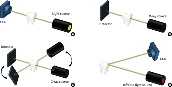
Artificial external force was applied to the extracted tooth to induce a crack. Cracked tooth images were acquired by OCT and conventional crack detection techniques. The images acquired from the respective technologies were compared, and crack lines were automatically detected through image processing. Intraoral X-ray imaging, trans-illumination, and CBCT are conventional in vivo techniques that are in widespread clinical use for the detection of cracks (Figure 1).
 | Figure 1The 4 imaging techniques used in the study. (A) Trans-illumination. A strong light is shone on one side of a tooth and the tooth image is acquired on the other side to identify cracks. (B) An intraoral X-ray is used to acquire a 2-dimensional image of a tooth using radiation. (C) CBCT is used to reconstruct a 3-dimensional image through image processing after obtaining radiographs from various angles. (D) OCT is used to acquire a cross-sectional image of a tooth using an infrared ray of 1,310 nm.
CBCT, cone-beam computed tomography; OCT, optical coherence tomography; CCD, charge-coupled detector.
|
Trans-illumination provides the most information regarding cracks and simply determines whether a crack is present [21]. Light passes through the tooth until it reaches a crack. Light is reflected when the refractive index changes inside the tooth because of a fracture. As a result, the bright and dark regions of the tooth are divided by the fracture line. The measurement equipment consists of a light source (MTS-100H, Microtek System, Minneapolis, MN, USA) and a charge-coupled device (KP-HD20A, Hitaci Kokusai Electric Inc., Tokyo, Japan). The position of the light source was appropriately adjusted to illuminate the tooth, following which the image was acquired.
Intraoral X-ray imaging has the advantage of obtaining more detailed images than panoramic CT or CBCT [22]. However, the lesion may not be seen depending on the angle of view and the fracture site may not appear depending on the angle. The intraoral X-ray imaging equipment consists of an X-ray generator (D-0711S, Instrumentarium Imaging, Tuusula, Finland) and a detector (RIOsensor, Raymedical, Hwaseong, Korea). The cracked tooth was photographed for 0.32 seconds at an X-ray tube voltage of 60 kVp and a current of 7 mA.
CBCT images of teeth were obtained with a bench-top CBCT system consisting of an X-ray generator (SourceRay SB-80-500, Source-Ray Inc., New York, NY, USA) and a detector (FLAATZ 330N, DRTECH, Seongnam, Korea). The CBCT images of teeth were obtained at 80 kVp, 0.3 mA, and 1 second with an isotropic voxel size of 129 μm. The rotation step was 1.125° and 320 projection images were used for reconstruction. The volumetric images were reconstructed by using the filtered back projection with a ramp filter.
Measurement of OCT images
The OCT apparatus used in this study was a multipurpose commercially available system (Oz-tec Co., Daegu, Korea) with a swept-laser source central wavelength of 1,310 nm, which is optimal for soft oral tissues, and a spectral bandwidth of 100 nm. The sweep source frequency was 50 kHz, with an average output power of up to 8 mW. The system captured 500 frames per second with an axial resolution in air of 7.56 μm/pixel and a lateral resolution of 10.03 μm/pixel.
The OCT images were stored as raw files and subsequently analyzed using software package (MATLAB, MathWorks, Natick, MA, USA). To improve the quality of OCT images, 100 images were averaged to obtain the final images. Next, the local adaptive threshold was used to improve the contrast of the edges, and a straight line was detected by a Hough transform. The contrast distribution of the background was not uniform in the tooth images. In degraded images with considerable background noise or variation in contrast and illumination, many pixels could not be easily classified as the foreground or background. In such cases, binarization with local thresholding is more appropriate, and this technique was applied in our experiment. We used a locally adaptive thresholding technique to remove the background by using the local mean and mean deviation.
After the local adaptive thresholding, the Hough transform was applied to detect tooth cracks from the acquired OCT images. The Hough transform is a feature extraction technique that detects straight lines, circles, or parametric curves. Pixels of an image from the Cartesian coordinate space are mapped to the polar coordinate space by the Hough transform. The polar coordinate representation of straight lines is obtained using the following equation.
In this equation, x and y represent a position in the Cartesian coordinate space, and ρ and θ represent a position in the polar coordinate space.
RESULTS
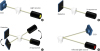
Six tooth samples were cracked in this study. The black arrow indicates a representative induced tooth crack in Figure 2A. However, it was difficult to observe a fine crack line with the naked eye, as shown in Figure 2B.
 | Figure 2Crack-induced tooth sample. (A) Black arrow indicates the crack. (B) Cracks are difficult to observe with the naked eye.
|
Figure 3A shows the trans-illumination image of a tooth with a circular crack lesion. However, the circular crack could not be found in the images shown in Figure 3B and C using radiation. Nonetheless, the OCT image presented in Figure 3D clearly shows a circular crack lesion confined to the enamel region. Severe light discontinuity in the longitudinal direction of the tooth can be observed in Figure 3E, and we assumed that it was a deep fracture. However, no deep fractures were detected in intraoral radiography, as shown in Figure 3F. CBCT radiography was used to assess the sample from various angles, and clearly showed that it was not a deep fracture, but a split tooth, as shown in Figure 3G. In the OCT image of Figure 3H, we confirmed that the deep fracture extended to the enamel and the dentin region. However, the penetration depth of OCT was insufficient to confirm that this was a divided tooth. Finally, several fractures were observed at the end of the tooth in Figure 3I and J, but no fracture was found in the CBCT image of Figure 3K. However, OCT easily detected crack lines in the enamel region, as shown in Figure 3L.
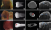 | Figure 3Results of 4 imaging techniques. The red line indicates the cross-section of CBCT and the OCT scan line. Red and blue arrows indicate crack lines. The blue circle is a false-positive crack line. E, D, and the DEJ are clearly distinguished in the OCT images. (A, E, I) Trans-illumination images. (B, F, J) Intraoral radiography images. (C, G, K) CBCT images. (D, H, L) OCT images.
CBCT, cone-beam computed tomography; E, enamel; D, dentine; DEJ, dentine-enamel junction; OCT, optical coherence tomography.
|
Figure 4 showed the result of applying an algorithm to identify the crack line in 6 tooth samples automatically through image processing using OCT. Green bars indicate the area estimated to be a crack. In all samples, crack lines were detected automatically by image processing in the region estimated to contain a fracture.
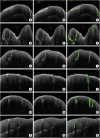 | Figure 4(A, D, G, J, M, and P) Single OCT images showing high noise. (B, E, H, K, N, and Q) OCT images were acquired 100 times, indicating that the noise was improved by image averaging. (C, F, I, L, O, and R) After averaging, crack lines were detected with the Hough transform.
OCT, optical coherence tomography.
|
DISCUSSION
We compared 4 clinical techniques that can be used in vivo and confirmed that SS-OCT could automatically diagnose a crack line by processing the image appropriately. CTS can be classified into 3 types according to the progress of the crack: craze line, crack, and split tooth [38]. Of these, a craze line is a fracture confined to the enamel only. It occurs naturally or secondarily by external injury and has no symptoms such as pain. Apart from aesthetic reasons, treatment is not needed. A fracture is a crack that extends to the dentin and root. Symptoms range from the absence of symptoms to severe pain. A split tooth is a fracture that extends from one side of the tooth to the other and divides the tooth into 2 parts [23].
Trans-illumination displays most cracks, including craze lines [7], but information cannot be obtained regarding the depth of the crack, and the reliability of the test is suboptimal because the degree to which a crack is displayed varies depending on the angle of light irradiation. Because the molar teeth in Figure 3A were thicker than those in Figure 3E, the penetration of light was poor and the cracks were not visible. The blue circle in Figure 3E may be identified as a crack even if it is not a crack. Due to the structure and color of the teeth, there is a high probability of this being a false positive. This technique is effective for easily irradiating light into teeth and confirming suspected fracture sites at various angles, but it is not suitable for the diagnosis of fractures because it does not provide information about depth.
Intraoral X-ray imaging is a technique in which 2-dimensional images of teeth are acquired using X-rays. However, information about the depth of the fracture cannot be calculated from images obtained with 2-dimensional sensors. Therefore, to derive the depth information, several images must be acquired in various directions, but this method is often impossible because of the position of the teeth and the intraoral structure. In addition, radiation exposure causes safety problems. Another problem is that the observation of the lesion area depends on the position and angle of the X-ray source and the sensor; therefore, the diagnostic reliability can be degraded. In addition, studies have reported that only 35.7% of cases with vertical root fractures are observed radiologically, and it has been established that fracture lines are usually detected when the radiation beam is irradiated within 4° of the fracture surface [24].
CBCT acquires tooth images from all directions and reconstructs 3-dimensional information regarding teeth; therefore, it can be used to diagnose root fractures because it can confirm the depth and structure of fractures. However, it is impossible to confirm the microstructure of teeth due to the low resolution and the patients' need to be treated with a medical examination based on the doctor's opinion after the initial diagnosis [10]. In general, for techniques that use radiation, the treatment process cannot be measured in real time.
OCT provides superior resolution (approximately 10 μm/pixel) in comparison with CBCT or intraoral X-ray imaging, without a risk of radioactivity exposure. The OCT equipment is smaller than the existing radiation equipment. If a handheld probe is developed in the future, it might be possible to perform a procedure and monitor it simultaneously, improving the reliability of the treatment. Using a wavelength of 1,310 nm, OCT can acquire tomographic information from the tooth surface to the boundary between the enamel and dentin and filter out clinically insignificant craze lines. However, it is difficult to confirm fractures in the deepest parts of teeth, such as root fractures, using OCT. After performing OCT image enhancement, the Hough transform algorithm can be used to detect the crack line automatically.
Table 1 summarizes the applicability of these diagnostic techniques based on experimental findings. Trans-illumination can detect all types of crack lines, but cannot discriminate the type of crack because it provides no depth information. Since the resolution of intraoral X-ray imaging and CBCT is over 100 μm/pixel [10], the detection of craze lines is difficult. Furthermore, with regard to crack lines, the detection of split teeth depends on the size of the cracks. Intraoral radiography and CBCT are ideal alternatives for the diagnosis of root fractures [25]. SS-OCT is suitable for the detection of early-stage cracks such as craze lines and crack lines. However, other techniques are needed to distinguish a split tooth, because doing so requires a cross-section 2 or 3 mm from the tooth surface.
Table 1
Applications of diagnostic technologies

| Type | Trans-illumination | Intraoral X-ray | CBCT | SS-OCT |
|---|---|---|---|---|
| Craze lines | ▬ | X | X | ○ |
| Crack lines | ▬ | ▬ | ◐ | ○ |
| Split tooth | ▬ | ◐ | ◐ | ◐ |
| Vertical root fracture | X | ○ | ○ | X |
○, diagnosed; X, not diagnosed; ◐, depends on size of cracks; ▬, presence or absence of cracks can be checked, but the type of cracks cannot be determined; CBCT, cone-beam computed tomography; SS-OCT, swept-source optical coherence tomography.
![]()
Figure 4 shows the results of automatic crack detection based on OCT images. By adjusting the threshold value, which affects the selection of the intersection point of the curve in the Hough transform, the fracture line can be determined based on the image. Figure 4A-F and P-R shows a crack in the enamel region of the tooth. Figure 4G-I shows the state of the crack up to the dentine-enamel junction, and Figure 4J-O shows a case where the fracture occurred as far as the dentine. OCT images require filters to reduce noise; averaging, Gaussian, and median filters are typically used. The current study used an averaging filter, which improved image noise, but reduced the image information, causing images to become blurred. Therefore, an edge enhancement filter based on a high-pass filter was used as a preprocessing technique to preserve the information of the boundary area that was lost by image processing. In this study, we first removed the base noise by averaging the images obtained by repeatedly measuring the same region, and used an edge enhancement filter and a moving average filter together to increase the degree of contrast in and around the crack area. However, it was not easy to detect cracks in areas where the contrast between the crack and the background was small, as in the preprocessing image of Figure 4B, E, H, K, N, and Q. For accurate crack detection, additional image processing techniques that emphasize the difference between cracks and the surrounding area are needed.
We were able to distinguish structural cracks, craze lines, and split lines in tooth cracks using SS-OCT images, and to automatically detect the position of various cracks in the OCT images. Therefore, the detection capability of SS-OCT images may make SS-OCT a useful diagnostic tool for CTS.
 XML Download