Journal List > J Periodontal Implant Sci > v.47(1) > 1108020

Thoma, Cha, and Jung: Treatment concepts for the posterior maxilla and mandible: short implants versus long implants in augmented bone

Abstract

The aim of this narrative review is to describe treatment options for the posterior regions of the mandible and the maxilla, comparing short implants vs. longer implants in an augmented bone. The dental literature was screened for treatment options enabling the placement of dental implants in posterior sites with a reduced vertical bone height in the maxilla and the mandible. Short dental implants have been increasingly used recently, providing a number of advantages including reduced patient morbidity, shorter treatment time, and lower costs. In the posterior maxilla, sinus elevation procedures were for long considered to be the gold standard using various bone substitute materials and rendering high implant survival rates. More recently, implants were even placed without any further use of bone substitute materials, but the long-term outcomes have yet to be documented. Vertical bone augmentation procedures in the mandible require a relatively high level of surgical skill and allow the placement of standard-length dental implants by the use of autogenous bone blocks. Both treatment options, short dental implants, and standard-length implants in combination with vertical bone augmentation procedures, appear to result in predictable outcomes in terms of implant survival rates. According to recent clinical studies comparing the therapeutic options of short implants vs. long implants in augmented bone, the use of short dental implants leads to a number of advantages for the patients and the clinician.

Graphical Abstract

jpis-47-2-abf001

INTRODUCTION

Implant therapy is considered a predictable treatment option rendering high implant and prosthesis survival rate when using fixed dental prostheses for replacing single- and multi-unit gaps [12]. These long-term favorable treatment outcomes have mostly been reported for implants placed in native bone or with only minor concomitant bone regenerative procedures and for implants with standard lengths and diameters. In the past years, implant lengths and diameters have continuously decreased. This shift was supported by the development of more favorable surface structures and more solid titanium alloys used for fabricating dental implants [345]. Moreover, patient demands for more minimally invasive surgical procedures, fewer complications, lower treatment costs, and less treatment time have resulted in a series of publications using dental implants with reduced dimensions, in terms of both diameter and length [678]. The primary goal following implant placement is to achieve osseointegration, a solid anchorage of the endosseous part of the implant within the bony envelope. Various methods have been developed to measure osseointegration and implant stability, including resonance frequency analysis (RFA) and histologic analyses [910]. On the basis of these outcome measures, bone formation and implant integration appear to follow a certain pattern that starts with the early phase of the bone resorption process [11], followed by a phase of bone apposition along the implant surface [12]. This results in dental implants demonstrating increasing RFA values and increasing bone-to-implant contact values over time [1314]. Thus far, threshold values for determining and predicting successful long-term outcomes have not been reported for either of these methods. One might, however, assume that an increase in the dimension of dental implants would result in clinically superior outcomes. However, this has not been documented thus far. In contrast, upon successful osseointegration and under healthy peri-implant conditions, attempts to break osseointegration have failed in preclinical experiments [1516]. These observations further support the decision to reduce implant dimensions and adapt them to the clinical situation. Therefore, the aim of the present narrative review is to describe treatment options in the posterior regions of the mandible and the maxilla, comparing short implants with long implants in augmented bone, and to propose a treatment concept based on current evidence.

SHORT DENTAL IMPLANTS

Short dental implants were defined as implants with an endosseous component of ≤8 mm [17]. These implants were predominantly introduced and clinically used for circumventing more extensive primary bone augmentation procedures such as lateral sinus augmentation [18]. However, the survival rates, according to the latter study, were lower than those of standard-length implants in identical clinical situations. Further, the development of new implant surfaces resulted in an increasing number of publications using short dental implants for a number of indications, thereby expanding the treatment options for fully and partially edentulous patients. From a clinical point of view and from a patient's perspective, short dental implants offer a number of clinical advantages: less skill necessary to perform the surgical intervention, less morbidity by avoiding more extensive bone augmentation procedures, easier removal in case of failure, and predominantly, an increased number of sites available for implant therapy. In contrast, clinicians might be worried about disadvantages including a high crown-to-implant ratio and a relatively high rate of biological and technical complications associated with potential overload. According to preclinical [192021] and clinical studies [22], as well as recent systematic reviews [72324252627], none of these possible limitations are clinically relevant. Exceptions include slightly increased failure rates of short implants in the posterior maxilla with soft bone present in comparison with the mandible [27] and a slightly higher rate of technical complications [28]. Moreover, longer-term studies have demonstrated that short implants exhibit implant survival rates and biological outcomes similar to those of long implants [293031].

SINUS ELEVATION

The maxillary posterior edentulous area presents a challenge to clinicians during the placement of dental implants because of the lack of bone height resulting from the resorption of the alveolar ridge and pneumatization of the maxillary sinus floor. This has led to the development of sinus augmentation techniques intended to increase the vertical bone dimension in the posterior maxilla in the cranial direction, allowing the placement of dental implants. Considering the residual ridge height, the most appropriate surgical technique and grafting protocol may be selected on the basis of the proposed treatment guidelines [32].
Sinus augmentation via a crestal or a lateral approach is routinely performed either primarily or simultaneously with implant placement, and is generally considered to result in predictable outcomes. Systematic reviews have demonstrated an overall implant survival rate well over 90% after sinus augmentation procedures [333435]. A recent systematic review reported that the implant survival rate after a minimum of 3 years of loading was 93.7% and 97.2% for sites augmented using lateral and crestal approaches, respectively [36]. Moreover, complication rates associated with sinus augmentation, such as membrane perforation, hemorrhage, and infection, were relatively low [37]. Despite successful outcomes, long healing periods needed to achieve an adequate level of new bone formation is a shortcoming for both clinicians and patients [3839].

VERTICAL AUGMENTATION

In case of a severely resorbed alveolar ridge in the posterior mandible, the available bone height for implant placement is often limited by the proximity of the inferior alveolar nerve. Vertical ridge augmentation could be an option for increasing the ridge dimension in order to place a standard-length implant. Moreover, this reduces the crown height of the implant prosthesis, which would otherwise result in an unfavorable crown-to-implant ratio, as well as the proximity of the restoration margin to the oral vestibulum, causing difficulties in maintaining proper oral hygiene [40].
Various surgical techniques have been developed for vertical ridge augmentation, including block bone grafting, distraction osteogenesis, and guided bone regeneration [41]. Among them, the autogenous block bone graft has been well documented and is still considered the gold standard [42]. A recent systematic review reported that the survival and success rates of implants placed in reconstructed ridges with autogenous bone blocks were similar to those of implants placed in native bone [43].
In contrast, unwanted volumetric resorption of an autogenous block bone graft is a concern for clinicians. The volume stability of the autogenous block bone remains controversial and has been reported to vary among clinical studies [444546]. These differences can be explained by the heterogeneity of the study designs, including different donor sites, different healing periods, different implantation times, and the use of barrier membranes. According to a review of studies with follow-up periods of at least 4 to 5 years, the resorption rate was the highest during the first year of grafting and appeared to stabilize afterwards [47]. As alternative candidates to the autogenous block bone, allogeneic or xenogeneic block grafts have been applied. However, scientific evidence for these materials is lacking, as the available data are limited to case series [48].
Alongside the many advantages of vertical augmentation, an increased rate of surgical complications and enhanced patient morbidity is associated with this type of procedure [49].

POSTERIOR MAXILLA: SHORT IMPLANTS VS. SINUS ELEVATION AND LONG IMPLANTS

In the posterior maxilla, a recent systematic review [50] included 8 clinical randomized controlled clinical trials (RCTs) with a follow-up period of up to 18 months after loading with final reconstructions. Based on the 5 studies reporting on long-term observation periods (16–18 months) [5152535455], a mean implant survival rate of 99.0% (95% confidence interval [CI], 96.4%–99.8%) for short implants and of 99.5% (95% CI, 97.6%–100.0%) for long implants in the augmented sinus was reported. Similar data were calculated with survival rates ranging between 97% and 100% for both groups on the restorative level. When discussing different treatment options with the patient, a clinician is confronted with challenges related to intraoperative, perioperative, and postoperative morbidity. On the basis of this information, a final decision is made to undergo a specific therapy. The most frequent technical complications included screw loosening in the reconstructions for both short and long implants, but few differences were found between the 2 treatment concepts. Further complications were predominantly observed because of the surgical interventions (i.e., membrane perforations). Short dental implants were responsible for 33% of the complications, whereas a sinus elevation procedure increased the risk of complications by 100%. This corresponds to as much as a 3 times higher risk of having an intraoperative complication when using long dental implants than when using short ones. These data are supported by observations that short dental implants offer further benefits in terms of less morbidity, lower costs, and a relatively short treatment time [555657]. At least on a short-term basis, short implants might be more appropriate, offering a variety of benefits in terms of patient-reported outcome measures, and still offer survival rates similar to those of long implants in the augmented sinus. A summary of the study characteristics is presented in Table 1.
Table 1

Summary of the RCTs comparing the treatment options of short implants vs. standard-length implants in combination with vertical bone augmentation procedures

jpis-47-2-i001
Region Author Year Results
Maxilla Esposito et al. [51] 2011 5-mm short implants achieved similar if not better results to those of longer implants placed in an augmented bone.
Maxilla Gulje et al. [52] 2014 6-mm implants and 11-mm implants combined with sinus floor elevation surgery were equally successful.
Maxilla and mandible Pistilli et al. [53] 2013 6-mm long implants with a conventional diameter of 4 mm achieved similar if not better results than longer implants placed in an augmented bone.
Maxilla and mandible Pistilli et al. [54] 2013 5-mm implants achieved results similar to those of longer implants placed in an augmented bone.
Maxilla Thoma et al. [55] 2015 Short implants may be more favorable in terms of short-term patient morbidity, treatment time, and treatment costs.
Maxilla and mandible Esposito et al. [56] 2012 Short implants might be a preferable choice to bone augmentation, particularly in posterior mandibles.
Maxilla and mandible Felice et al. [57] 2009 There was no statistically significant difference in patient preferences, as patients found both short and long implants acceptable.
Mandible Esposito et al. [61] 2014 5-mm short implants achieved results similar to those of longer implants in augmented bone.
Mandible Felice et al. [62] 2014 The prognosis of short implants was as good as that of long implants placed vertically in augmented mandibles.
RCTs, randomized controlled clinical trials.

POSTERIOR MANDIBLE: SHORT IMPLANTS VS. VERTICAL RIDGE AUGMENTATION AND LONG IMPLANTS

For the posterior mandible with a limited alveolar ridge height, 3 options exist: primary vertical ridge augmentation and subsequent implant installation, simultaneous implant placement with vertical ridge augmentation, and the use of short implants [57585960]. As in the case of the posterior maxilla, different treatment modalities were evaluated in recent RCTs [53546162]; however, the overall number of studies is limited. According to a recent systematic review [63], only 4 studies could be included, reporting on 135 patients who underwent restorations with 328 implants. The survival rates on the implant and prosthetic level have not been reported to be different in any study. However, an analysis of the rate of complications revealed certain differences. In the group with primary bone augmentation, even if some of the grafting procedures failed, all the short implants could be placed. This was reflected in a calculation of the overall number of complications in patients. In 56 out of 85 patients, complications were reported for the augmented group, whereas in the short dental implants group, only 18 patients were affected. The main complications included paresthesia of the mandibular nerve, predominantly in the augmented group [63]. Clinical recommendations favoring one procedure over the other have to be interpreted with caution, since the scientific evidence is based on few patients and research groups.

CURRENT TREATMENT CONCEPT

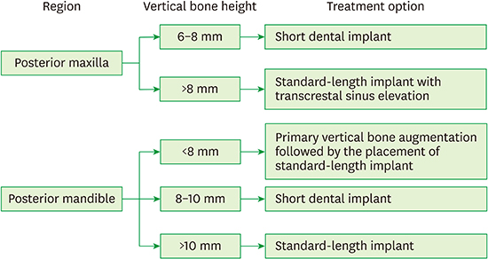
The clinical decision between these options (short dental implants or primary bone augmentation, followed by the placement of long dental implants) is based on a number of parameters. These parameters predominantly include scientific evidence, the surgical skill and experience of the surgeons and, increasingly, the patient's preferences. In order to support the clinician in the decision-making process and to inform the patient more extensively on the available treatment options, systematic reviews have summarized the available literature on high-level evidence from RCTs for the posterior mandible and maxilla (for an overview see: [5063]). For the posterior maxilla, short dental implants are the preferred option in cases with a vertical bone height of 6–8 mm (Figure 1). A transcrestal sinus elevation approach is chosen for a vertical ridge dimension exceeding 8 mm and if standard-length implants are the preferred option (Figure 2). In the posterior mandible, primary vertical bone augmentation using one of the abovementioned treatment options should be performed, followed by the placement of standard-length implants (Figure 3). Short dental implants are predominantly recommended in cases with a remaining ridge height of 8–10 mm, allowing the surgeon to place a 6-mm implant and including a safety distance to the alveolar nerve of 2 mm (Figure 4). In cases where more than 10 mm of the bone is available, standard-length implants are recommended (Figure 5). The decision-making process for the posterior maxilla and mandible is presented in Figure 6.
Figure 1
(A) A treatment option for the posterior maxilla with a vertical bone height of 6–8 mm. (B, C) A short dental implant is recommended.
jpis-47-2-g001
Figure 2
(A) A treatment option for the posterior maxilla with a vertical bone height of more than 8 mm. (B, C) A transcrestal sinus elevation approach can be chosen for a vertical ridge dimension exceeding 8 mm and if standard-length implants are the preferred option.
jpis-47-2-g002
Figure 3
(A) A treatment option for the posterior mandible with a remaining ridge height of less than 8 mm. (B) Primary vertical bone augmentation should be performed, (C) followed by the placement of standard-length implants.
jpis-47-2-g003
Figure 4
(A) A treatment option for the posterior mandible with a remaining ridge height of 8–10 mm. (B, C) A short dental implant is recommended.
jpis-47-2-g004
Figure 5
(A) A treatment option for the posterior mandible with a vertical bone height of more than 10 mm. (B, C) Standard-length implants are recommended.
jpis-47-2-g005
Figure 6
Decision-making process for the posterior maxilla and mandible.
jpis-47-2-g006

CONCLUSION

Short dental implants as well as standard-length implants in combination with vertical bone augmentation procedures appear to result in predictable outcomes in terms of implant survival rates. However, according to recent clinical studies comparing these therapeutic options, the use of short dental implants appears to have a number of advantages for the patients and the clinician.

Notes

Funding This research was supported by Basic Science Research Program through the National Research Foundation of Korea (NRF) funded by the Ministry of Science, ICT & Future Planning (NRF-2014R1A1A1A05002953).

Author Contributions Conceptualization: Daniel Stefan Thoma; Data curation: Jae-Kook Cha; Formal analysis: Jae-Kook Cha; Funding acquisition: Ui-Won Jung; Investigation: Daniel Stefan Thoma, Ui-Won Jung; Methodology: Daniel Stefan Thoma; Writing - original draft: Daniel Stefan Thoma, Jae-Kook Cha, Ui-Won Jung; Writing - review & editing: Daniel Stefan Thoma, Ui-Won Jung.

Conflict of Interest No potential conflict of interest relevant to this article was reported.

References

1. Jung RE, Zembic A, Pjetursson BE, Zwahlen M, Thoma DS. Systematic review of the survival rate and the incidence of biological, technical, and aesthetic complications of single crowns on implants reported in longitudinal studies with a mean follow-up of 5 years. Clin Oral Implants Res. 2012; 23:Suppl 6. 2–21.
2. Pjetursson BE, Thoma D, Jung R, Zwahlen M, Zembic A. A systematic review of the survival and complication rates of implant-supported fixed dental prostheses (FDPs) after a mean observation period of at least 5 years. Clin Oral Implants Res. 2012; 23:Suppl 6. 22–38.
crossref
3. van Velzen FJ, Ofec R, Schulten EA, Ten Bruggenkate CM. 10-year survival rate and the incidence of peri-implant disease of 374 titanium dental implants with a SLA surface: a prospective cohort study in 177 fully and partially edentulous patients. Clin Oral Implants Res. 2015; 26:1121–1128.
crossref
4. Annunziata M, Guida L. The effect of titanium surface modifications on dental implant osseointegration. Front Oral Biol. 2015; 17:62–77.
crossref
5. Bahrami B, Shahrbaf S, Mirzakouchaki B, Ghalichi F, Ashtiani M, Martin N. Effect of surface treatment on stress distribution in immediately loaded dental implants--a 3D finite element analysis. Dent Mater. 2014; 30:e89–97.
crossref
6. Mendonça JA, Francischone CE, Senna PM, Matos de Oliveira AE, Sotto-Maior BS. A retrospective evaluation of the survival rates of splinted and non-splinted short dental implants in posterior partially edentulous jaws. J Periodontol. 2014; 85:787–794.
crossref
7. Monje A, Suarez F, Galindo-Moreno P, García-Nogales A, Fu JH, Wang HL. A systematic review on marginal bone loss around short dental implants (<10 mm) for implant-supported fixed prostheses. Clin Oral Implants Res. 2014; 25:1119–1124.
crossref
8. Lee JS, Kim HM, Kim CS, Choi SH, Chai JK, Jung UW. Long-term retrospective study of narrow implants for fixed dental prostheses. Clin Oral Implants Res. 2013; 24:847–852.
crossref
9. Manresa C, Bosch M, Echeverría JJ. The comparison between implant stability quotient and bone-implant contact revisited: an experiment in Beagle dog. Clin Oral Implants Res. 2014; 25:1213–1221.
crossref
10. Shin SY, Shin SI, Kye SB, Hong J, Paeng JY, Chang SW, et al. The effects of defect type and depth, and measurement direction on the implant stability quotient value. J Oral Implantol. 2015; 41:652–656.
crossref
11. Al-Juboori MJ, AbdulRahaman SB, Hassan A. The correlation between crestal bone resorption and implant stability during healing period using resonance frequency analysis. Implant Dent. 2013; 22:351–355.
crossref
12. Gehrke SA, Maté Sánchez de Val JE, Fernández Domínguez M, de Aza Moya PN, Gómez Moreno G, Calvo Guirado JL. Effects on the osseointegration of titanium implants incorporating calcium-magnesium: a resonance frequency and histomorphometric analysis in rabbit tibia. Clin Oral Implants Res. Forthcoming. 2016.
crossref
13. Dagher M, Mokbel N, Jabbour G, Naaman N. Resonance frequency analysis, insertion torque, and bone to implant contact of 4 implant surfaces: comparison and correlation study in sheep. Implant Dent. 2014; 23:672–678.
14. Degidi M, Perrotti V, Piattelli A, Iezzi G. Mineralized bone-implant contact and implant stability quotient in 16 human implants retrieved after early healing periods: a histologic and histomorphometric evaluation. Int J Oral Maxillofac Implants. 2010; 25:45–48.
15. Isidor F. Histological evaluation of peri-implant bone at implants subjected to occlusal overload or plaque accumulation. Clin Oral Implants Res. 1997; 8:1–9.
crossref
16. Isidor F. Loss of osseointegration caused by occlusal load of oral implants. A clinical and radiographic study in monkeys. Clin Oral Implants Res. 1996; 7:143–152.
crossref
17. Renouard F, Nisand D. Impact of implant length and diameter on survival rates. Clin Oral Implants Res. 2006; 17:Suppl 2. 35–51.
crossref
18. Renouard F, Nisand D. Short implants in the severely resorbed maxilla: a 2-year retrospective clinical study. Clin Implant Dent Relat Res. 2005; 7:Suppl 1. S104–10.
crossref
19. Gotfredsen K, Berglundh T, Lindhe J. Bone reactions adjacent to titanium implants subjected to static load of different duration. A study in the dog (III). Clin Oral Implants Res. 2001; 12:552–558.
crossref
20. Gotfredsen K, Berglundh T, Lindhe J. Bone reactions adjacent to titanium implants with different surface characteristics subjected to static load. A study in the dog (II). Clin Oral Implants Res. 2001; 12:196–201.
crossref
21. Gotfredsen K, Berglundh T, Lindhe J. Bone reactions adjacent to titanium implants subjected to static load. A study in the dog (I). Clin Oral Implants Res. 2001; 12:1–8.
22. Blanes RJ, Bernard JP, Blanes ZM, Belser UC. A 10-year prospective study of ITI dental implants placed in the posterior region. II: influence of the crown-to-implant ratio and different prosthetic treatment modalities on crestal bone loss. Clin Oral Implants Res. 2007; 18:707–714.
crossref
23. Annibali S, Cristalli MP, Dell’Aquila D, Bignozzi I, La Monaca G, Pilloni A. Short dental implants: a systematic review. J Dent Res. 2012; 91:25–32.
24. Atieh MA, Zadeh H, Stanford CM, Cooper LF. Survival of short dental implants for treatment of posterior partial edentulism: a systematic review. Int J Oral Maxillofac Implants. 2012; 27:1323–1331.
25. Neldam CA, Pinholt EM. State of the art of short dental implants: a systematic review of the literature. Clin Implant Dent Relat Res. 2012; 14:622–632.
crossref
26. Sun HL, Huang C, Wu YR, Shi B. Failure rates of short (≤ 10 mm) dental implants and factors influencing their failure: a systematic review. Int J Oral Maxillofac Implants. 2011; 26:816–825.
27. Telleman G, Raghoebar GM, Vissink A, den Hartog L, Huddleston Slater JJ, Meijer HJ. A systematic review of the prognosis of short (<10 mm) dental implants placed in the partially edentulous patient. J Clin Periodontol. 2011; 38:667–676.
28. Quaranta A, Piemontese M, Rappelli G, Sammartino G, Procaccini M. Technical and biological complications related to crown to implant ratio: a systematic review. Implant Dent. 2014; 23:180–187.
29. Sahrmann P, Naenni N, Jung RE, Held U, Truninger T, Hämmerle CH, et al. Success of 6-mm implants with single-tooth restorations: a 3-year randomized controlled clinical trial. J Dent Res. 2016; 95:623–628.
crossref
30. Mertens C, Meyer-Bäumer A, Kappel H, Hoffmann J, Steveling HG. Use of 8-mm and 9-mm implants in atrophic alveolar ridges: 10-year results. Int J Oral Maxillofac Implants. 2012; 27:1501–1508.
31. Deporter D, Pharoah M, Yeh S, Todescan R, Atenafu EG. Performance of titanium alloy sintered porous-surfaced (SPS) implants supporting mandibular overdentures during a 20-year prospective study. Clin Oral Implants Res. 2014; 25:e189–95.
crossref
32. Jensen SS, Terheyden H. Bone augmentation procedures in localized defects in the alveolar ridge: clinical results with different bone grafts and bone-substitute materials. Int J Oral Maxillofac Implants. 2009; 24:Suppl. 218–236.
33. Del Fabbro M, Corbella S, Weinstein T, Ceresoli V, Taschieri S. Implant survival rates after osteotome-mediated maxillary sinus augmentation: a systematic review. Clin Implant Dent Relat Res. 2012; 14:Suppl 1. e159–68.
crossref
34. Pjetursson BE, Tan WC, Zwahlen M, Lang NP. A systematic review of the success of sinus floor elevation and survival of implants inserted in combination with sinus floor elevation. J Clin Periodontol. 2008; 35:Suppl. 216–240.
crossref
35. Silva LD, de Lima VN, Faverani LP, de Mendonça MR, Okamoto R, Pellizzer EP. Maxillary sinus lift surgery-with or without graft material? A systematic review. Int J Oral Maxillofac Surg. 2016; 45:1570–1576.
crossref
36. Del Fabbro M, Wallace SS, Testori T. Long-term implant survival in the grafted maxillary sinus: a systematic review. Int J Periodontics Restorative Dent. 2013; 33:773–783.
crossref
37. Boffano P, Forouzanfar T. Current concepts on complications associated with sinus augmentation procedures. J Craniofac Surg. 2014; 25:e210–2.
crossref
38. Fugazzotto PA. GBR using bovine bone matrix and resorbable and nonresorbable membranes. Part 1: histologic results. Int J Periodontics Restorative Dent. 2003; 23:361–369.
39. Wang F, Zhou W, Monje A, Huang W, Wang Y, Wu Y. Influence of healing period upon bone turn over on maxillary sinus floor augmentation grafted solely with deproteinized bovine bone mineral: a prospective human histological and clinical trial. Clin Implant Dent Relat Res. Forthcoming. 2016.
crossref
40. Mecall RA, Rosenfeld AL. Influence of residual ridge resorption patterns on implant fixture placement and tooth position. 1. Int J Periodontics Restorative Dent. 1991; 11:8–23.
41. McAllister BS, Haghighat K. Bone augmentation techniques. J Periodontol. 2007; 78:377–396.
crossref
42. Sanz M, Vignoletti F. Key aspects on the use of bone substitutes for bone regeneration of edentulous ridges. Dent Mater. 2015; 31:640–647.
crossref
43. Aloy-Prósper A, Peñarrocha-Oltra D, Peñarrocha-Diago M, Peñarrocha-Diago M. The outcome of intraoral onlay block bone grafts on alveolar ridge augmentations: a systematic review. Med Oral Patol Oral Cir Bucal. 2015; 20:e251–8.
44. Johansson B, Grepe A, Wannfors K, Aberg P, Hirsch JM. Volumetry of simulated bone grafts in the edentulous maxilla by computed tomography: an experimental study. Dentomaxillofac Radiol. 2001; 30:153–156.
crossref
45. Nkenke E, Neukam FW. Autogenous bone harvesting and grafting in advanced jaw resorption: morbidity, resorption and implant survival. Eur J Oral Implantology. 2014; 7:Suppl 2. S203–17.
46. Restoy-Lozano A, Dominguez-Mompell JL, Infante-Cossio P, Lara-Chao J, Espin-Galvez F, Lopez-Pizarro V. Reconstruction of mandibular vertical defects for dental implants with autogenous bone block grafts using a tunnel approach: clinical study of 50 cases. Int J Oral Maxillofac Surg. 2015; 44:1416–1422.
crossref
47. Keestra JA, Barry O, Jong L, Wahl G. Long-term effects of vertical bone augmentation: a systematic review. J Appl Oral Sci. 2016; 24:3–17.
crossref
48. Waasdorp J, Reynolds MA. Allogeneic bone onlay grafts for alveolar ridge augmentation: a systematic review. Int J Oral Maxillofac Implants. 2010; 25:525–531.
49. Raghoebar GM, Meijndert L, Kalk WW, Vissink A. Morbidity of mandibular bone harvesting: a comparative study. Int J Oral Maxillofac Implants. 2007; 22:359–365.
50. Thoma DS, Zeltner M, Hüsler J, Hämmerle CH, Jung RE. EAO Supplement Working Group 4 - EAO CC 2015 Short implants versus sinus lifting with longer implants to restore the posterior maxilla: a systematic review. Clin Oral Implants Res. 2015; 26:Suppl 11. 154–169.
crossref
51. Esposito M, Pellegrino G, Pistilli R, Felice P. Rehabilitation of postrior atrophic edentulous jaws: prostheses supported by 5 mm short implants or by longer implants in augmented bone? One-year results from a pilot randomised clinical trial. Eur J Oral Implantology. 2011; 4:21–30.
52. Guljé FL, Raghoebar GM, Vissink A, Meijer HJ. Single crowns in the resorbed posterior maxilla supported by either 6-mm implants or by 11-mm implants combined with sinus floor elevation surgery: a 1-year randomised controlled trial. Eur J Oral Implantology. 2014; 7:247–255.
53. Pistilli R, Felice P, Cannizzaro G, Piatelli M, Corvino V, Barausse C, et al. Posterior atrophic jaws rehabilitated with prostheses supported by 6 mm long 4 mm wide implants or by longer implants in augmented bone. One-year post-loading results from a pilot randomised controlled trial. Eur J Oral Implantology. 2013; 6:359–372.
54. Pistilli R, Felice P, Piattelli M, Gessaroli M, Soardi E, Barausse C, et al. Posterior atrophic jaws rehabilitated with prostheses supported by 5 x 5 mm implants with a novel nanostructured calcium-incorporated titanium surface or by longer implants in augmented bone. One-year results from a randomised controlled trial. Eur J Oral Implantology. 2013; 6:343–357.
55. Thoma DS, Haas R, Tutak M, Garcia A, Schincaglia GP, Hämmerle CH. Randomized controlled multicentre study comparing short dental implants (6 mm) versus longer dental implants (11-15 mm) in combination with sinus floor elevation procedures. Part 1: demographics and patient-reported outcomes at 1 year of loading. J Clin Periodontol. 2015; 42:72–80.
crossref
56. Esposito M, Cannizzaro G, Soardi E, Pistilli R, Piattelli M, Corvino V, et al. Posterior atrophic jaws rehabilitated with prostheses supported by 6 mm-long, 4 mm-wide implants or by longer implants in augmented bone. Preliminary results from a pilot randomised controlled trial. Eur J Oral Implantology. 2012; 5:19–33.
57. Felice P, Checchi V, Pistilli R, Scarano A, Pellegrino G, Esposito M. Bone augmentation versus 5-mm dental implants in posterior atrophic jaws. Four-month post-loading results from a randomised controlled clinical trial. Eur J Oral Implantology. 2009; 2:267–281.
58. Simion M, Jovanovic SA, Tinti C, Benfenati SP. Long-term evaluation of osseointegrated implants inserted at the time or after vertical ridge augmentation. A retrospective study on 123 implants with 1–5 year follow-up. Clin Oral Implants Res. 2001; 12:35–45.
crossref
59. Simion M, Dahlin C, Rocchietta I, Stavropoulos A, Sanchez R, Karring T. Vertical ridge augmentation with guided bone regeneration in association with dental implants: an experimental study in dogs. Clin Oral Implants Res. 2007; 18:86–94.
crossref
60. Rocchietta I, Fontana F, Simion M. Clinical outcomes of vertical bone augmentation to enable dental implant placement: a systematic review. J Clin Periodontol. 2008; 35:Suppl. 203–215.
crossref
61. Esposito M, Pistilli R, Barausse C, Felice P. Three-year results from a randomised controlled trial comparing prostheses supported by 5-mm long implants or by longer implants in augmented bone in posterior atrophic edentulous jaws. Eur J Oral Implantology. 2014; 7:383–395.
62. Felice P, Cannizzaro G, Barausse C, Pistilli R, Esposito M. Short implants versus longer implants in vertically augmented posterior mandibles: a randomised controlled trial with 5-year after loading follow-up. Eur J Oral Implantology. 2014; 7:359–369.
63. Nisand D, Picard N, Rocchietta I. Short implants compared to implants in vertically augmented bone: a systematic review. Clin Oral Implants Res. 2015; 26:Suppl 11. 170–179.
crossref
TOOLS
ORCID iDs

Daniel Stefan Thoma
https://orcid.org/http://orcid.org/0000-0002-1764-7447

Jae-Kook Cha
https://orcid.org/http://orcid.org/0000-0002-6906-7209

Ui-Won Jung
https://orcid.org/http://orcid.org/0000-0001-6371-4172

Similar articles