

 PDF
PDF Citation
 Print
Print


INTRODUCTION
With the advent of the prostate-specific antigen (PSA) screening era, an increasing number of newly diagnosed prostate cancer (PCa) cases tend to be assessed as lower-risk disease.1 Nevertheless, between 20% and 35% of PCa patients in the United States present with clinically high-risk disease.123 In these high-risk patients, survival outcomes are heterogeneous, and there is no consensus on the optimal management strategy.4
In the last decade, radiotherapy (RT) administered in combination with androgen deprivation therapy (ADT) has been the strategy of choice for the management of localized high-risk PCa. However, with the advancement of minimally invasive surgical techniques, surgery has gained acknowledgement as a feasible alternative.5 In a recent study using propensity-score matched analysis to compare oncological outcomes of patients with localized or locally advanced PCa treated with radical prostatectomy (RP) and RT, the two treatments demonstrated comparable cancer-specific survival (CSS) for all risk groups defined by the National Comprehensive Cancer Network (NCCN).6 Moreover, robot-assisted radical prostatectomy (RARP) was demonstrated to be a curative treatment option for a subset of high-risk patients with long-term undetectable PSA levels. Another advantage of surgical treatment is the opportunity to perform precise pathological evaluation and use postoperative nomograms, which facilitate the initiation of appropriate and timely adjuvant therapy.7 Indeed, 14% to 37% of high-risk PCa patients treated with RP demonstrate favorable pathologic results and better oncological outcomes than those noted among other high-risk patients.8910
Patients with high-risk PCa treated with RP may achieve undetectable PSA levels postoperatively. Compared to patients with a low or intermediate risk, a higher proportion of patients with a high-risk will experience biochemical recurrence (BCR), although the clinical course of patients with BCR is highly variable.11 Specifically, some patients experience rapid clinical progression to metastases, whereas BCR may pose no threat throughout the remaining life span of other patients.12 Studies have reported time to BCR as a significant prognosticator of cancer-specific mortality (CSM). However, few data are available regarding the relevance of time to BCR in patients with high-risk PCa undergoing RP.1314
The purpose of the present study was to confirm the feasibility of RARP for high-risk PCa and to investigate the effect of time to disease progression on metastasis and mortality rates in patients with high-risk PCa who achieved undetectable PSA following RARP. To address these issues, we investigated metastasis-free survival (MFS), CSS, and overall survival (OS) outcomes, as well as the prognosticators of these survival endpoints.
Go to : 
METHODS
Patient selection
We retrospectively reviewed the records of 342 patients treated with RARP and pelvic lymph node dissection between August 2005 and June 2011 at a single tertiary institution. We identified 251 (73.4%) patients with high-risk PCa who achieved undetectable PSA postoperatively, defined as < 0.01 ng/mL by ultrasensitive assay. High-risk PCa was defined as clinical stage ≥ T3, biopsy Gleason score ≥ 8, and/or PSA levels ≥ 20 ng/mL, according to the NCCN guidelines.15 PCa staging was determined according to the 7th American Joint Committee on Cancer TNM system, with the definition of distant metastasis based on either demonstrable metastatic deposits on imaging (bone scan, computerized tomography, magnetic resonance imaging, or positron emission tomography) or pathologic confirmation of PCa in tissue samples collected from outside the prostatic fossa. The following exclusion criteria were applied: 1) incomplete clinical data; 2) loss to follow-up; 3) unknown cause of death.
Prognostic factors and outcome variables
All patients had complete clinical and pathological data including age, body mass index (BMI), preoperative PSA levels, clinical stage, biopsy Gleason score, pathological stage with Gleason score, positive surgical margin status, tumor volume, seminal vesicle invasion status, lymph node invasion status, prostatic intraepithelial neoplasia grading, and time to BCR. These patients were further stratified into two groups according to time to BCR dichotomized at 60 months. BCR was defined as the first of two or more consecutive increases in PSA levels of > 0.2 ng/mL noted later than 3 months following surgery. Time to metastasis was defined as the time period from the date of histological diagnosis to the time of metastasis detection. OS was determined as the time elapsed between the date of histological diagnosis and the date of death. For all patients, the survival status and the cause of death were obtained from the National Cancer Registry Database or from the electronic medical records of the treating institution.
Study endpoints
The primary endpoints were the rates of MFS, CSS, and OS, whereas the secondary endpoints included the prognosticators of these survival endpoints.
Statistical analysis
Demographic characteristics of patients and tumors were compared using descriptive statistics including median and interquartile range (IQR). Appropriate comparative tests such as the Student's t-test and the χ2 test, were used to compare continuous and categorical variables, respectively. Kaplan-Meier curves were used to estimate MFS and OS. Univariate and multivariate analyses were performed using Cox-proportional hazards regression models in order to adjust for potential confounders in predicting survival. Variables considered potential predictors for the purpose of multivariate modeling were selected by univariate analysis. Results regarding prognosticators were expressed in terms of hazard ratio (HR) with 95% confidence intervals (CIs). Statistical analysis was performed using SPSS version 18 (SPSS Inc., Chicago, IL, USA). All tests were two-sided, with statistical significance set at P < 0.05.
Ethics statement
This retrospective study was approved by the Institutional Ethics Committee after review of the protocol and procedures employed (approval No. 2014-0112).
Go to :
RESULTS
Patient demographics
Table 1 summarizes the demographic and clinical characteristics of 251 patients with high-risk PCa who achieved undetectable PSA levels postoperatively. The median age and PSA levels were 66.5 years (IQR, 63.0–71.0 years) and 8.7 ng/mL (IQR, 6.1–18.3 ng/mL), respectively.
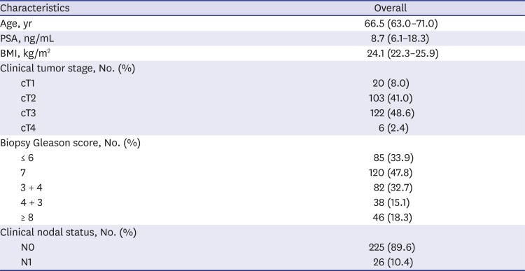
Table 1
Preoperative characteristics of patients with high-risk PCa who achieved undetectable PSA following robot-assisted radical prostatectomy
![]()
Postoperative characteristics
The patients were followed-up for a median of 75.9 months (IQR, 59.4–85.8 months) and stratified into two groups according to the time to BCR (dichotomized at 60 months). The incidence of pathological N1 disease was higher in patients who experienced BCR at ≤ 60 months than in those who did not (P = 0.044), but no differences between the two groups were noted regarding other postoperative tumor characteristics (Table 2).
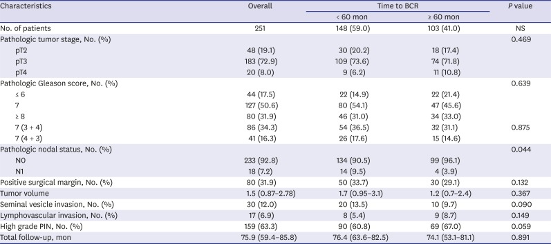
Table 2
Postoperative pathological outcomes of patients with high-risk PCa who achieved undetectable PSA following robot-assisted radical prostatectomy
PCa = prostate cancer, PSA = prostate-specific antigen, BCR = biochemical recurrence, PIN = prostatic intraepithelial neoplasia.
![]()
Oncological outcomes
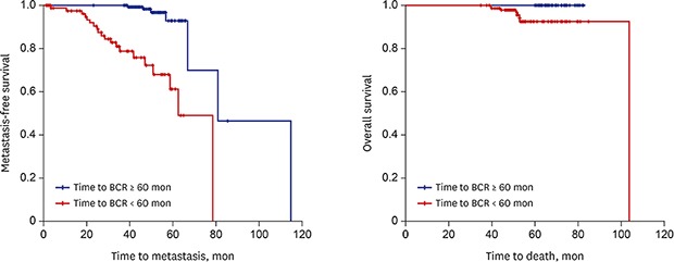
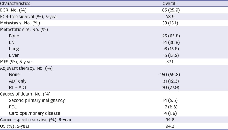
The oncological outcomes of our cohort are summarized in Table 3. BCR was observed in 65 (25.9%) patients, with an overall 5-year BCR-free survival rate of 73.9%. Metastasis occurred in 38 (15.1%) patients, mostly to the bones (65.8%), followed by the lymph nodes (36.8%), lungs (15.8%), and liver (13.2%). Overall, 101 (40.2%) patients received adjuvant treatment with either ADT or RT plus ADT. The 5-year MFS, CSS, and OS rates were 87.1%, 94.8%, and 94.3%, respectively. Patients who exhibited BCR at ≤ 60 months after RARP exhibited significantly lower MFS and OS rates compared to those of patients who did not (log-rank P < 0.001; Figs. 1 and 2). The most common cause of death was second primary malignancy, followed by PCa and cardiopulmonary disease. Among the 342 high-risk patients reviewed in our study, 186 (54.4%) patients achieved cure, defined as persistent undetectable PSA levels at the median follow-up.
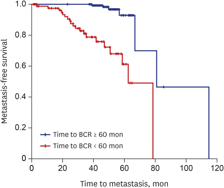 | Fig. 1Kaplan-Meier curves for MFS rate according to time to BCR (log-rank P < 0.001).MFS = metastasis-free survival, BCR = biochemical recurrence.
|
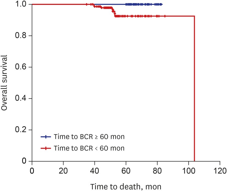 | Fig. 2Kaplan-Meier curves for OS rate according to time to BCR (log-rank P < 0.001).OS = overall survival, BCR = biochemical recurrence.
|
Table 3
Oncological outcomes of patients with high-risk PCa who achieved undetectable PSA following robot-assisted radical prostatectomy
PCa = prostate cancer, PSA = prostate-specific antigen, BCR = biochemical recurrence, LN = lymph node, MFS = metastasis-free survival, ADT = androgen deprivation therapy, RT = radiotherapy, OS = overall survival.
![]()
Perioperative prognosticators of MFS and CSS
Univariate Cox-regression analyses revealed pathologic Gleason score (≥ 8 vs. < 7) (HR, 2.227; 95% CI, 1.197–4.412; P = 0.011), pathologic node stage (HR, 2.883; 95% CI, 1.523–5.459; P = 0.001), seminal vesicle invasion (HR, 5.656; 95% CI, 2.003–15.97; P = 0.001), shorter time to BCR (HR, 0.956; 95% CI, 0.939–0.973; P < 0.001) as an independent predictor of a higher risk of metastasis (), while a shorter time to BCR (HR, 0.956; 95% CI, 0.925–0.989; P = 0.009) and metastasis (HR, 0.857; 95% CI, 0.813–0.903; P < 0.001) was revealed to be an independent predictor of a higher risk of CSM (Table 5).
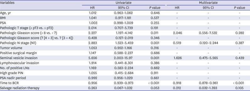
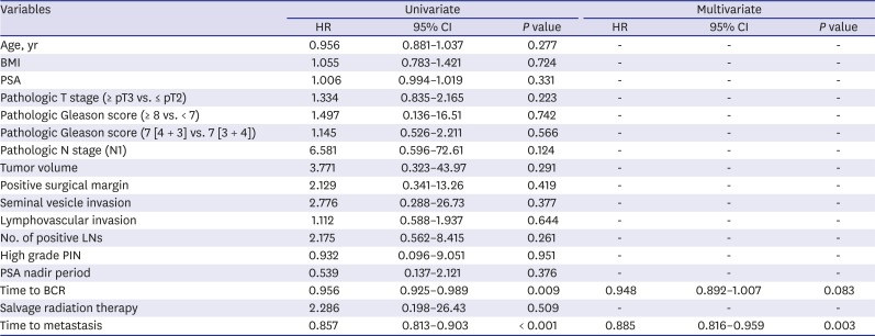
Table 4
Association of perioperative factors with the risk of metastasis among patients with high-risk PCa who achieved undetectable PSA levels following radical prostatectomy
PCa = prostate cancer, PSA = prostate-specific antigen, CI = confidence interval, HR = hazard ratio, BMI = body mass index, LN = lymph node, PIN = prostatic intraepithelial neoplasia, BCR = biochemical recurrence.
![]()
Table 5
Association of perioperative factors with the risk of CSM among patients with high-risk PCa who achieved undetectable PSA levels following radical prostatectomy
CSM = cancer-specific mortality, PCa = prostate cancer, PSA = prostate-specific antigen, HR = hazard ratio, CI = confidence interval, BMI = body mass index, LN = lymph node, PIN = prostatic intraepithelial neoplasia, BCR = biochemical recurrence.
![]()
Multivariate Cox-regression analyses revealed longer time to BCR as an independent predictor of a lower risk of metastasis (HR, 0.918; 95% CI, 0.878–0.961; P < 0.001) (Table 4), while a longer time to metastasis was revealed to be an independent predictor of a lower risk of CSM (HR, 0.885; 95% CI, 0.816–0.959; P = 0.003) (Table 5).
Go to :
DISCUSSION
RP is one of the most commonly used treatments for patients with localized PCa and has been proven to provide favorable prognosis in a subset of high-risk patients.161718 In the present study, we confirmed that RP may confer undetectable PSA levels postoperatively, and, consequently, a chance for cure in a substantial proportion of patients. Pompe et al.19 examined oncologic outcome of high-risk or very high-risk patients who underwent RP. The authors reported that despite the relatively poor prognosis of patients with high-risk PCa, RP results in favorable 5 and 8 years MFS, CSM-free survival and OS rates. At the same time, our data suggest that RP alone may not be sufficient for local control of PCa, and that maximal oncological control is rather achieved by timely administration of multidisciplinary adjuvant treatment, even beyond 5 years postoperatively. Amling et al.20 suggested patients undergoing RP should be subjected to long-term follow-up to allow the option of early intervention should progression occur due to a significant number of patients, including those with organ confined cancers which may exhibit disease progression even after 5 years.
We observed that 73.9% of patients achieved undetectable PSA levels postoperatively, which adds to the literature regarding the feasibility of RP for the treatment of high-risk disease. Specifically, RP provides several distinct advantages over ADT or RT. First, accurate pathological staging following RP enables timely administration of adjuvant therapy, at the same time avoiding morbidities associated with unnecessary adjuvant treatment.21 Recent studies have shown that up to 35% of patients are inaccurately staged, and up to 50% of patients originally identified to have high-risk disease demonstrate lower-risk disease on final pathological review.1122 Second, RP may provide superior local cancer control compared to that allowed by other treatment modalities. Boorjian et al.16 evaluated the long-term survival outcomes of high-risk PCa treated with RP or external beam RT with or without adjuvant ADT, and observed that patients treated with RT had a significantly increased risk of all-cause mortality compared to that noted among patients who received RP. Third, RP offers better health-related quality of life (HRQoL). Indeed, a long-term assessment of HRQoL in men receiving RT and brachytherapy showed that their prostate-specific HRQoL scores continued to decline, whereas those of RP patients remained relatively stable or gradually improved.23 Fourth, men undergoing RP are less likely to require ADT, and have significantly longer ADT-free survival compared to the values noted in patients undergoing external beam RT.5 Lastly, primary tumors have been suggested to play a significant role in tumor shedding and cytokine/growth factor production. In this respect, the advantage of RP is that it provides definitive tumor debulking, which may improve overall outcomes.24
In our study, patients were stratified according to the time to BCR, dichotomized at 60 months. The NCCN guideline recommends closed follow-up for up to 5 years, followed by annual follow-up for subsequent years.15 Our results indicate that BCR may occur even after 60 months in high-risk patients. Therefore, we investigated the subgroup of patients who would benefit from observation beyond 5 years. We found a marginal difference between the two groups regarding pathologic nodal status. However, there were no differences in pathologic features well known to affect oncological outcomes, namely tumor stage, tumor grade, and positive surgical margin status. In particular, there was no significant difference in pathologic Gleason score between 7 (4 + 3) and 7 (3 + 4). However, we did find differences in MFS and OS rates according to time to BCR. Specifically, patients who exhibited BCR within < 60 months after surgery had significantly lower MFS and OS rates. Briganti et al.13 evaluated the role of time to BCR in CSM and observed that patients who experienced BCR within 3 years from surgery had significantly higher CSM rates than those noted among patients who developed BCR at a later time. Freedland et al.14 also found that the time to BCR following surgery was significantly associated with CSM, and that stratifying patients according to BCR dichotomized at 3 years after surgery provided the best risk stratification approach. The difference between the cutoff period reported by Freedland et al.14 (3 years) and that observed in our study (60 months, i.e., 5 years) may be presumed to arise from the difference in the study cohorts, and specifically because we only included patients who achieved undetectable PSA levels following RP. We also noted that the time to metastasis was a significant prognosticator of the risk of CSM, in accordance with the observations of Pound et al.25, who reported that, after the development of metastatic disease, the actuarial median time until death due to PCa was 5 years, and that time to metastasis was an important determinant of CSM.
Our findings have important clinical implications. In our study, 41% of patients experienced BCR later than 5 years following RARP. The NCCN guideline recommends that PSA levels should be monitored every 6 to 12 months until 5 years postoperatively, and annually thereafter.15 Our observation that a substantial proportion of patients are likely to experience BCR later than 5 years after surgery suggests that the monitoring interval beyond 5 years postoperatively should be shortened for high-risk PCa patients even if PSA levels remain undetectable beyond this period, in order to enable early detection of disease progression and timely administration of multidisciplinary adjuvant treatment. Finally, our findings indicate that the time from RP to BCR is a prognosticator for MFS and OS and may therefore be utilized as a proxy for decision making regarding the length of monitoring intervals (in months).
Our study has several limitations. First, its retrospective nature precludes exclusion of potential selection bias regarding the indication for surgical treatment, which was made at the discretion of the treating surgeon. Second, there was discrepancy regarding the monitoring intervals, adjuvant treatment protocol, and treatment duration. Third, patients included in our study received RARP performed by different surgeons, albeit at the same institution, which may have affected the time to BCR and postoperative outcomes such as positive surgical margin or pathologic nodal status. Indeed, Klein et al.26 reported that the surgeon's experience, independent of surgical volume, affects BCR. However, we consider this aspect as an inherent bias of data collected in the clinical setting, which, in turn, may reflect everyday clinical practice in general. Finally, the median follow-up period was 75.9 months, which is relatively short to draw definitive conclusions regarding MFS or CSS.
In conclusion, a substantial proportion of high-risk PCa patients may achieve undetectable PSA levels following RP. Nevertheless, the risk of BCR and metastasis is relatively high. We confirmed that time to BCR and metastasis were direct or indirect prognosticators of survival rates, and may therefore, be utilized as a proxy for decision making regarding monitoring intervals. Considering that a substantial proportion of patients with postoperatively undetectable PSA are likely to experience BCR later than 60 months after surgery, close monitoring even beyond this period is warranted to enable early detection of disease progression and timely administration of multidisciplinary adjuvant treatment.
Go to :
 XML Download