

 PDF
PDF Citation
 Print
Print


INTRODUCTION
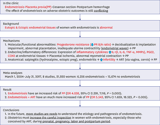
Placenta previa (PP) is one of the diseases in which the placenta covers the internal os of the cervix and causes obstetric hemorrhagic complications during pregnancy and cesarean section.1 The incidence of this disease is about 0.3%–0.5%, and it is an important disease because it increases maternal and perinatal morbidity and mortality from massive bleeding.2 The risk factors for PP include a previous history of cesarean section, increasing maternal age, multiple pregnancy, and smoking, and recent reports have indicated that assisted reproductive technology (ART) is also a cause.345 However, the pathogenesis of PP has not been clearly understood until recently. Most cases of PP should be delivered by caesarean section, not by vaginal delivery. Endometriosis is a chronic reproductive disease characterized by the presence of endometrial glands outside the uterus, mainly on the ovaries and peritoneum. The prevalence of endometriosis in women of reproductive age is about 6%–10%.6 Endometriosis causes two main symptoms, pain and infertility. It is well accepted that endometrium of women with endometriosis is abnormal. Additionally, these abnormal endometrium may be the causes of decidual impairment and abnormal placentation during pregnancy.7 Endometriosis has been linked with increased peritoneal inflammation with a higher concentration of cytokines, angiogenic factors, and growth factors. Other harmful factors include poorer oocyte quality, progesterone receptor resistance, molecular and functional abnormalities in eutopic endometrium, and anatomical distortion of uterine posterior walls, fallopian tubes and ovaries in women with endometriosis.89 So, all of these processes may have negative impacts on pregnancy outcomes and fertility. However, studies on the relationship between PP and endometriosis have led to conflicting results, with some studies reporting a significantly increased risk of PP,1011121314 and others showing no statistical significance, though in small sample sizes.15 Many women with endometriosis tend to have conceived with ART, though it is unclear whether the causes of the increase in PP are due to endometriosis, ART, or both.1617 The objective of our study was to examine whether pregnant women with endometriosis are associated with an increased risk of PP.
METHODS
We developed a search strategy to use Medical Subject Heading (MeSH) terms and free key words and text words related to “endometriosis”, “pregnancy”, “adverse pregnancy outcomes”, “bad pregnancy outcomes”, “placenta previa”, “ART”, “in vitro fertilization (IVF)”, "intracytoplasmic sperm injection (ICSI)”, and “placental problems”.
We searched these terms using the PubMed MEDLINE database, the Korea education and research information service (KERIS), Scopus, and Google Scholar from March 1, 2004 through July 31, 2017 without language restrictions. The inclusion studies included a prospective cohort study, a retrospective cohort study, a retrospective case control study, a large national population-based cohort study, a retrospective secondary analysis, and double blinded, multicentric, observational and cohort studies, and a placebo-controlled, randomized clinical trial on the relations between endometriosis and pregnancy prognosis, while publications in abstract form alone were excluded. Data abstraction was completed by two independent investigators. Each investigator independently abstracted data from each study and analyzed these data separately.
Inclusion and exclusion criteria
The endometriosis group had pelvic endometriosis confirmed histologically and visually during the surgical procedure, or in two or more repetitive ultrasound examinations or magnetic resonance imaging (MRI) scans before pregnancy or after the operation. All patients who were clinically suspected to have pelvic endometriosis but with an absence of imaging or surgical confirmation were excluded from the endometriosis group. In this study, ART that was conducted by conventional IVF or ICSI was included. The gamete intrafallopian transfer (GIFT) women were excluded. The unexposed group included women who did not have a previous surgical or clinical diagnosis of endometriosis, and who did not have any ultrasonographic signs of endometriosis. Women with malignancies, autoimmune diseases, endocrine diseases, and cardiovascular diseases were also excluded. Donor oocyte and embryo recipients were excluded. Miscarriages and terminations of pregnancy at < 20 weeks gestation were not registered as births. Late termination ≥ 20 weeks gestation and neonatal death (within 28 days of birth) were included.
Definitions
PP is defined as placental tissue completely obstructing internal os or within 20 mm. PP was confirmed at delivery. Confirmation of the diagnosis of PP was based on transvaginal ultrasonography with empty bladder performed prior to delivery. If the PP was not confirmed prior to delivery and there was a sudden unexpected birth, the diagnosis was confirmed at the time of delivery.
Statistical analysis
Differences were reviewed, and further resolved by a common review of the entire data set. Data abstracted included the number of study patients, the number of patients in endometriosis groups and control groups, and adverse pregnancy outcomes. When possible, authors of included trials were contacted for missing data. The risk of bias in each included study was assessed using the criteria outlined in the Risk of Bias AssessmentTool for Non-randomized studies (RoBANS). The following seven domains related to risk of bias were assessed in each included trial since there is evidence that these issues are associated with biased estimates of treatment effect: 1) random sequence generation, 2) allocation concealment, 3) blinding of participants and personnel, 4) blinding of outcome assessment, 5) incomplete outcome date, 6) selective reporting, and 7) other bias. The judgments of review authors were categorized as having a low, high, or unclear risk of bias.
Meta-analyses were performed with random effects models according to the heterogeneity of studies and we attempted to estimate the mean of a distribution of true effects. The completed analyses were then compared, and any differences were resolved with a review of the entire data set and independent analysis. Statistical heterogeneity between the studies was assessed using I2 statistics. Heterogeneity was considered to be statistically significant if the I2 value was more than 50%. Publication bias was evaluated by inspection of the funnel plot, Duval and Tweedie's trim and fill, and Begg and Mazumdar's rank correlation. Kendall's tau value and Q value were adjusted to see a bias in publications. All effect sizes were calculated through Comprehensive Meta-analysis (CMA) ver.2.0 (Biostat Inc., Englewood, NJ, USA). Meta-analyses were estimated with odds ratio (OR) and 95% confidence interval (CI) using random effect analysis according to the heterogeneity of studies. A P value less than 0.05 was considered to be statistically significant. The meta-analysis was performed following the Preferred Reporting Items for Systematic Reviews and Meta-analyses (PRISMA) statement.
RESULTS
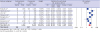
Fig. 1 shows a flow diagram of how we identified relevant studies. A total of 246 articles were identified by searching the PubMed, the Embase, the Cochrane Library, and the KERIS. We excluded 45 duplicated articles and an additional 182 articles that did not satisfy the selection criteria. The full texts of the remaining 19 articles were reviewed, and 11 additional articles were excluded for reasons shown in Fig. 1. The remaining 8 articles were included in the final analysis. The summary of characteristics of the enrolled studies included in these meta-analyses are in order. All of their RoBANS were a low and unclear risk of bias, and they were evaluated as good studies in the assessment of risk of bias (Table 1).
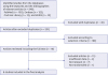 | Fig. 1Flow diagram of identification of relevant studies.KERIS = Korea Education and Research Information Service.
|
Table 1
Summary of characteristics of enrolled studies included meta-analyses
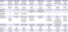
| Descriptions | Healy et al.10 | Takemura et al.11 | Lin et al.12 | Tatsuya et al.13 | Benaglia et al.14 | Li et al.15 | Saraswat et al.16 | Berlac et al.17 |
|---|---|---|---|---|---|---|---|---|
| Study design | Retrospective cohort study | Retrospective cohort study | Retrospective cohort study | Retrospective cohort study | Retrospective case control study | Retrospective cohort study | National population based cohort study | National cohort study |
| Type of conceivement | 1,265 ART+En+, 5,465 ART+En− | 44 ART+En+, 305 ART+En− | 249 non-ART+En+, 249 non-ART+En− | 92 ART+En+, 512 ART+En− | 239 ART+En+, 239 ART+En− | 1 ART+En+, 74 non-ART+En+, 300 non ART+En− | ART not distinguished | ART not distinguished |
| Site, year | Australia, 1991–2004 | Japan, 2004–2008 | China, 1995–2013 | Japan, 2000–2014 | Italy, 2008–2014 | China, 2011–2013 | Scotland, 1981–2010 | Denmark, 1997–2014 |
| Endometriosis surgery before pregnancy (type & time) | NA | 44 (type & time: NA) | 249 (type & time: NA) | 92 (type: adhesiotomy, coagulation, cystectomy, time: NA) | 186 (78%) (type: cystectomy, removal of deep peritoneal nodules, time: NA) | 75 (type & time: NA) | 4,232 (type & time: NA) | 60 (type & time: NA) |
| Percentage of previous cesarean section (%) | NA | 0% ART+En+, 2.6% ART+En− | 0% non-ART+En+, 0% non-ART+ En− | NA | NA | NA | NA | NA |
| Percentage of nulliparity (%) | NA | 100% ART+En+, 97.4% ART+En− | 100% non-ART+En+, 100% non-ART+ En− | 84.8% ART+En+, 77.6% ART+En− | 90% ART+En+, 84% ART+En− | 100% En+, 58% En− | 72% En+, 52% En− | 67% En+, 54% En− |
| Primary outcomes | Obstetric hemorrhage (APH, PP, PA, PPH) | PP | Adverse obstetric outcomes (preterm labor, PP, cesarean section, PIH, SGA) | Adverse obstetric outcomes (preterm labor, PP, SGA) | Preterm birth | Adverse obstetric outcomes (PPH, preterm labor, PP, SGA, GDM) | Adverse obstetric outcomes (miscarriage,ectopic pregnancy, stillbirths, PIH, APH, PPH, preterm labor, cesarean section) | The risk of obstetrical & neonatal complications |
| Diagnostic methods of endometriosis | NA | 44 Histopathology on surgery | 249 Histologically & visually on surgery | 92 Histopathology on laparoscopic surgery | 186 Histopathology on surgery, 53 transvaginal sonography | 75 Histopathology on laparoscopic surgery | 4,232 Surgically (laparoscopy or laparotomy)confirmed | 60 Histopathology on surgery |
| Sample size (No.) (En+/En−) | 6,730 (1,265/5465) | 349 (44/305) | 498 (249/249) | 604 (92/512) | 478 (239/239) | 375 (75/300) | 10,939 (4,232/6,707) | 1,957 (60/1,897) |
| Adjusted RR/OR (95% CI) | 1.700 (1.220–2.404) | 13.347 (3.939–45.224) | 4.517 (1.233–16.544) | 12.095 (3.563–41.061) | 4.800 (1.369–16.824) | 1.616 (0.307–8.499) | 2.240 (1.518–3.306) | 5.700 (4.395–7.392) |
| Risk of bias | Unclear | Low | Low | Low | Low | Low | Unclear | Unclear |
NA = not available, No = number of patients, En+ = endometriosis group, En− = control group, RR = risk ratio, OR = odds ratio, CI = confidence interval, ART = assisted reproductive technology, APH = antepartum hemorrhage, PP = placenta previa, PA = placental abruption, PPH = postpartum hemorrhage, PIH = pregnancy induced hypertension, SGA = small for gestational age, GDM = gestational diabetes mellitus.
![]()
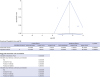
Meta-analyses were performed with random effects models according to the heterogeneity of eight studies (endometriosis) as we attempted to estimate the mean of a distribution of true effects. According to Kendall's tau without continuity correction, the tau was −0.214, the Z value for tau was 0.742, the P value (1-tailed) was 0.228, and the P value (2-tailed) was 0.457. Also, as a result of Duval and Tweedie's trim and fill, the point estimate was shown, and the Q value was 45.053. It was difficult to see if there was a bias in publications because the adjusted values and observed values of the studies were the same. Figure of the funnel plot for assessing publication bias for the risk of PP and endometriosis (endometriosis) is shown; it looks apparent that the funnel plot is symmetric (Fig. 2). Data from eight effect sizes from eight studies involving 21,930 patients were enrolled. These meta-analyses showed that women with endometriosis (endometriosis) have an increased risk of PP (OR, 4.038; 95% CI, 2.291–7.116; P = 0.000, Fig. 3).
 | Fig. 2Funnel plot for assessing publication bias for the PP risk in women with endometriosis (endometriosis).PP = placenta previa.
|
 | Fig. 3Forest plot of eight studies. The result of the PP risk in women with endometriosis (endometriosis).PP = placenta previa, OR = odds ratioo, CI = confidence interval.
|
Meta-analyses were performed with random effects models according to the heterogeneity of four studies (endometriosis + ART) as we attempted to estimate the mean of a distribution of true effects. According to Kendall's tau without continuity correction, the tau was 0.000, the Z value for tau was 0.000, the P value (1-tailed) was 0.500, and the P value (2-tailed) was 1.000. Also, as a result of Duval and Tweedie's trim and fill, point estimate was shown, and the Q value was 19.282. Figure showed funnel plot for assessing publication bias for the risk of PP conceived by ART (endometriosis + ART); it looked apparent that the funnel plot was also symmetric (Fig. 4). It was difficult to see if there was a bias in publications. Data from four effect sizes from four studies involving 8,161 patients who conceived with ART were included. These results showed that women with endometriosis who conceived with ART (endometriosis+ ART) have a substantially increased risk of PP (OR, 5.543; 95% CI, 1.659– 18.523; P = 0.005, Fig. 5).
DISCUSSION
Endometriosis is a common and painful disease affecting women of reproductive age. While the underlying pathophysiology is still largely unknown, much advancement has been made in understanding the progression of the disease. A recent meta-analyses by Kim et al.18 demonstrated that primiparous singleton women with endometriosis at pregnancy have an increased risk of preterm birth (OR, 1.473; 95% CI, 1.216–1.785). In a study comparing the complications of pregnancy and delivery in pregnant women with endometriosis, it is important to distinguish between whether an accurate laparoscopic diagnosis was given before or after pregnancy and whether it was pregnancy through ART such as IVF/ET or ICSI. A retrospective analysis by Jeon et al.19 found that complete cul-de-sac obliteration is the independent factor of spontaneous pregnancy failure in women with endometriosis following laparoscopic surgery. In the process of embryo implantation in the endometriosis, inadequate uterine contractility called ‘endometrial waves’ observed in the sub-endometrial layer are thought to be the causes of PP.20 Population studies have found annual incidence rates of endometriosis ranging from 0.1–0.3.21 This reflects an increase in the diagnosis of endometriosis. According to the study on progesterone resistance in endometriosis conducted by Chae et al.22 in 2016, it may be caused by proinflammatory conditions in the pelvic peritoneal microenvironment. The biochemical materials associated in implantation include local and systemic inflammatory cytokines such as interleukin-6, interleukin-1ß, and tumor necrosis factor-α. Additionally, the levels of prostaglandin E2, cyclooxygenase (COX)-2 and various cytokines are highly elevated in endometriotic tissue relative to normal endometrium. The hypermethylation of progesterone receptor (PR)-B promotor and PR isoform A are also more expressed than PR-B as well as in endometriosis.23 According to a retrospective analysis by Yang et al.,24 endometriosis itself has worse IVF parameters regardless of previous cyst enucleation. In 2012, Vercellini et al.25 retrospectively assessed pregnancy outcomes in 419 women who achieved a first spontaneous singleton pregnancy following surgery for endometriosis, and stratified the results obtained by endometriosis localization. In this study, an almost six-fold increase in risk of PP has been found in women with rectovaginal endometriosis compared to all women with ovarian and peritoneal lesions (OR, 5.81; 95% CI, 1.53–22.03). Because a fixed abnormal anatomical uterine position owing to dense pelvic adhesions may theoretically reduce the efficacy of myometrial contraction, particularly in women with rectovaginal endometriosis, the risk of PP increases. In a small case–control study involving women with and without endometriosis matched for parity and ART procedures conducted by Kortelahti et al.,26 no difference in PP incidence was found. However, the study likely had a very limited statistical power, as the number of placental abnormalities reported was very low. Conversely, two retrospective cohort studies conducted by Healy et al.10 and Kuivasaari-Pirinen et al.27 comparing the pregnancy outcomes of ART singleton pregnancies with those of natural pregnancies found higher rates of PP in the ART groups and in the various subgroups. The limitation of Healy et al.'s study10 was no information of previous section history, percentage of nulliparity, and diagnostic methods of endometriosis. Although the study was conducted on only ART pregnancies and may cause bias in the final statistics, it was included in this analysis due to a large number of samples. Kuivasaari-Pirinen et al.27 showed that mothers with endometriosis and those in the male factor infertility group have a greater incidence of PP than the reference group. One of the questions of this study is why ART increases the risk of PP. This study is consistent with other studies in that PP is increased in ART pregnancies. The underlying mechanism for this effect is not clear. In assisted fertilization, drugs are utilized to induce multiple follicular development. Fertilization and embryo development take place outside the body, and embryos enter the uterine cavity through the cervix and vagina by mechanical means. With ART, embryos are placed in the uterine cavity by the transcervical route using a catheter. This procedure may induce uterine contraction, possibly due to the release of prostaglandins after mechanical stimulation of the internal cervical os. It is conceivable that these mechanically induced abnormal uterine contractions could lead to higher frequencies of implantation in the lower uterine segment and thereby increase the risk of PP. And, the normal frequency and amplitude of uterine contractions are altered in women with endometriosis, which may cause abnormal uterine dysperistalsis in endometriotic tissue leading to abnormal embryo implantation resulting in PP.28 These comorbidities in women with endometriosis who conceived with ART may increase the incidence of PP, not by ART itself.
In 2016, Tatsuya et al.13 tried to exclude the negative effects of ART on pregnancy by studying only women who conceived with ART. They also retrospectively investigated whether the severity of endometriosis was associated with increased complications of pregnancy conceived with ARTs. Endometriosis was classified according to revised American Society for Reproductive Medicine (rASRM) staging.29 They found that the frequencies of PP was significantly increased in women with laparoscopically diagnosed endometriosis (OR, 12.1; 95% CI, 3.6–41.1).13 The association between ovarian or rectovaginal endometriosis and the increased risks of PP involves the relationship between the severity of endometriosis and increased complications of pregnancy. The prevalence of bad pregnancy outcomes in the first pregnancy has been reported to increase compared to the next pregnancy, so this study has a strength in that it examined only nulliparous women.30
We selected studies of women who underwent pre-pregnancy surgery, except for the study of Healy and others in 2010. The study of Healy et al did not show how endometriosis was diagnosed. And, while the study of Benaglia et al.14 in 2016 found that only 78% of women had surgery before conception, the other six had surgery all before pregnancy. And the percentage of nulliparity was from 52% to 100%. The diagnostic methods of endometriosis were histology except for 53 transvaginal sonography in the study of Benaglia et al.14 in 2016. Especially, both multiparity and previous cesarean section history have been known as important risk factors of PP. But, most of the studies included in our meta-analysis did not show previous C-section rates. It is assumed that it will be similar to the percentage of nulliparity. These factors may cause risk of bias in our analysis.
We would like to highlight the important findings and the significance of our analyses. The first strength of our study was that we analyzed the most recent studies available. The second strength of our study was that we performed the meta-analyses by separating the ART plus endometriosis group, taking into account that ART may affect PP development. The limitations of our study are that seven out of the eight studies used in the analysis indicated that endometriosis increased the incidence of PP, and the only other study was not significant. Because it is almost certain that PP increases in women with endometriosis, these analyses focused on how much it increases. The “unexposed” cohort can include some women with endometriosis undiagnosed by surgery or through imaging methods. These inclusion criteria thus tend to underestimate the risk of complications in women with endometriosis. Future research should focus on the use of fresh embryos and FETs and the type of infertility (tubal, ovulatory disorder, and male infertility) used in ART, as well as endometriosis localization (rectovaginal lesions, ovarian endometriomas, and peritoneal implants) and comparisons of the severity of endometriosis (classification of as rASRM stage I, II, III, and IV), followed by correction of maternal age and the study period, which will lead to better studies.
In conclusion, our study demonstrates that women with endometriosis have an increased risk of PP. We must increase international effort to understand the etiology and pathogenesis of endometriosis. Also, obstetrics should try to reduce complications in women with endometriosis from the time of conception to the postpartum period.
 XML Download