

 PDF
PDF ePub
ePub Citation
 Print
Print


Degenerative lumbar foraminal stenosis is a common cause of lumbar radiculopathy, accounting for approximately 8%–11% of lumbar degenerative diseases requiring surgical procedures.1) The exiting root is gradually subjected to compression by osseous hypertrophy and ligamentous structures around the canal.23) Currently, two major surgical treatment options are available for this disease: decompression with fusion and simple decompression. Conventional surgical methods for foraminal stenosis are currently divided into total facetectomy with lumbar fusion and microscopic foraminotomy preserving facet.456) Microscopic decompression surgery preserving the facet joints was introduced by Wiltse and Spencer7) and has been developed by several authors. This approach allows foraminal decompression while minimally violating the foraminal area.4) Foraminotomy via the Wiltse approach is considered a gold standard for stenosis of the foraminal or extraforaminal area, and the success rate is reported to be approximately 80%.8910) However, the Wiltse approach may lead to incomplete surgery due to limited visualization. Some studies have reported unfavorable results, including postoperative neurologic symptoms and complications using such technique.891011) Recently, studies on spinal surgery using the unilateral biportal endoscopic technique have been reported by several authors.121314) To date, few studies reported the results of over a year of follow-up after farlateral decompression using unilateral biportal endoscopic technique in foraminal stenosis. This study was to evaluate the clinical and radiological outcomes of far-lateral decompression using unilateral biportal technique.
METHODS
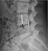
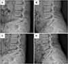
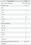
We conducted this study in compliance with the principles of the Declaration of Helsinki. The study was a retrospective medical chart review with approval by the Institutional Review Board of Andong Hospital (IRB No. 2018-004). All patients who underwent unilateral biportal endoscopic far-lateral decompression (UBEFLD) for lumbar foraminal stenosis provided a signed informed consent forms before the surgery. All surgeries were performed by a single surgeon (JEK). Clinical outcomes, including Oswestry disability index (ODI), modified Macnab criteria, visual analogue scale (VAS), and duration of surgery, and complication rate were analyzed in patients who were treated with UBEFLD. Patient demographic data were obtained by chart review and patient-based clinical outcome questionnaires were collected in the outpatient clinic. We compared and analyzed the radiographs of patients preoperatively and 1 year postoperatively. Plain radiographs obtained in flexion and extension posture preoperatively and postoperatively were compared and analyzed to confirm disc height index (DHI), foraminal height index (FHI),15) percent of slip, and intervertebral angle (IVA) (Fig. 1). We evaluated 31 patients diagnosed with lumbar spinal foraminal stenosis and treated in our institution with UBEFLD. Patients with moderate or severe foraminal stenosis were included, and they were all unresponsive to conservative treatment for over 6 weeks, which required surgery. Detailed diagnoses are descripted in Table 1.
Statistical Analysis
All statistical analyses were performed using the SPSS ver. 18.0 (SPSS Inc., Chicago, IL, USA). Values are presented as mean and standard deviation. Patient data were analyzed using the paired t-test. The p < 0.05 was considered statistically significant.
Operational Technique
Basic setup
Basic spine surgery instruments, 0° and 30° angled 4-mm diameter endoscopes (Arthrex, Naples, FL, USA), commonly used in joint arthroscopic surgery, a radiofrequency catheter, a 4.2-mm diameter arthroscopic burr, and a shaver, were used during the surgery (Fig. 2).
Surgical approach to the foraminal area
Two portals were created to perform this surgery. Water was infused through the endoscope through the viewing portal, and the working portal had an additional purpose as a portal for water outflow. The proximal and distal portals were created 2 cm lateral from the pedicle level on the C-arm anteroposterior image. Each incision for the portals were 0.8 cm in length, which was adequate for instrument and endoscope insertion. For the left side foramen, the proximal and distal portals were used as the viewing and working portals, respectively, and vice versa for the right foramen. After the endoscope insertion through the viewing portal, we secured a space for the lower transverse process around the lateral surface of the facet joint. A radiofrequency catheter or a shaver was used to secure the space, and a radiofrequency catheter was used to control active bleeding.
Decompression of foraminal stenosis
After a sufficient working space was obtained, the cranial 50% of the superior articular process of the thickened facet joint was removed using an arthroscopic burr or an osteotome. After removing the superior articular process, the ligamentum flavum around the foramen was removed using a curette and a Kerrison punch. After completion of flavectomy, nerve root and epidural fat were identified. If herniated disc material was found preoperatively, additional discectomy was performed usually from the axilla of the root. Surgery was confirmed to be completed after achieving an amount of free space concordant with the diameter of the nerve root in the foraminal zone, and then a drain tube was inserted.
RESULTS
A total of 31 patients (14 males and 17 females) were enrolled in our institution. The mean age was 70.5 ± 8.9 years (range, 51 to 89 years). Exact estimated blood loss was not recordable due to continuous water irrigation throughout the procedure. The mean follow-up period was 14.8 ± 1.6 months. The mean duration of surgery for one level was 48.7 ± 13.9 minutes (Table 1).
Clinical Results
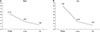
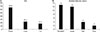
The mean VAS for the back improved from 5.13 ± 0.8 preoperatively to 2.61 ± 0.76 at 3 months postoperatively; it was 1.52 ± 1.02 (p < 0.01) at 1-year follow-up. The VAS for the leg improved from 7.87 ± 0.88 preoperatively to 2.55 ± 1.02 at 3 months postoperatively; it was 1.45 ± 1.28 (p < 0.01) at 1-year follow-up (Fig. 3). The mean ODI significantly improved from 66.81 ± 7.45 preoperatively to 24.14 ± 6.11 at 3 months postoperatively; it was 17.39 ± 1.20 (p < 0.01) at 1-year follow-up. Among the patients, 80% reported improvement based on the Macnab criteria; the recorded outcomes were excellent, good, fair, and poor in 13 patients (42%), 12 (39%), 4 (13%), and 2 (6%), respectively (Fig. 4). Complications such as dura tear or hematoma did not occur intraoperatively or postoperatively. One patient diagnosed with foraminal stenosis due to spondylolytic spondylolisthesis preoperatively experienced recurrence of symptom even after far-lateral decompression. She underwent transforaminal interbody fusion via the unilateral biportal endoscopic technique.
Radiological Results
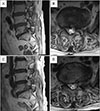
The IVA increased significantly from 6.24° ± 4.27° to 6.96° ± 3.58° at 1 year postoperatively (p = 0.306). The dynamic IVA slightly decreased from 6.27° ± 3.12° to 6.04° ± 2.41°, but the difference was not statistically significant (p = 0.375). The preoperative percentage of slip was 3.41% ± 5.24%, with a slip of 6.01% ± 1.43% at the 1-year follow-up (p = 0.227), which was not significantly changed. The preoperative dynamic percentage of slip was 2.90% ± 3.37%; it was 3.13% ± 4.11% (p = 0.720) at 1 year postoperatively, which did not show significant difference. The DHI changed from 34.78% ± 9.54% preoperatively to 35.05% ± 8.83% postoperatively, which was not statistically significant (p = 0.837). In addition, the FHI slightly decreased from 55.15% ± 9.45% preoperatively to 54.56% ± 9.86% postoperatively, but the results were not statistically significant (p = 0.705) (Table 2). Pre- and postoperative magnetic resonance images and radiological changes as well as intraoperative photographs of one of the patients are shown in Fig. 5.
DISCUSSION
Lumbar foraminal stenosis is a relatively common disease that accounts for approximately 8%–11% of degenerative lumbar spines.110) The surgical goal of treatment for symptomatic lumbar foraminal stenosis is alleviation of symptoms through proper neural decompression while preserving the original anatomy and biomechanics of the spine. Total facetectomy or lumbar fusion is known as a conventional treatment method.716) The success rate of open microforaminotomy has been reported to be 58%–80%.8910) Total facetectomy cannot eliminate the concern of instability, and adjacent segmental degeneration must be considered if decompression with fusion surgery is performed for foraminal stenosis.19) Moreover, muscle injury due to excessive dissection of the paraspinal muscle was reported to be related to muscle atrophy.17) Several studies have reported that a large dead space due to open spinal surgery increases the infection rate or contributes to scarring on neural structures.1819) The limitation of operation field in the paraspinal approach may cause incomplete decompression.2) Recent advances in optics and endoscopy devices have allowed better vision and more precise surgery, and good results of decompression surgery in foramen or extraforaminal stenosis using endoscopy have been reported. Recent developments in optical technology and instruments have enabled us to obtain delicate procedures and better operation fields. One portal endoscopy-based foraminal decompression was reported by several authors.220) The lengths of hospital stay and surgery were reported to be less than those in open foraminal decompression, and the success rate was reported to be 73%–100%.2)
The review conducted at 3 months and 1 year postoperatively showed significant improvement in clinical outcomes, including VAS and ODI. These results show that this technique is efficient in decompressing the exiting root. The result of our series was similar to data shown in other studies on conventional open foraminotomy and microscopic foraminotomy.81011162122) Our study demonstrated 80.6% of successful outcomes based on the modified Macnab criteria. Studies on conventional open foraminotomy demonstrated a success rate of 76.9%–80.6%.10162122) Though studies investigating microscopic foraminal decompression reported a success rate of approximately 83%, some involved statistically insignificant case numbers.4) Studies involving foraminal decompression using one portal endoscopic technique, including percutaneous endoscopic lumbar discectomy (PELD), also demonstrated a success rate of 73%–83.3%.23242526) Studies on foraminal decompression using the one portal technique have reported several neurologic complications, including foot drop postoperatively.23242526) Our cases showed no occurrence of complications postoperatively. In addition, several complications such as weakness, hematoma, and seroma were reported in the study of conventional open foraminal decompression, and duration of surgery was relatively longer (127–156 minutes) than that in our study (48.7 ± 13.9 minutes).
Few studies have reported the radiological change and postoperative instability after partial facetectomy. According to the study by Haufe and Mork,27) translation or sagittal rotation did not occur after endoscopic total facetectomy in severe foraminal stenosis because such surgery minimizes tissue damage and protects the ligamentous structure. Kiapour et al.28) reported in the biomechanical study that no instability occurred in graded partial facetectomy, except for total facetectomy. A recent study by Youn et al.29) about one portal endoscopic partial facetectomy supports this theory. All 25 patients who underwent uniportal endoscopic partial facetectomy did not show radiological progression of instability at 2 years of follow-up. No definite postoperative instability occurred or progressed in our study at the last follow-up, similar to the aforementioned study (Fig. 6). UBEFLD is performed by limited facet destruction and can avoid excessive resection of the bone under a magnified operation field. The absence of lumbar instability after surgery is speculated to be due to UBEFLD technique, since it is less invasive and minimizes destruction of posterior elements including facet joints based on several reports including our study. Our study confirmed that UBEFLD is a good surgical option to decompress the stenosis while preserving the intrinsic stability.
The UBEFLD could reduce surgery-related complications, including neurologic symptoms, especially problems with open surgery, but it is as effective as the conventional open or microscopic foraminotomy in relieving neurological symptoms and improving patients' quality of life. There are several factors to account for such positive results. First, triangulation technique with endoscopy was used, and thus accessing the lesion from various angles was possible, enabling complete decompression of the exiting nerve root. Second, muscle damage was minimal. Unlike in open surgery, there are few potential risks of muscle injury because the procedure was performed percutaneously with a small incision. Third, decompression was performed under a magnified arthroscopic field, reducing excessive facet injuries and exiting nerve injury.
UBEFLD is slightly different from one portal endoscopic foraminal decompression. First, it can reduce exiting nerve injury because a working cannula, which is used during one portal endoscopic foraminal decompression, is not necessary. Unlike one portal endoscopic technique, the floating-type biportal endoscopic technique does not need cannula insertion and allows entry of the various surgical instruments through a separate portal from the endoscope. Thus, it allows wider working angle compared to one portal technique. Second, the approach to far-lateral stenosis, caused by enlarged superior facet, is limited by one portal technique, whereas the biportal arthroscopic technique can simply solve it by using an osteotome, which is not an easy procedure in one portal technique, due to difficult insertion of surgical instruments. Third, to achieve successful decompression, the superior articular process in the foramen must be completely removed until the ligamentum flavum is exposed, and the exiting root must be fully decompressed from the entrance of the foramen to the extraforaminal area. In particular, accessing the L5–S1 foramen by open microscopic or one portal technique is difficult due to the iliac crest. However, in unilateral biportal endoscopic decompression, a wide area can be accessed through triangulation by switching the proximal and distal portals as viewing and working portals. Finally, the C-arm is introduced only during level confirmation intraoperatively; thus, radiation exposure is relatively lower than open surgery. Recent studies have reported that PELD using one portal endoscopy has a higher mean radiating dose than minimally invasive open discectomy.30) UBEFLD is assumed to be associated with lesser radiation exposure than PELD, as there is no chance of radiating exposure after level confirmation, similar to minimally invasive open discectomy.
There are a few limitation of this study. Firstly, it is a single-group study with a relatively small number of patients and no control group. The follow-up period is short because the surgical technique was introduced very recently. We are currently constantly following up on patients that underwent UBEFLD and will include more patients who fulfill the inclusion criteria.
In conclusion, UBEFLD is an effective minimally invasive surgical technique that produces good results without causing postoperative spinal instability and neurologic complications of the lumbar exiting root. It could be a minimally invasive alternative method that can effectively decompress foraminal stenosis.


 XML Download