

 PDF
PDF Citation
 Print
Print


INTRODUCTION
The coordinated activities of the neurons, as measured in an electroencephalogram (EEG), are synchronized with various cognitive states, such as wakefulness, perception, attention, and memory.12 In clinics, the EEG is used to diagnose epilepsy, observe sleep patterns, and estimate the depth of anesthesia.345 In addition to the cognitive and clinical studies, EEG recording capability has been incorporated into consumer devices for daily activities,6 and is used in brain-computer interface applications due to its non-invasiveness and high temporal resolution.78 For EEG recordings in daily life, increasing the quality of the EEG signal is important, as is improving the convenience of measuring the signal. In particular, the skin-electrode impedance significantly influences the signal-to-noise ratio of the measured signal.910 At the same time, the electrode should be unobtrusive and easy to install for daily use, otherwise the applications will be limited to clinical settings.11
The adoption of the electrode suitable for recording EEG in daily life facilitates the out-of-the laboratory EEG recoding. In laboratories and clinics, a wet silver-silver chloride electrode is a commonly used biopotential recording electrode due to its reliability and robustness.1213 The electrode adheres well to the skin and enables the stable acquisition of signals with relatively few motion artifacts.14 On the other hand, the use of an electrolytic paste is uncomfortable for the user, time consuming to install, and hinders self-installation,6 which makes the wet electrode unfeasible for daily life EEG recording applications. On the contrary, dry electrodes use no electrolytic gel or paste to mediate the electrode and skin interface.12 The use of a dry electrode is convenient for users, because it does not require the tedious processes of applying the paste and then cleaning the residues after the recording.15 However, dry electrodes show a high skin-electrode impedance due to the capacitive coupling with the skin and the poor adherence to the skin. These characteristics make the dry electrode more prone to motion artifacts. For these reasons, it is more challenging to use a dry electrode than a wet electrode on ambulatory patients or freely moving users.16 The need for improved performance and comfort led recent studies to develop electrodes with different characteristics, including a flexible thin film electrode using micro-robot and replica molding techniques,17 carbon nanotube electrodes,9 and micromachined spiked electrodes.15
A nanoporous platinum electrode, namely L2 phase electro-deposited nanoporous platinum (L2-ePt), has been previously reported as a way to increase the effective surface area of the electrode to facilitate electrochemical reactions for glucose oxidation, O2 reduction, and H2O2 reduction in an electrochemical glucose sensor.18 The L2-ePt electrode has a three-dimensional nanoporous surface, and is fabricated by mixing a reverse micelle (L2) solution and platinum nanoparticles. Park et al.19 showed that the surface roughness can be controlled through varying the amount of charge used during electroplating. The nanoporous surface structure of the L2-ePt increases the effective surface area, which subsequently reduces the impedance of the electrode. Furthermore, the L2-ePt electrode was utilized in neural cell stimulation and extracellular potential recording applications due to its large effective surface area, biocompatibility, and highly controllable fabrication characteristics.1820 With the above-mentioned properties suitable for recording biosignals, however, no study has used the L2-ePt electrode in EEG recording applications.
The current study proposes the L2-ePt electrode as a new electrode for conveniently capturing high-quality EEG signals. The fabricated L2-ePt electrodes were compared to commonly used clinical electrodes, such as gold cup electrodes and flat platinum (FlatPt) electrodes. Initially, the increased effective surface area of the fabricated L2-ePt electrode was quantitatively confirmed through an electrochemical technique called cyclic voltammetry, and the effect of surface roughness was further observed through the electrode impedances of the L2-ePt electrodes in a saline solution. Then, the electrode impedance in the saline solution was compared with that for the FlatPt electrodes. Finally, the skin-electrode impedance and EEG signals, including the alpha rhythm, were acquired from the head using the L2-ePt electrode, and captured signals were compared to the simultaneously recorded EEG signals that were captured using a gold cup electrode and a FlatPt electrode.
METHODS
Fabrication
An electrochemical analyzer (Model CH660; CH Instruments Inc., Bee Cave, TX, USA) was used for bulk electrolysis (the electro-deposition process) and cyclic voltammetry (electrode cleansing and surface area calculations). During the cyclic voltammetry procedures, an Ag/AgCl (3 M KCl) electrode and a platinum wire were used as a reference and counter electrode, respectively. The active electrode was FlatPt foil for the electro-deposition process, and nanoporous platinum electrodes during the cyclic voltammetry procedures.
According to previous research on the fabrication of L2-ePt electrodes, nanoporous platinum layers were electrochemically deposited onto platinum foil surfaces (Fig. 1A). Initially, a mixture of t-octylphenoxypolyethoxyethanol (Triton X-100; 50 wt%), a 0.3 M NaCl aqueous solution (45 wt%), and hexachloroplatinic acid (HCPA; 5 wt%) was prepared and then heated to 60°C to ensure thorough mixing. A substrate platinum foil was prepared by connecting a wire to the non-submerging part of the foil. The submerging part of the platinum foil was inserted into the homogeneous mixture, and then the electrochemical deposition process took place by applying a constant potential at −0.2 V versus the Ag/AgCl (0.17 mA was used to maintain the constant voltage). During this process, the temperature was maintained at a constant 41°C using a water jacket connected to a thermostat. In order to remove the surfactant (Triton X-100) from the fabricated electrode surface, the electrode was placed in distilled water for 3–4 days and the water was replaced every hour.18
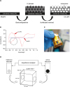
Fig. 1
Schematic diagrams of experimental design. (A) L2-ePt fabrication process including electro-deposition and surfactant removal steps. (B) A cyclic voltammogram of the FlatPt (blue) and the L2-ePt with roughness factor of 200 (red). The circled area indicates the enlarged hydrogen adsorption area of the L2-ePt. (C) Image of a fabricated L2-ePt electrode. (D) Electrode impedance measurement setup using a box filled with 0.9% NaCl solution.
L2-ePt = L2 phase electro-deposited nanoporous platinum, FlatPt = flat platinum.
Surface roughness measurement
Before measuring the surface area of the L2-ePt electrode, a cleansing process was carried out using cyclic voltammography. The electrode was placed in a 1.0 M sulfuric acid solution and a cycling potential was applied between +1.2 V and −0.22 V versus the Ag/AgCl, as mentioned in Park et al.19 The process was halted when successive cyclic voltammograms produced similar results. To quantitatively determine the surface area of the fabricated electrodes, the roughness factors (Rf) were calculated. The definition of the Rf is the ratio of the effective surface area to the geometric area of an electrode.18 In order to determine the effective surface area of the fabricated L2-ePt electrodes, cyclic voltammetry was performed using a 1 M H2SO4 solution with an Ag/AgCl electrode and a platinum wire as a reference and a counter electrode, respectively. The surface area of the electrode was determined from the hydrogen adsorption peaks of the cyclic voltammograms (scan rate 0.2 V s−1) in the 1.0 M sulfuric acid solution. For the calculation of the effective surface area (Equation 1), hydrogen adsorption area (in μC) was determined from the cyclic voltammogram by manually marking the starting and the ending peaks (Fig. 1B). The conversion factor of 210 μC cm2 was used to convert the units from the cyclic voltammogram into cm2.1820

A total of eight L2-ePt electrodes were fabricated with varying Rfs (Rf 100, Rf 200, and Rf 300) by applying a constant voltage of −0.2 V while varying the final charge value during the electro-deposition process. Although the charge values were empirically determined, a final charge value of 0.5 C cm−2 produced electrodes with an Rf of 100 (approximately 1-hour process), 1.0 C cm−2 produced an Rf of 200 (approximately 2 hours process), and charge values higher than 1.5 C cm−2 resulted in an Rf of 300 (approximately 3 hours process). To observe the nanoporous structure of the fabricated electrode (Fig. 1C), a field emission scanning electron microscope (FESEM) was used (AURIGA39-37; Carl Zeiss, Oberkochen, Germany).
The electrical impedances were measured in order to analyze the impedance characteristics of the L2-ePt electrodes and the FlatPt electrodes. As shown in Fig. 1D, a transparent acryl box was constructed (30 × 20 × 30 mm) with two circular holes (6 mm diameter) that were on opposite sides. A 0.9% NaCl solution was used as medium between the two electrodes, which were placed at the two openings in the box. At one end of the box, a reference electrode (Ag/AgCl electrode; 3M, Maplewood, MN, USA) was placed, while at the other end, an electrode of interest, either the FlatPt or the L2-ePt electrode, was placed. Then, the electrodes were connected to an impedance analyzer (6440A; Wayne Kerr, Bognor Regis, UK) for impedance measurements from 20 Hz to 1,000 Hz.17 With this procedure, the electrode impedances of the L2-ePt electrodes with different Rfs were compared.
Skin-electrode impedance measurement
The skin-electrode impedance was measured at 30 Hz using an impedance analyzer (F-E5GH; Grass Technologies, Warwick, RI, USA) on ten subjects. 30 Hz was used as a benchmark for EEG signal recording since the value is within the range of the EEG frequency spectrum, and it is the standard skin-electrode impedance monitoring frequency when recording EEG.21 All impedance measurements were carried out with current less than 1 μA, which is in a safe current range for recording the skin-electrode impedance.22 The L2-ePt electrode, the FlatPt electrode, and the gold cup electrode (Grass Technologies) were simultaneously attached to the occipital part of the head. To maintain stable contact, a felt pad (Emotive, San Francisco, CA, USA), moistened with saline solution, was used as a medium between the electrodes and the skin.
EEG measurement
EEG signals were obtained from all ten subjects. The occipital region was prepared using the skin prep gel, and the electrodes were affixed in their stationary positions. Similar to the skin-electrode impedance measurement, the felt pad was used between the electrode and the skin for both stable contact and enhanced utilization of the nanoporous surface. The signals were acquired using an L2-ePt electrode, a FlatPt, and a gold cup electrode that were positioned as close to each other as possible. In order to see the dominant alpha rhythm (8–13 Hz), eye-closed and eye-open cycles were repeated sequentially for 20 seconds each for an overall time of two minutes. Before the analog-digital conversion (ADC) process, the EEG was band-pass filtered using a cutoff frequency of 1 to 100 Hz, and the sampling rate was 1,000 Hz. The measured EEG signals were bandpass filtered for the 1–30 Hz frequency region in the MATLAB R2013a software (MathWorks, Natick, MA, USA). The correlation coefficients were calculated between the gold cup electrodes and the FlatPt electrodes, and between the gold cup electrodes and the L2-ePt electrodes.
Statistical analysis
SPSS version 21 IBM software (IBM, Armonk, NY, USA) was used for the statistical analyses. The electrode impedance in solution experiment results were assessed with the independent sample t-test, and the skin-electrode impedance experiment utilized a one-way analysis of variance (ANOVA) and Tukey post hoc test to compare the gold cup electrode, the FlatPt electrode, and the L2-ePt electrode. For the EEG analysis, a Wilcoxon signed-rank test was used to assess the differences in the measured EEG signal correlations.
RESULTS
Surface roughness
An L2-ePt electrode was electrochemically deposited on a platinum metal substrate. The electrode surface morphologies of the L2-ePt and FlatPt electrodes were observed from the FESEM images (Fig. 2), and the images showed that the L2-ePt electrode had homogeneous nanoporous structures with 10 nm pores, while the FlatPt electrode had a distinguishable flat surface different from that of the L2-ePt electrodes. From the cyclic voltammograms of the L2-ePt and FlatPt, the area under the hydrogen atom adsorption peaks showed that the L2-ePt electrodes had an enlarged surface area when compared to that of the FlatPt electrode. The Rf calculated from the cyclic voltammograms confirmed that the surface area of the L2-ePt electrodes increased, while that of the FlatPt electrode remained nearly the same (Table 1).
Fig. 2
Surface roughness images of the FlatPt electrode and the L2-ePt electrode. (A) FESEM image showing surface morphology of the FlatPt. (B) FESEM image of the L2-ePt surface. (300,000 × magnification, scale bar = 100 nm).
FlatPt = flat platinum, L2-ePt = L2 phase electro-deposited nanoporous platinum, FESEM = field emission scanning electron microscope.

Table 1
Rf of the electrodes and corresponding impedances

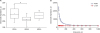
The electrode impedances of the fabricated electrodes were analyzed using an impedance analyzer in saline solution (0.9% NaCl solution). The electrode impedance comparison between the L2-ePt electrodes with different Rfs (Fig. 3A and Table 1) at 30 Hz shows significant differences between the groups (F [2, 36] = 6.9; P = 0.003). The impedances of the Rf 100 L2-ePt electrodes were significantly different from those of the Rf 200 L2-ePt electrodes (P = 0.003), but otherwise when the Rf 100 L2-ePt electrodes compared to the impedance of the Rf 300 L2-ePt electrodes (P = 0.69). The electrode impedance of the Rf 200 L2-ePt electrodes and the Rf 300 electrodes did not show significant difference (P = 0.088). The electrode impedance of the FlatPt electrode was significantly smaller than those of the L2-ePt electrodes (P < 0.001). After the Rf comparison, the Rf 200 L2-ePt electrodes were used for the rest of the experiment due to their lowest mean electrode impedance. In addition, Fig. 3B shows the average electrode impedances of the FlatPt electrodes and the L2-ePt electrodes across the frequencies between 20 Hz and 1,000 Hz. For the entire frequency range, the FlatPt electrodes were significantly different from the L2-ePt electrodes (P < 0.001).
Fig. 3
Electrode impedance comparison of electrodes with different Rfs. (A) Boxplot showing electrode impedances of three L2-ePt groups (Rf 100, Rf 200, and Rf 300). The impedances were measured at 30 Hz. (B) Average electrode impedance in solution measured by the FlatPt electrode (empty triangle) and the L2-ePt electrode with Rf of 200 (filled diamond). Error bars indicate standard deviation.
Rf = roughness factor, L2-ePt = L2 phase electro-deposited nanoporous platinum, FlatPt = flat platinum.
aP < 0.05.
Skin-electrode impedance
The skin-electrode impedances measured from the head (Fig. 4 and Table 2) showed that there were significant differences on the hairy occipital part of the head (F [2, 18] = 8.64; P = 0.002). The L2-ePt electrode with impedance of 10.0 ± 6.03 kΩ had significant difference from the FlatPt electrode with impedance of 26.10 ± 11.83 kΩ, P = 0.003. The gold cup electrode with impedance of 12.23 ± 2.78 kΩ was significantly different from that of the FlatPt electrode, P = 0.015.
Fig. 4
The skin-electrode impedance of the gold cup electrode, the FlatPt electrode, and the L2-ePt electrode (Rf = 200) on the occipital region of the head. The impedance was measured at 30 Hz. Error bars indicate standard deviation.
FlatPt = flat platinum, L2-ePt = L2 phase electro-deposited nanoporous platinum, Rf = roughness factor.
aP < 0.05; bP < 0.005.
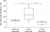
Table 2
Impedance and EEG results by electrode types

EEG: alpha rhythm
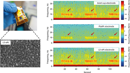
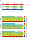
The eye-closed segments of the raw EEG and a spectrogram of the data displayed noticeable alpha waves compared to the eye-open segments (Fig. 5). As shown in Table 2, a Wilcoxon signed-rank test revealed that the correlation coefficients between the signals acquired by the gold cup electrodes and those of the FlatPt electrodes (r = 0.82 ± 0.15), and the correlation coefficients between the signals obtained by the gold cup electrodes and those of the L2-ePt electrodes (r = 0.94 ± 0.053), were significantly different (Z = −2.19; P = 0.029).
Fig. 5
Raw EEG signals and spectrograms during alpha rhythm recording sessions. (A) Raw EEGs during eye-open and closed sessions. (B) Spectrogram of the gold cup electrode, the FlatPt electrode, and the L2-ePt electrode during eye-open and eye-closed sessions, and the arrow points to the alpha rhythm.
EEG = electroencephalogram, FlatPt = flat platinum, L2-ePt = L2 phase electro-deposited nanoporous platinum.
DISCUSSION
The L2-ePt electrode has been studied as a way to detect electrochemical materials or as a pH sensor.23 However, no study has investigated the L2-ePt electrode for EEG recording applications, and the electrode characteristics as an EEG recording material remained unknown. Thus, this study examined the surface roughness through the electrode impedance comparisons, and then we assessed the skin-electrode impedance and EEG signal quality of the L2-ePt to provide comparative guidelines for use when considering the efficacy of the L2-ePt electrode.
The L2-ePt electrode has multiple beneficial properties as an EEG sensor. Since the sensor material contacting the skin is prone to corrosion, which leads to electrochemical noise and signal degradation, the electrode needs to be electrochemically stable. According to Fonseca et al.,24 metals such as aluminum and stainless-steel undergo corrosion or copper which induce allergic reactions. But, Park et al.25 reported that the L2-ePt electrode exhibits neither corrosion nor allergic reactions when exposed under saline conditions. Also, as reported in previous studies, the mechanical stability of the L2-ePt electrode was shown by applying 12.6 mN with a 5 μm diameter diamond indenter. As a result, the L2-ePt electrode displayed an intact electro-deposition layer without peeled off residues.1823 In addition to electrochemical, mechanical stability, and biocompatibility,20 the quantitatively controllable fabrication of the electrodes is one of the advantages of the L2-ePt electrode since it is possible to produce uniform and quality controlled electrodes. The confirmation of the uniform surface through cross-section scanning electron microscope (SEM) was restricted due to limitation in uniformly slicing the platinum foil (0.25 mm thick), and possible effect of the surfactant residues in the deeper layers. However, previous studies support the fabrication of the uniform nanoporous thin film of the L2-ePt when compared to the FlatPt surface.26 When plated on a much thinner substrate, the cross-section SEM revealed that the nanoporous layer was formed with uniform thickness and free of surfactant residues.18
This study was in agreement with the previous study where Boo et al.26 described the relationship between the pore size and the thickness of the electrical double layer. When the thickness of the double layer is larger than the pore radius, the double layer is formed outside the pores because the equipotential line cannot follow along the pore surface. Furthermore, Park et al.18 noted that the larger roughness might not necessarily mean more electrochemical activity, but there might be an ultimate porosity. They found that from Rf 200 and onward, the electrical activity makes a plateau. Consequently, the larger electrode impedance of the Rf 300 than that of the Rf 200 from this study may be ascribed to the non-linear relationship between the roughness and the electrical activity at the surface.
In this study, the impedance in solution was influenced by the nanoporous surface that increased the effective contact area between the surface and the medium, and consequently reduced the impedance. This decrease in the impedance improved the electrical conductance of the electrode for measuring accurate electrical changes that occur under the electrode. Park et al.23 reported that high impedance could be a source of noise, which distorts or weakens the true neural signals. Moreover, only signals that are in very close proximity to the surface of the electrode are recorded and ones that are far away are degraded when the electrical impedance is high. The electrode impedance of the L2-ePt electrode when measured in the electrolytic solution was significantly lower than that of the FlatPt electrode. These results suggest that the nanoporous surface plays a major role in reducing the electrode impedance of the L2-ePt electrodes.
The skin-electrode impedance significantly influences the performance of EEG recording. One of the factors affecting the skin-electrode impedance is the effective surface area, where larger contact area reduces the skin-electrode impedance by lowering the reactance.272829 In this respect, the lowest skin-electrode impedance of the L2-ePt electrode from our study can be credited to the high surface capacitance and reduced impedance due to the increased roughness of the L2-ePt electrode surface.23 Also, when used with the moistened-pad, the uniform nanopores of the L2-ePt which facilitated electrical charge transfer between the moisture and the metal, could have further reduced the skin-electrode impedance. According to Han et al.,29 particles trapped in the pores require a longer time to react than particles on the flat surface. Consequently, the electron transfer can be enhanced.
However, it is important to note that the reduction in skin-electrode impedance cannot be solely explained by the increased effective surface area. As reported by Rosell et al.,30 skin impedance is another factor dominating the skin-electrode impedance. Because of the human-to-human differences in the skin, it has been reported that the impedance of the skin varies across different individuals. However, when the L2-ePt electrode was used with the moistened-pad, the individual differences were observed to be reduced. The moisture held in the pad may have penetrated the stratum corneum, which would thereby reduce the effect of the skin impedance.31 The decrease in the skin-electrode variance between individuals might be due to the less influential effect of the stratum corneum.
In terms of employing the developed L2-ePt electrode in daily life EEG recording, the level of skin-electrode impedance and convenience should be considered. As many EEG studies have noted, a recommended range of the skin-electrode impedance is below 10 or 20 kΩ.323334 When compared to the skin-electrode impedances of commercially available electrodes, such as Ag/AgCl electrode (in a range of a few tens of kΩ)35 or gold cup electrodes (12 kΩ in this study) with conductive paste, that of the developed L2-ePt electrode (10 kΩ in this study) falls within in a practical range. The L2-ePt electrode with moistened pad is conceptually similar to the “wet” electrode because the skin and the electrode of both conventional electrodes and the L2-ePt electrode is mediated by moisture and gel, respectively. However, the proposed method maintains the advantage and overcomes the disadvantage of the “wet” electrode by providing low skin-electrode impedance without the use of sticky conductive paste that dirty the hair and cause inconvenience for the user, which has been one of the major hurdles for daily life EEG recording.
In addition to the impedance properties of the electrode over the relevant frequency ranges, an EEG was recorded using the L2-ePt electrode to compare its signal recording performance to that of the clinically-used gold cup electrode. The improved performance of the L2-ePt electrode compared to the FlatPt electrode can be attributed to the increased active surface area due to the nanoporous surface. The correlation value of the L2-ePt reflects the effect of the decreased skin-electrode impedance on increasing the signal-to-noise level.23 As the enhanced electrode impedance performance due to the interaction between the L2-ePt and solution suggests, moisture was introduced in order to lower the skin-electrode impedance. Furthermore, the action of attaching and detaching the L2-ePt electrode on the head was facilitated by a simple contact on the scalp. The similar correlations of the signals acquired by the clinically used gold cup electrode and the L2-ePt electrode show that the L2-ePt electrodes can acquire high-quality signal. With respect to EEG signal quality comparison, our result is comparable to the previous study by Lin et al.,27 where their dry foam electrode showed 96.14% and 90.12% EEG correlation on the forehead and hairy sites, respectively. In another study, a flexible silicone-based dry electrode showed 92% correlation from the forehead.36 Therefore, the L2-ePt electrodes can be employed in a variety of EEG applications where it would be cumbersome and inconvenient to use electrodes with adhesive paste.6 Consequently, the L2-ePt electrode can be used for EEG recordings over the hair to provide both convenience and high-quality signal acquisition capability. The characteristics of the L2-ePt electrode will be especially useful in brain-computer interface applications where user comfort and excellent signal acquisition is important.
However, costly platinum metal was used as the substrate metal for the L2-ePt electrode. Despite their price disadvantage, the stable metals, like platinum or gold, are used in commercially available EEG recording electrodes. Like the commercial gold or platinum electrodes, the L2-ePt electrode provides biocompatibility, mechanical stability, and electrochemical stability when recording EEG.
In this study, the possibility and benefits of using nanoporous surface electrodes as EEG recording electrodes have been introduced. Using a nanoporous structure, the L2-ePt electrode exhibited distinguishable surface morphology as well as lower electrode impedance, as opposed to those of the untreated FlatPt electrode. Furthermore, the skin-electrode impedance measurements illustrated that nanoporous electrodes are reliable for recording EEG frequencies with lower impedance than the FlatPt electrode, and similar impedances to the gold cup electrode. Finally, for the EEG recording experiment, the L2-ePt electrodes performed with similar signal quality as the gold cup electrode. In short, this study suggests that the nanoporous platinum electrode holds promise as a new method for EEG recording in many applications, including brain-computer interface.
 XML Download