Journal List > J Korean Med Sci > v.33(4) > 1107740

Lee, Lee, Lee, and Yu: Adhesion of Acanthamoeba on Cosmetic Contact Lenses

Abstract

Background

This study aimed to evaluate the adhesion of Acanthamoeba trophozoites on cosmetic contact lenses (CLs) with and without CL care multipurpose solution (MPS) treatment.

Methods

Acanthamoeba lugdunensis L3a trophozoites were inoculated onto disks trimmed from CLs: 1-day Acuvue moist, 1-day Acuvue define, Acuvue 2, and Acuvue 2 define. After 18-hour inoculation, the number of adherent trophozoites was counted under phase contrast microscopy. The effects of MPS, Opti-Free Express, soaking CLs for 6 hours, on Acanthamoeba adhesion were analyzed. Scanning electron microscopic examination was performed for assessment of Acanthamoeba attached on the lens surface.

Results

Acanthamoeba trophozoites showed greater adhesion to cosmetic CL (P = 0.017 for 1-day CL and P = 0.009 for 2-week CL) although there was no significant difference between the types of cosmetic CL. On all lenses, the number of adherent Acanthamoeba was significantly reduced after treatment with MPS (P < 0.001 for 1-day Acuvue moist, P = 0.046 for 1-day Acuvue define, P < 0.001 for Acuvue 2, and P = 0.015 for Acuvue 2 define), but there was still significant difference between conventional and cosmetic CLs (P = 0.003 for 1-day CL and P < 0.001 for 2-week CL, respectively). More attachment of Acanthamoeba was observed on colored area and the acanthopodia of Acanthamoeba was placed on the rough surface of colored area.

Conclusion

Acanthamoeba showed a greater affinity for cosmetic CL and mostly attached on colored area. Although MPS that contained myristamidopropyl dimethylamine reduced the adhesion rate, there was a significant difference between conventional and cosmetic CLs.

Graphical Abstract

jkms-33-e26-abf001.jpg

INTRODUCTION

Acanthamoeba keratitis occurs in contact lens (CL) users of all lens types, and can culminate in severe vision loss. The demand for cosmetic CLs continues to increase, particularly in teenage girls and young women all over the world, accounting for 13% of CL use in 2012, because they can be used to drastically alter the appearance of the eye.12 For these reasons, the possibility of an increasing incidence of Acanthamoeba keratitis in cosmetic CL users should be considered. It has been reported that complications that can arise when wearing CL occur more frequently and are more serious in patients using cosmetic CLs than conventional CLs.3
Adhesion of Acanthamoeba to CLs may play an important role in the pathogenesis of Acanthamoeba keratitis and depend on different parameters. Although many studies have reported the importance of hydrophobicity and surface roughness for microbial adhesion, analyses were mostly limited to noncolored conventional CLs.45678910 Recent studies have shown that the relative risk of developing CL related microbial keratitis is 16.5 times higher in cosmetic CL wearers than in conventional CL wearers, and suggested noncompliance as one of the most key factors.34567891011 However, none of the studies have analyzed the characteristics of cosmetic CLs related to the adhesion of Acanthamoeba.
CL care multipurpose solutions (MPSs) for soft CLs comprise an aqueous liquid medium, an antimicrobial component, a surfactant, a buffer component, a viscosity-inducing component, a tonicity component, and a chelating component.12 Although most CL wearers use MPSs because of their simplicity and convenience, several studies131415161718 conducted on the disinfecting efficacy of MPS against Acanthamoeba in conventional CLs showed that some MPSs had limited efficacies against Acanthamoeba trophozoites. It is thus important to determine the effects of MPSs on Acanthamoeba adhesion to cosmetic as well as conventional CLs.
Therefore, we investigated Acanthamoeba trophozoite adhesion in both cosmetic and conventional CLs and the efficacy of MPS disinfection against Acanthamoeba. We also used scanning electron microscopy (SEM) to examine adhesion patterns of Acanthamoeba trophozoites on the CL surface.

METHODS

CLs

CLs were purchased from commercial sources and used for the adhesion assay. The properties of conventional (1-day Acuvue moist and Acuvue 2; Johnson & Johnson Vision Care, Inc., Jacksonville, FL, USA) and cosmetic (1-day Acuvue define and Acuvue 2 define; Johnson & Johnson Vision Care, Inc.) CLs are summarized in Table 1. All CLs were placed at the bottom of a 48-well plate (Falcon; BD Biosciences, Franklin Lakes, NJ, USA) with the convex side up.
Table 1

The properties of CLs

jkms-33-e26-i001
Lenses Material Recommended replacement Dk/t Water content, % Base curve/diameter, mm Center thickness, mm Color-tinting method
1-day Acuvue moist EtafilconA 1 day 25.5 × 10⁻⁹ 58 8.5/14.2 0.084 NA
1-day Acuvue define EtafilconA 1 day 25.5 × 10⁻⁹ 58 8.5/14.2 0.084 Sandwich
Acuvue 2 EtafilconA 2 wk 25.5 × 10⁻⁹ 58 8.3/14.0 0.084 NA
Acuvue 2 define EtafilconA 2 wk 25.5 × 10⁻⁹ 58 8.3/14.0 0.084 Sandwich
CL = contact lens, Dk = measure of oxygen permeability, NA = not applicable.

MPSs

The MPS (Opti-Free Express; Alcon, Fort Worth, TX, USA) was purchased from commercial sources and were used within its expiration date. The composition is myristamidopropyl dimethylamine (MAPD, Aldox) 0.0005%, polyquaternium (Polyquad) 0.001%, and poloxamine.

Preparation of Acanthamoeba trophozoites

Although Acanthamoeba castellanii and Acanthamoeba polyphaga are common causative agents of Acanthamoeba keratitis in many countries,192021 this study examined Acanthamoeba lugdunensis, the most frequently isolated type of Acanthamoeba, from CL storage cases in Korea,222324 which were isolated from patients with Acanthamoeba keratitis and identified by riboprinting and 18S rDNA sequence analyses as previously described.21252627 Acanthamoeba trophozoites were cultured in a culture flask (Falcon; BD Biosciences) with peptone-yeast extract/glucose (PYG) medium (20.02 g of Bacto Proteose Peptone and 1.00 g of yeast extract in 950 mL of pure water, 50.0 mL of 2 MD(+)glucose, 10.0 mL of 0.4 M MgSO4·7H2O, 8.0 mL of 0.05 M CaCl2, 34.0 mL of 0.1 M sodium citrate 2H2O, 10.0 mL of 0.005 M Fe(NH4)2(SO4)2·6H2O, 10.0 mL of 0.25 M NaHPO4·7H2O, and 10.0 mL of 0.25 M KH2PO4) at room temperature. Prior to an experiment, the medium was exchanged by rigorously shaking the culture bottles, removing the medium, and adding new medium. For the experiments, the number of Acanthamoeba applied to a substrate was determined by counting trophozoites beforehand using a Neubauer hemocytometer.

Adhesion assay of Acanthamoeba trophozoites to lenses

Lenses were placed in a 48-well plate filled with 1 mL of PYG. Acanthamoeba of 2 × 10⁴ were added to each well containing a CL, followed by incubation for 18 hours. In the experiments, PYG medium was used to ensure that the Acanthamoeba was in a healthy state. After washing each well twice with 2 mL of phosphate buffered saline, the total number of trophozoites adhered to lenses was counted using a phase contrast microscope. Three lenses of each type were used in this experiment. Sixteen images per sample were taken on arbitrary positions and the number of trophozoites attached to each lens was counted.

Adhesion assay of MPS-treated Acanthamoeba to lenses

After soaking CLs for 6 hours with Opti-Free Express solution for testing, Acanthamoeba trophozoites of 2 × 10⁴ in PYG were inoculated into a 48-well plate containing a lens. The total number of Acanthamoeba adhered to lenses was counted as described in the previous section. Three lenses were used for each type of lenses.

SEM examination of adherent Acanthamoeba

The lens was subsequently fixed with 2.5% glutaraldehyde (Electron Microscopy Sciences, Fort Washington, PA, USA) in 100 mM sodium cacodylate buffer (pH 7.4) for 1 hour at 4°C, and then postfixed with 1% OsO4 in the same buffer for 1 hour at 4°C. After standard graded dehydration with ethanol (50%, 70%, 80%, 85%, 90%, 95%, and 100%), the specimens were critically point-dried with liquid CO2 using a Samdri 780 apparatus (Tousimis Research Corp., Rockville, MD, USA), and coated with a thin layer (30 nm) of gold in an Ion sputtering device (JFC-1100; JEOL Corp., Tokyo, Japan). Specimens were examined using a field emission-scanning electron microscope with electron backscattered diffraction (SUPRA 40 VP; Carl Zeiss, Jena, Germany).

Statistical analysis

Statistical analysis of the number of adherent Acanthamoeba trophozoites to lenses was performed using analysis of variance with the Scheffe's test or non-paired t-test via SPSS (SPSS Inc., Chicago, IL, USA).

RESULTS

Adhesion of Acanthamoeba trophozoites to CLs with MPSs

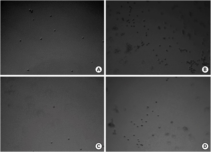
After 18-hour inoculation, the number of adherent trophozoites on CLs was counted under phase contrast microscopy. We observed the adherence of trophozoites to cosmetic as well as conventional CLs and adhesion to cosmetic CLs was greater than that to conventional CLs (Figs. 1 and 2). There was no significant difference between 1-day and 2-week CLs in either conventional or cosmetic CLs.
Fig. 1
Phase contrast microscopic images of adherent Acanthamoeba trophozoites to CLs. (A) 1-day Acuvue moist, (B) 1-day Acuvue define, (C) Acuvue 2, and (D) Acuvue 2 define (original magnification × 100). More adhesion was observed to cosmetic CLs than to conventional CLs.
CL = contact lens.
jkms-33-e26-g001
Fig. 2
The relationship of Acanthamoeba trophozoites adherence among CLs treated with and without MPS. Although Opti-Free Express significantly reduced the numbers of adherent trophozoites in all lenses, there were significant differences between conventional and cosmetic CLs regardless of Opti-Free Express treatment in both 1-day and 2-week CLs.
CL = contact lens, MPS = multipurpose solution.
aP < 0.001 for 1-day Acuvue moist, P = 0.046 for 1-day Acuvue define, P < 0.001 for Acuvue 2, and P = 0.015 for Acuvue 2 define.
jkms-33-e26-g002
Treatment with Opti-Free Express significantly reduced the number of adherent trophozoites in all CLs examined (P < 0.001 for 1-day Acuvue moist, P = 0.046 for 1-day Acuvue define, P = 0.0003 for Acuvue 2, and P = 0.015 for Acuvue 2 define), whereas there was still significant differences between conventional and cosmetic CLs even after Opti-Free Express treatment (Fig. 2).

Morphology of attached Acanthamoeba by SEM

SEM was performed for comparison of the lens surface of CLs. We found that the colored area was rougher than the uncolored area of cosmetic CLs (Fig. 3) and more Acanthamoeba trophozoites adhered to the surface of colored area (Fig. 4). The surface of cosmetic CLs to which amoebae adhere better is rough, which increases the contact surface with trophozoites, allowing firm attachment of acanthopodias (Fig. 5A and B).
Fig. 3
SEM image of cosmetic CL surface (original magnification × 1,000). Colored area (C) of cosmetic CL was rougher than the noncolored area (N).
SEM = scanning electron microscopy, CL = contact lens.
jkms-33-e26-g003
Fig. 4
SEM images of Acanthamoeba trophozoites adhesion (original magnification × 100). More Acanthamoeba trophozoites adhesion (arrows) was found on the colored areas (C) than on noncolored areas (N) of both (A) 1-day Acuvue define and (B) Acuvue 2 define.
SEM = scanning electron microscopy.
jkms-33-e26-g004
Fig. 5
SEM images of Acanthamoeba trophozoites adhesion at high magnification (original magnification × 3,000). (A, B) The acanthopodia (arrows) of Acanthamoeba trophozoites firmly attached to the colored rough area. (C) Acanthamoeba trophozoites treated with MPS showed a relatively round or shrunken shape like cystic form.
SEM = scanning electron microscopy, MPS = multipurpose solution.
jkms-33-e26-g005
Acanthamoeba treated with MPS attached firmly at the surface of cosmetic CL and was observed to be relatively round or a shrunken cystic shape, although the trophozoite of Acanthamoeba has a tendency of showing an elongated or slender shape (Fig. 5C).

DISCUSSION

Cosmetic CLs not only improve visual acuity but also change the appearance of the eye. Currently, many teenagers are exposed to cosmetic CLs and start to use them without proper education, and complications such as microbial keratitis occur more frequently and are more serious in patients using cosmetic CLs than in those using conventional CLs.3 Acanthamoeba keratitis is a rare but potentially blinding infection associated with CL wear. CL use can have a negative impact on the eye's natural defenses and favor microbial colonization and survival.28 A critical step before adhesion and invasion of the cornea is the adhesion to the CL. Although previous studies have demonstrated the ability of Acanthamoeba to stick to soft CL surfaces, little is known about their adherence properties to cosmetic CLs.
Numerous risk factors for CL related microbial keratitis have been described. Despite the progress in the quality of both CL materials2930 and sterile packaging,2931 the number of CL related microbial keratitis cases has remained roughly constant.332 The smoothness of and electrical charge on the CL surface have previously been shown to be related to bacterial adhesion. In particular, an increased surface roughness caused more bacterial adhesion.933 Bruinsma et al.34 reported that the surface roughness was increased by CL wearing time, which was an important predictive factor for Pseudomonas aeruginosa adhesion to Etafilcon A lenses. Chan et al.35 mentioned that surface pigments could increase the surface roughness of cosmetic CL and Ji et al.36 found that significantly more Staphylococcus aureus and P. aeruginosa adhered to cosmetic CL than to conventional CL mainly on the colored surface rather than on the noncolored areas, which correlated with the surface roughness of CL. They concluded that bacterial adhesion and proliferation may be related to the increased microbial keratitis rate in cosmetic CL wearers because cosmetic CLs have more irregular and rougher surfaces than conventional CLs. In accordance with these studies, the adherence of Acanthamoeba trophozoites on cosmetic CL in this study was more than on conventional CL and might be related to the irregular surface of the cosmetic CL.
On SEM, the surface of cosmetic CL was more irregular and rougher compared with that of conventional CL. The mean lens surface roughness measured by atomic force microscopy was significantly higher for cosmetic CL than for conventional CL.36 We confirmed that the noncolored surface of cosmetic CL was smoother and flatter than colored area, and the attachment of Acanthamoeba trophozoites to colored rough area was more than to noncolored smooth area. The attachment occurred through acanthopodia and a cystic pattern of Acanthamoeba trophozoites after MPS treatment was observed because Acanthamoeba changed its shape to survive under the poor condition, as we previously showed in the study of silicone hydrogel CLs.37
Opti-Free Express, which was the most effective MPS in reducing attachment of Acanthamoeba on the silicone hydrogel CL surfaces,37 was used in this study, and significantly reduced the numbers of adherent trophozoites in all lenses. However, the significant differences between conventional and cosmetic CLs even after Opti-Free Express treatment were still observed. In addition, there were also differences in the number of Acanthamoeba between 1-day and 2-week CLs with and without MPS treatment although they were not statistically significant. The colored area of 1-day lens was much rougher than that of 2-week lens and more Acanthamoeba trophozoites were observed in 1-day CLs than 2-week CLs. It may be argued that the adherence of bacteria to 1-day lenses is not of concern as they should be discarded after use. However, previous studies have reported that 1-day CLs did not reduce the risk of CL associated microbial keratitis.3038 Hence, the importance of microbial adherence to daily disposable cosmetic CL should not be underestimated.
In summary, Acanthamoeba showed a greater affinity for the cosmetic CLs, interestingly on the colored surface, than on the conventional CLs, because cosmetic CLs have more irregular and rougher surfaces than conventional CLs, which could facilitate Acanthamoeba adhesion on the surface through acanthopodia. An MPS containing MAPD significantly reduced the adhesion rate on both conventional and cosmetic CLs, but there were still significant differences between these lenses even after MPS treatment. Awareness of increased propensity for Acanthamoeba adherence of cosmetic CLs would be useful for users as well as eye care professionals, and cosmetic CL wearers should be educated on the risks.

Notes

Funding: This research was supported by Basic Science Research Program through the National Research Foundation of Korea (NRF) funded by the Ministry of Education (Grant No. NRF-2017R1D1A1B03032153).

Disclosure: The authors have no potential conflicts of interest to disclose.

Author Contributions: Conceptualization: Lee JE, Yu HS. Data curation: Lee SM, Lee DI. Formal analysis: Lee SM, Lee DI. Investigation: Lee SM, Lee DI. Writing - original draft: Lee JE.

References

1. Efron N, Morgan PB, Woods CA. International Contact Lens Prescribing Survey Consortium. International survey of contact lens prescribing for extended wear. Optom Vis Sci. 2012; 89(2):122–129. PMID: 22179218.
2. Efron N, Morgan PB, Woods CA. International Contact Lens Prescribing Survey Consortium. An international survey of daily disposable contact lens prescribing. Clin Exp Optom. 2013; 96(1):58–64. PMID: 22853742.
3. Sauer A, Bourcier T. French Study Group for Contact Lenses Related Microbial Keratitis. Microbial keratitis as a foreseeable complication of cosmetic contact lenses: a prospective study. Acta Ophthalmol. 2011; 89(5):e439–e442. PMID: 21401905.
4. Willcox MD. Microbial adhesion to silicone hydrogel lenses: a review. Eye Contact Lens. 2013; 39(1):61–66. PMID: 23266589.
5. Shen EP, Tsay RY, Chia JS, Wu S, Lee JW, Hu FR. The role of type III secretion system and lens material on adhesion of Pseudomonas aeruginosa to contact lenses. Invest Ophthalmol Vis Sci. 2012; 53(10):6416–6426. PMID: 22918630.
6. Dutta D, Cole N, Willcox M. Factors influencing bacterial adhesion to contact lenses. Mol Vis. 2012; 18:14–21. PMID: 22259220.
7. Tang H, Cao T, Liang X, Wang A, Salley SO, McAllister J 2nd, et al. Influence of silicone surface roughness and hydrophobicity on adhesion and colonization of Staphylococcus epidermidis . J Biomed Mater Res A. 2009; 88(2):454–463. PMID: 18306290.
8. Giraldez MJ, Serra C, Lira M, Real Oliveira ME, Yebra-Pimentel E. Soft contact lens surface profile by atomic force microscopy. Optom Vis Sci. 2010; 87(7):E475–E481. PMID: 20473237.
9. Geoghegan M, Andrews JS, Biggs CA, Eboigbodin KE, Elliott DR, Rolfe S, et al. The polymer physics and chemistry of microbial cell attachment and adhesion. Faraday Discuss. 2008; 139:85–103. PMID: 19048992.
10. Desrousseaux C, Sautou V, Descamps S, Traoré O. Modification of the surfaces of medical devices to prevent microbial adhesion and biofilm formation. J Hosp Infect. 2013; 85(2):87–93. PMID: 24007718.
11. Kim JH, Song JS, Hyon JY, Chung SK, Kim TJ. A survey of contact lens-related complications in Korea: the Korean Contact Lens Study Society. J Korean Ophthalmol Soc. 2014; 55(1):20–31.
12. Dutot M, Reveneau E, Pauloin T, Fagon R, Tanter C, Warnet JM, et al. Multipurpose solutions and contact lens: modulation of cytotoxicity and apoptosis on the ocular surface. Cornea. 2010; 29(5):541–549. PMID: 20418717.
13. Borazjani RN, Kilvington S. Efficacy of multipurpose solutions against Acanthamoeba species. Cont Lens Anterior Eye. 2005; 28(4):169–175. PMID: 16332501.
14. Kobayashi T, Gibbon L, Mito T, Shiraishi A, Uno T, Ohashi Y. Efficacy of commercial soft contact lens disinfectant solutions against Acanthamoeba . Jpn J Ophthalmol. 2011; 55(5):547–557. PMID: 21748273.
15. Lonnen J, Heaselgrave W, Nomachi M, Mori O, Santodomingo-Rubido J. Disinfection efficacy and encystment rate of soft contact lens multipurpose solutions against Acanthamoeba . Eye Contact Lens. 2010; 36(1):26–32. PMID: 20009947.
16. Shoff M, Rogerson A, Schatz S, Seal D. Variable responses of Acanthamoeba strains to three multipurpose lens cleaning solutions. Optom Vis Sci. 2007; 84(3):202–207. PMID: 17435534.
17. Beattie TK, Seal DV, Tomlinson A, McFadyen AK, Grimason AM. Determination of amoebicidal activities of multipurpose contact lens solutions by using a most probable number enumeration technique. J Clin Microbiol. 2003; 41(7):2992–3000. PMID: 12843032.
18. Kilvington S, Heaselgrave W, Lally JM, Ambrus K, Powell H. Encystment of Acanthamoeba during incubation in multipurpose contact lens disinfectant solutions and experimental formulations. Eye Contact Lens. 2008; 34(3):133–139. PMID: 18463477.
19. Siddiqui R, Khan NA. Biology and pathogenesis of Acanthamoeba. Parasit Vectors. 2012; 5:6. PMID: 22229971.
20. Molmeret M, Horn M, Wagner M, Santic M, Abu Kwaik Y. Amoebae as training grounds for intracellular bacterial pathogens. Appl Environ Microbiol. 2005; 71(1):20–28. PMID: 15640165.
21. Yu HS, Kong HH, Kim SY, Hahn YH, Hahn TW, Chung DI. Laboratory investigation of Acanthamoeba lugdunensis from patients with keratitis. Invest Ophthalmol Vis Sci. 2004; 45(5):1418–1426. PMID: 15111597.
22. Kong HH, Shin JY, Yu HS, Kim J, Hahn TW, Hahn YH, et al. Mitochondrial DNA restriction fragment length polymorphism (RFLP) and 18S small-subunit ribosomal DNA PCR-RFLP analyses of Acanthamoeba isolated from contact lens storage cases of residents in southwestern Korea. J Clin Microbiol. 2002; 40(4):1199–1206. PMID: 11923331.
23. Lee SM, Choi YJ, Ryu HW, Kong HH, Chung DI. Species identification and molecular characterization of Acanthamoeba isolated from contact lens paraphernalia. Korean J Ophthalmol. 1997; 11(1):39–50. PMID: 9283153.
24. Yu HS, Choi KH, Kim HK, Kong HH, Chung DI. Genetic analyses of Acanthamoeba isolates from contact lens storage cases of students in Seoul, Korea. Korean J Parasitol. 2001; 39(2):161–170. PMID: 11441503.
25. Chung DI, Yu HS, Hwang MY, Kim TH, Kim TO, Yun HC, et al. Subgenus classification of Acanthamoeba by riboprinting. Korean J Parasitol. 1998; 36(2):69–80. PMID: 9637824.
26. Gast RJ, Ledee DR, Fuerst PA, Byers TJ. Subgenus systematics of Acanthamoeba: four nuclear 18S rDNA sequence types. J Eukaryot Microbiol. 1996; 43(6):498–504. PMID: 8976608.
27. Stothard DR, Schroeder-Diedrich JM, Awwad MH, Gast RJ, Ledee DR, Rodriguez-Zaragoza S, et al. The evolutionary history of the genus Acanthamoeba and the identification of eight new 18S rRNA gene sequence types. J Eukaryot Microbiol. 1998; 45(1):45–54. PMID: 9495032.
28. Shovlin JP, Argüeso P, Carnt N, Chalmers RL, Efron N, Fleiszig SM, et al. 3. Ocular surface health with contact lens wear. Cont Lens Anterior Eye. 2013; 36(Suppl 1):S14–S21. PMID: 23347571.
29. Dart JK, Radford CF, Minassian D, Verma S, Stapleton F. Risk factors for microbial keratitis with contemporary contact lenses: a case-control study. Ophthalmology. 2008; 115(10):1647–1654. 1654.e1–1654.e3. PMID: 18597850.
30. Sankaridurg P, Lazon de la Jara P, Holden B. The future of silicone hydrogels. Eye Contact Lens. 2013; 39(1):125–129. PMID: 23266592.
31. Keay L, Edwards K, Stapleton F. Referral pathways and management of contact lens-related microbial keratitis in Australia and New Zealand. Clin Experiment Ophthalmol. 2008; 36(3):209–216. PMID: 18412588.
32. Singh S, Satani D, Patel A, Vhankade R. Colored cosmetic contact lenses: an unsafe trend in the younger generation. Cornea. 2012; 31(7):777–779. PMID: 22378117.
33. Giraldez MJ, Resua CG, Lira M, Oliveira ME, Magariños B, Toranzo AE, et al. Contact lens hydrophobicity and roughness effects on bacterial adhesion. Optom Vis Sci. 2010; 87(6):E426–E431. PMID: 20375748.
34. Bruinsma GM, Rustema-Abbing M, de Vries J, Stegenga B, van der Mei HC, van der Linden ML, et al. Influence of wear and overwear on surface properties of etafilcon A contact lenses and adhesion of Pseudomonas aeruginosa. Invest Ophthalmol Vis Sci. 2002; 43(12):3646–3653. PMID: 12454031.
35. Chan KY, Cho P, Boost M. Microbial adherence to cosmetic contact lenses. Cont Lens Anterior Eye. 2014; 37(4):267–272. PMID: 24440107.
36. Ji YW, Cho YJ, Lee CH, Hong SH, Chung DY, Kim EK, et al. Comparison of surface roughness and bacterial adhesion between cosmetic contact lenses and conventional contact lenses. Eye Contact Lens. 2015; 41(1):25–33. PMID: 25536530.
37. Lee GH, Lee JE, Park MK, Yu HS. Adhesion of Acanthamoeba on silicone hydrogel contact lenses. Cornea. 2016; 35(5):663–668. PMID: 26938330.
38. Stapleton F, Keay L, Edwards K, Naduvilath T, Dart JK, Brian G, et al. The incidence of contact lens-related microbial keratitis in Australia. Ophthalmology. 2008; 115(10):1655–1662. PMID: 18538404.
TOOLS
ORCID iDs

Seung-Mok Lee
https://orcid.org/0000-0003-0006-1498

Ji-Eun Lee
https://orcid.org/0000-0002-4927-2854

Da-In Lee
https://orcid.org/0000-0001-7507-7086

Hak-Sun Yu
https://orcid.org/0000-0002-6696-7981

Similar articles