

 PDF
PDF Citation
 Print
Print


INTRODUCTION
The hand bones and the extrinsic and intrinsic hand muscles are fundamental structures of the hand. Their anatomy is too complicated to be recognized solely by books or plastic models, so medical students dissect the cadaver hand. However, cadaver dissection is not always feasible; this restriction is prominent, especially during clinical classes, such as orthopedics and plastic surgery. Computer-assisted software tools to substitute for cadavers are becoming increasingly important.12
Virtual dissection software, including three-dimensional (3D) models, is being used to study the hand anatomy. However, these software packages have problems: In many cases, the 3D models used have been manually drawn, so they are not representative of the real features of the hand.3 3D models made from computed tomographs (CTs) or magnetic resonance images (MRIs) do not reveal the true tissue color or minute structures (e.g., small muscles) of the hand.4 On-line software can be interrupted due to communication errors;5 costly software has limited usage in developing countries.
The Visible Korean team, including the authors, has prepared not only the sectioned images6 and the outlined images78 of a whole male cadaver but also the surface models9 and the volume models10 from the two-dimensional (2D) images. 2D images and 3D models of the hand can be extracted from those of the whole body to focus on the hand morphology.11
The purpose of this study was to present software tools to visualize a variety of hand images that facilitate learning of the hand anatomy. Therefore, software was designed to browse pairs of sectioned and outlined images and to browse a series of peeled volume models; a portable document format (PDF) file was developed to browse surface models. All of these software tools, made from the same cadaver hand, are downloadable free of charge and usable off-line.
METHODS
In the Visible Korean project, 8,590 horizontally sectioned images (intervals 0.2 mm; pixel size 0.2 mm; resolution 3,040 × 2,008 pixels; color depth 24 bit) were acquired from an entire male cadaver (33 years old; stature 1.64 m; weight 55 kg). We selected 736 sectioned images involving only the right hand; excessive margins were cropped to reduce the resolution to 479 × 587 pixels. The subject did not remain in an anatomical position: The palms faced the trunk, while the fingers were naturally flexed.6
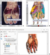
We used outlined images in which contours of the skin, hand muscles, and hand bones had been traced manually.78 The outlined images' intervals were 1 mm; therefore, there were 148 outlined images. To reduce the intervals of the outlined images from 1 mm to 0.2 mm, the outlines of the structures were interpolated. The outlines were then filled with different colors according to the individual components (Fig. 1A, right column).
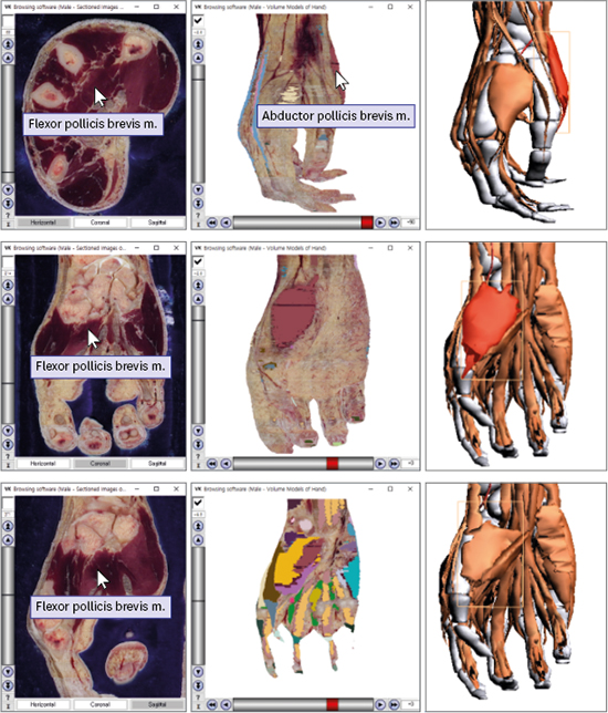
 | Fig. 1Browsing software of the sectioned images (left column) and color-filled images (right column) of the hand. The planes are horizontal (A), coronal (B), and sagittal (C). The color-filled images enable the names of the hand muscles and bones to appear automatically.
|
Using the sectioned images and color-filled images of the hand, vertical planes, 3D volume and surface models were generated. All of these image data were the sources of the two browsing software packages and one PDF file.
Browsing software of sectional planes
The 736 horizontal sectioned images of the hand (resolution 479 × 587) were stacked to produce 479 coronal planes and 587 sagittal planes (Fig. 1, left column). The procedure was repeated with color-filled images to produce the corresponding horizontal, coronal, and sagittal planes (Fig. 1, right column).
Three sets of the sectioned images (horizontal intervals 1 mm; coronal and sagittal intervals 0.2 mm) in the form of portable network graphics (PNG) files were loaded in browsing software that was upgraded from the previous version.1213 The equivalent color-filled images were loaded so that they could appear instead of the sectioned images (Fig. 1).
Browsing software of volume models
The original sectioned images of the hand were stacked to build a volume model (voxel size 0.2 mm). The model was peeled using distance-based organ peeling software made by the authors.1415 Peeling (thickness 0.4 mm) was continuously carried out to a maximum of 16 mm, so that 80 peeled volume models were prepared. Each model was rotated every 5 degrees to generate 72 captured views (PNG files).11
As preprocessing, the color-filled outlines were superimposed on the sectioned images. With the color-filled sectioned images, the same procedure of volume reconstruction, peeling, and rotation was repeated to acquire the corresponding captured views (Fig. 2, left column).11
 | Fig. 2Browsing software of volume models (left column) and PDF file of surface models (right column) of the hand. In the browsing software, the volume model (A) is switched to the color-filled volume model (B); the radial view is rotated to become the palmar view (C); the model is peeled further (D); and then magnified (E). The name of a selected muscle can be displayed any time. The comparable surface models are displayed in the PDF file.
PDF = portable document format.
|
PDF file of surface models
For the surface models, only the color-filled outlines (interval 1 mm), excluding the sectioned images, were saved as bitmap (BMP) files (Fig. 1A, right column). On Mimics version 17 software (Materialise, Leuven, Belgium), the color-filled outlines were stacked to achieve surface reconstruction of the hand muscles and bones. In each surface model, the accumulated outlines were removed and triangular surfaces were appropriately reduced in number.16
RESULTS
Two browsing software packages and one PDF file were downloadable from the homepage (http://anatomy.co.kr) for free by selecting the links: “Browsing software (Male — Sectioned hand),” “Browsing software (Male — Peeled hand),” and “PDF file of 3D models (Male — Hand).” The browsing software and the PDF file were compressed in ZIP files. Once the ZIP files were extracted, the browsing software packages could be operated without installation. After the decompression, the PDF file was readily readable using Adobe Acrobat Reader (Adobe Systems, Inc., San Jose, CA, USA).
All three software tools were operated off-line on a personal computer to show that the desired images could be promptly displayed. For the real-time operation, the sectional planes were saved in PNG format with a reduced file size (Fig. 1). Moreover, the volume models were processed to yield the peeled volume models in advance; their captured views were only exhibited in the browsing software (Fig. 2, left column).
Browsing software of sectional planes
Initially, in the browsing software, a sectioned image that was one of the raw data appeared. It could be replaced by the coronal or sagittal sectioned image by clicking the buttons in the horizontal direction. The sectioned images were browsed by using the scroll bar in the vertical direction or by inputting an image number. Any sectioned image could be swapped for the color-filled image by using the checkbox above the scroll bar (Fig. 1).
The selected image could be zoomed in or zoomed out by rolling the mouse wheel. The image could be shifted by moving the mouse with its left button pressed.
When the mouse pointer was located on an outlined muscle or bone in the color-filled images, the structure name was shown as a tool tip text beside the mouse pointer. The structure name was also displayed in the sectioned images because the sectioned images correspond to the color-filled images (Fig. 1).
Browsing software of volume models
In the browsing software, a volume model can continuously be rotated by 5 degrees with the scroll bar in the horizontal direction. The volume model could be continuously peeled at a 0.4 mm thickness with the scroll bar in the vertical direction; in the box just above the scroll bar, the peeling thickness (mm) was designated. Any volume model could be swapped for the color-filled volume model by using the checkbox (Fig. 2, left column).
The selected model could be zoomed in/out and shifted in the same manner. Not only in the color-filled volume models but also in the volume models of actual color, the name of the outlined structure was shown as a tool tip (Fig. 2, left column).
PDF file of surface models
In the PDF file, a list of the hand muscles and bones was displayed in the left top of the window; their selected surface models were exhibited in the right. At the bottom left, four bookmarks could be used to display the palmar, dorsal, radial, and ulnar views (Fig. 2, right column).
By clicking a surface model, its anatomical name was highlighted in the list. Reverse highlighting was also possible: By clicking an interesting hand structure in the list, the matching surface model was highlighted. The chosen surface model could be made to disappear and reappear (Fig. 2, right column).
The surface models could be freely rotated by moving the mouse with its left button pressed. They could be zoomed in and zoomed out either by rolling the mouse wheel or by moving the mouse with its right button pressed (Fig. 2, right column). Moreover, the models could be shifted by moving the mouse with both its left button and the keyboard Ctrl key pressed.
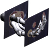
The surface models could be conveniently compared to the original sectioned images, which were embedded in the surface models (Fig. 3).
Combination of three software tools
Users who became familiar with the strong points of the three software tools were able to utilize them to the highest degree. An example was to observe the flexor digitorum superficialis, an extrinsic hand muscle.
In the browsing software of sectional planes, the details of the flexor digitorum superficialis could be obtained. In the carpal tunnel, the muscle was covered by the flexor retinaculum. In the fingers, the muscle's tendon was bifurcated within the synovial sheath (Fig. 4A).17
 | Fig. 4Observation of the flexor digitorum superficialis with the software tools. The tools are the browsing software packages of the sectional planes (A) and volume models (B) as well as the PDF file of the surface models (C).
PDF = portable document format.
|
In the browsing software of volume models, the depth of the flexor digitorum superficialis could be recognized; the muscle was more than 8 mm deep around the wrist, while it was approximately 4 mm deep at the palm and fingers (Fig. 4B).
In the PDF file of surface models, the locational relationship of the flexor digitorum superficialis could be grasped. The muscle intervened between the superficial group (flexor carpi radialis, palmaris longus, and flexor carpi ulnaris) and deep group (flexor digitorum profundus, flexor pollicis longus, and pronator quadratus) of the forearm muscles (Fig. 4C).17
DISCUSSION
In this study, three software tools for hand anatomy have been developed and are presented. The two browsing software packages and one PDF file each have their own strong points.
The browsing software involves the horizontal, coronal, and sagittal planes (Fig. 1) as well as the peeled volume models (Fig. 2, left column). The images are made from sectioned images of cadaver so that they show the real color of the hand in high resolution. For instance, the flexor digitorum superficialis' bifurcated tendons are identified (Fig. 4A), while they are hardly recognized on CTs or MRIs.
In the PDF file, the surface models of any desired structures can be chosen for display; the surface models can be rotated at arbitrary directions; the surface models can be made semitransparent to reveal hidden surface models. The Adobe Reader toolbar is available for diverse exploration of the surface models (Fig. 2, right column).
All three software tools present automatic labeling of the hand muscles and bones, even though they have different styles of labeling. Overall, the labeling on the basis of the color-filled images is superior to classical labeling because the use of color-filled images enables users to figure out the extent of each structure in the sectional planes (Fig. 1), peeled volume models, and surface models (Fig. 2).
The three tools are available to students for the self-learning of the hand clinics and hand anatomy. Examples are as follows. When CTs of the hand are interpreted, students may guess how a muscle appears on CT by using the browsing software of the sectional planes (Fig. 1A). For the same purpose, students can also use the PDF file of the surface models overlapped by the sectioned images (Fig. 3). When the hand of a cadaver is dissected, students can recognize how thick the subcutaneous tissue is using the browsing software of the volume models (Fig. 2, left column). When a hand is injured by trauma, such as laceration, students can anticipate which muscles are hurt by using the PDF file of the surface models (Fig. 2, right column).
The three tools are also practical for multimedia pedagogy of the hand anatomy. The following executions are to be considered. First, a lecture on hand anatomy is carried out by showing the images of the tools installed on the classroom computer. The tools successively display images in real time since they operate off-line on the computer. Otherwise, the captured images on the tools can be placed in lecture slides in advance.
Second, it is desirable to have students review the lecture using the same tools that are installed on every student's personal computer. Autonomic labeling is a robust function for reviewing the lecture individually (Figs. 1, 2, and 4).
Third, students can be evaluated by means of these tools. For testing, question numbers instead of structure names can be displayed in the browsing software. The lecturers may prefer a conventional tag examination. In such a preference, the images of the tools are printed out in colors and used for the tag examination.12
In the present study, software tools for viewing 2D planes and 3D models of the hand are released. The three types of tools, which involve the realistic images from a cadaver and the various functions, can be alternatively employed according to different goals.
 XML Download