

 PDF
PDF Citation
 Print
Print


INTRODUCTION
Hepatocellular carcinoma (HCC) is one of the most predominant and important malignancies in the world. It is the fifth most common cause of cancer in men and seventh in women, and is the third most common cause of cancer-related death worldwide.1 HCC possesses distinctive pathogenic features and its prognosis is affected by both the effect on the liver function and the overall tumor burden.2
According to the Barcelona Clinic Liver Cancer (BCLC) guideline, transarterial chemoembolization (TACE) is the standard treatment for intermediate HCC.3 However, there is increasing evidence that it may also be used in treatment of patients with early or advanced HCC.456 Nevertheless, the long-term prognosis of patients with TACE-treated HCC varies substantially because of the heterogeneous pathogenic nature, as well as tumor burden, liver function, and disease etiology. Therefore, identifying and predicting the patients who would benefit most from TACE is important for providers faced with the challenge of determining the most effective therapeutic strategy and assessing the prognosis.7
MicroRNAs (miRNAs) are endogenous, evolutionarily conserved, non-coding RNAs recognized as epigenetic factors regulating gene expression.8 Aberrant miRNA expression is a ubiquitous feature in numerous cancers including HCC.9 miRNAs play important roles in several aspects of carcinogenesis, including cell proliferation, cell cycle regulation, apoptosis, local invasion, and metastasis.10 Therefore, it has been theorized that several tumor-suppressing and oncogenic miRNAs may have potential applications in early diagnosis and prediction of prognosis in HCC.1112
miRNA-21 is considered a typical “onco-miRNA.” It inhibits the expression of phosphatases that downregulates AKT and mitogen-activated protein kinase (MAPK) signaling pathways.13 miRNA-21 can lead to upregulate cell proliferation. Therefore, miRNA-21 is associated with a wide variety of cancers, including liver cancer.14 A recent meta-analysis evaluated circulating miRNA-21 as a biomarker of various carcinomas and uncovered its potential as an early diagnostic tool.15 Inversely, miRNA-26a induces cell cycle arrest at the G1 phase in human HCC cells, in part through direct downregulation of cyclin D2 and cyclin E2.16 miRNA-26a is consistently downregulated in HCC.17 miRNA-29a-3p is also downregulated in HCC tissues, compared to adjacent non-cancerous liver tissues, and regulates HCC growth stems from modulation of the secreted protein acidic, rich in cysteine-AKT (SPARC-AKT) pathway.18
Even though these pathways are well studied, there is limited research regarding the prognostic role of levels of circulating miRNA-21, -26a, and -29a-3p in patients with HCC, especially with regard to TACE. The present study aimed at evaluating the association between pretreatment plasma levels of miRNA-21, -26a, and -29a-3p, treatment responses, and survival, in patients with TACE-treated HCC.
METHODS
Patients
A total of 1,024 patients underwent initial TACE for unresectable HCC, between February 2005 and December 2013 at Ajou University Hospital, Suwon, Korea. Of them, plasma samples from 198 patients were kept in the Ajou Human Bio-Resource Bank and were analyzed in this study. Patients were diagnosed with HCC if their tumor had a maximum diameter > 1 cm, and typical features of HCC (hypervascularity in the arterial phase and washout in the portal venous or delayed phase) were observed using dynamic computed tomography (CT) and/or magnetic resonance imaging (MRI). For tumors < 1 cm, ultrasonography was performed after 3 months.19 Patients were excluded from the study if 1) they had been previously treated with TACE or chemotherapy, or 2) they had undergone surgical resection or radiofrequency ablation within 6 months of their first TACE.
TACE procedure
TACE procedures were performed by 2 interventional radiologists. Doxorubicin (50 mg, ADM®; Dong-A Pharm. Co. Ltd., Seoul, Korea) and iodized oil (10 mL, Lipiodol Ultrafluide® emulsion; Laboratoire André Guerbet, Aulnay-sous-Bois, France) were injected into the tumor-feeding vessels per the typical TACE protocol followed by injection of gelatin sponge particles (Cutanplast®; Mascia Brunelli Spa, Viale Monza, Italy) until the blood flow was mostly obstructed.
Definitions
Liver cirrhosis was defined by histological evidence or ultrasonographic findings of: spleen size > 12 cm, portal vein diameter > 16 mm, or nodules within the hepatic parenchyma.20 Treatment outcomes after TACE were assessed using TACE refractoriness and liver transplantation (LT)-free survival. TACE refractoriness was defined as; 1) 2 or more consecutive insufficient responses of the treated tumor (viable lesion > 50%), even after changing chemotherapeutic agents, and/or revascularization of the feeding artery observed during post procedure evaluation CT/MRI at 1–3 months after selective TACE, 2) appearance of more than 2 consecutive intrahepatic lesions visualized during post procedure-evaluation CT, 3) increased vascular invasion, 4) extrahepatic spread, 5) continuous elevation of tumor markers, including immediately after TACE, and 6) technical failure of TACE.212223 If the patients received LT during the follow-up period, they were included in the analyses until the time of LT. Early TACE refractoriness was arbitrarily defined as TACE refractoriness within 1 year of the first TACE procedure.2223
Contrast-enhanced CT was performed 4 weeks after TACE, and every 3 months thereafter, to evaluate the tumor response. TACE was repeated every 6 to 8 weeks if viable tumors were detected on the follow-up imaging and if the liver function was adequate. Tumor response was estimated according to the modified Response Evaluation Criteria in Solid Tumors.24
miRNAs measurements
Pre-TACE plasma samples were collected and stored at −70°C until processing for miRNA analyses. Quantitative real-time polymerase chain reaction (qRT-PCR) was performed to identify and quantify the miRNA levels in plasma. Magnetic beads were utilized for miRNA extraction, using the TaqMan® miRNA ABC Purification Kit (Applied Biosystems, Carlsbad, CA, USA), according to the manufacturer's instructions. TaqMan® miRNA hsa-miRNA-specific primers (miRNA-29a-3p #002112; Applied Biosystems) and TaqMan® MicroRNA Reverse Transcription Kit (Applied Biosystems) were used to identify complementary DNA strands from the isolated miRNAs. An ABI PRISM 7300 instrument (Life Technologies, Foster City, CA, USA) and TaqMan® Universal Master Mix (Applied Biosystems) were used for polymerase chain reaction (PCR) amplification. The expression levels of miRNA were detected as Ct values, and the relative level of miRNA expression (fold change) was calculated by the 2(−ΔΔCt) method. miRNA-16 was selected as the internal control to normalize miRNA data. Samples from patients with HCC were compared with 5 reference samples obtained from patients with liver cirrhosis. The calculations used were as follows:
The mean was calculated after the qRT-PCR assays were performed in duplicate, using the same ABI PRISM 7300 instrument.
Statistical analysis
The results were reported as mean ± standard deviation. Hepatitis B virus (HBV) DNA levels were logarithmically adjusted for analysis. Significant cut-off values for each miRNA were determined with the Kaplan-Meier analysis with a log-rank test. miRNA levels were further analyzed as a panel (all 3 criteria met or not). Continuous variables were compared using independent-samples t-tests. Categorical data were compared using a Pearson χ2 test or Fisher's exact test. The cumulative probabilities of LT-free survival and overall TACE refractoriness were assessed by Kaplan-Meier analysis. The Cox proportional hazard model was used to identify factors associated with LT-free survival and overall TACE refractoriness. Binary logistic regression analysis was performed to identify factors associated with early TACE refractoriness, and variables that yielded P values < 0.05 in the univariate analyses were considered significant. miRNA performance, with respect to LT-free survival and overall TACE refractoriness, was assessed using an integrated area under the receiver operating characteristic curve (IAUC) plot and was compared by the bootstrapping method. IAUC was performed with R 3.1.1 (R Core Team [2014]; R: A language and environment for statistical computing; R Foundation for Statistical Computing, Vienna, Austria; URL: http://www.R-project.org/). Other statistical tests were performed using SPSS version 21.0 (SPSS Inc., Chicago, IL, USA). A P value < 0.05 was considered statistically significant.
RESULTS
Baseline characteristics of study population and HCC
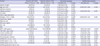
The mean age of the 198 patients included in the study was 58.6 ± 10.4 years and 75.8% of these patients were men. Most of the patients enrolled in this study had liver cirrhosis (94.9%) and HBV-associated HCC (99.0%). Among the patients with HBV-associated HCC, 67 (33.8%) patients were receiving oral antiviral therapy at the time of HCC diagnosis. Approximately one-half of all patients (44.4%) had a single tumor with a mean tumor size of 4.7 ± 3.2 cm. The percentage of patients with stage A, B, C, and D per the BCLC classification criteria was 44.9%, 28.3%, 25.3%, and 1.5%, respectively.3 The median follow-up duration was 22.3 months (range, 0.7–79.0 months). The baseline characteristics of the study population are summarized in Table 1.
Table 1
Baseline characteristics of study population
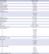
Values are presented as number (%), mean ± standard deviation, or median (range).
HBsAg = hepatitis B surface antigen, HBeAg = hepatitis B e antigen, ALT = alanine aminotransferase, INR = international normalized ratio, AFP = alpha-fetoprotein, HBV = hepatitis B virus, BCLC = Barcelona Clinic Liver Cancer, UICC = Union for International Cancer Control.
![]()
LT-free overall survival following TACE treatment and TACE outcomes
The 1-, 3-, and 5-year LT-free survival rates (95% confidence interval [CI]) were 65.9% (59.2–72.6), 40.6% (33.2–48.0), and 19.9% (11.5–28.3), respectively. During the follow-up period, 10 patients underwent LT. TACE refractoriness was evaluable in 7 of these transplant patients, of whom 2 exhibited TACE refractoriness before LT and 5 did not. Three of the 10 patients received LT prior to demonstrating TACE refractoriness and were categorized with the no-TACE refractoriness group. The cumulative LT-free survival rate is presented in the Supplementary Fig. 1A.
During the follow-up period, 59.6% of all patients demonstrated a complete response with repetitive TACE treatments. The mean number of TACE procedures needed to achieve a complete response was 1.49 (range, 1–6). Of these cases, 96 patients (81.4%) eventually showed tumor recurrence. The median time of recurrence was 10.7 (range, 1–37) months.
Among the 198 patients, TACE refractoriness was evaluated in 177 patients, as worsening liver function in 21 patients prevented evaluation. Of patients with TACE refractoriness, 118 patients (66.3%) became refractory during the follow-up period (Supplementary Fig. 1B). The 1-, 3-, and 5-year TACE refractoriness rates (95% CI) were 47.2% (39.8–54.6), 71.3% (63.5–79.1), and 80.4% (71.8–89.0), respectively. The median time of TACE refractoriness was 14.0 (0.77–73) months. Etiology of TACE refractoriness was as follows: incomplete necrosis of the intrahepatic lesion or new intrahepatic lesion development (45.6%), metastatic spread (42.2%), further vascular invasion (20.3%), and TACE failure due to technical problems (3.3%).
Predictive factors for LT-free survival
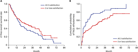
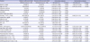
Predictive factors for LT-free survival were evaluated in all the 198 patients included in this study. In the univariate analysis, absence of liver cirrhosis, high Child-Pugh class (B/C vs. A), low albumin level, high bilirubin level, low international normalized ratio, low alpha-fetoprotein (AFP) level (< 400 ng/mL), low HBV DNA level, small tumor size (≤ 2 cm), and absence of macrovascular invasion represented positive predictive factors for LT-free survival. Multivariate Cox regression analyses revealed that large tumor size (hazard ratio [HR], 3.04; 95% CI, 1.51–6.11; P = 0.002) and macrovascular invasion (HR, 3.57; 95% CI, 2.32–5.48; P < 0.001) were the only independent risk factors for poor LT-free survival. Individual or combined miRNA-21, -26a, and -29a-3p levels could not statistically predict LT-free survival after a Cox regression analysis (Table 2 and Fig. 1A).
Table 2
Predictive factors of overall liver transplantation-free survival in HCC patients treated with TACE
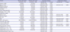
Values are presented as number (%) or HR (95% CI).
HCC = hepatocellular carcinoma, TACE = transarterial chemoembolization, HR = hazard ratio, CI = confidence interval, HBeAg = hepatitis B e antigen, ALT = alanine aminotransferase, INR = international normalized ratio, AFP = alpha-fetoprotein, HBV = hepatitis B virus, miRNA = microRNA.
aP < 0.05.
![]()
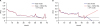
 | Fig. 1Cumulative probabilities of LT-free survival and TACE refractoriness according to plasma miRNA-21, 26a, and 29a-3p level. (A) Patients with high miRNA-21 (≥ 2.5), high miRNA-26a (≥ 1.5), and low miRNA-29a-3p (< 0.4) levels (all 3 criteria met) showed a shorter LT-free survival than those who did not; however, this was not statistically significant. (B) Patients with high miRNA-21 (≥ 2.5), high miRNA-26a (≥ 1.5), and low miRNA-29a-3p (< 0.4) levels (all 3 criteria met) had a higher probability of overall TACE refractoriness than those who did not.
LT = liver transplantation, TACE = transarterial chemoembolization, miRNA = microRNA.
|
Predictive factors for overall TACE refractoriness
Univariate and multivariate Cox regression analyses were used to investigate predictive factors for overall TACE refractoriness. In the univariate analysis, advanced age, hepatitis B e antigen positivity, cirrhosis, and a high platelet count were protective factors for overall TACE refractoriness. In patients with HCC, large tumor size (≥ 2 cm), macrovascular invasion, high AFP levels (> 400 ng/mL), high miRNA-21 levels (≥ 2.5), high miRNA-26a levels (≥ 1.5), and low miRNA-29a-3p levels (< 0.4) were found to be risk factors for overall TACE refractoriness (Table 3 and Fig. 1B). However, individual or combined miRNA-21, -26a, and -29a-3p levels did not demonstrate significant predictive power for overall TACE refractoriness. In multivariate analysis, large tumor size (HR, 2.43; 95% CI, 1.27–4.67; P = 0.007), macrovascular invasion (HR, 2.18; 95% CI, 1.28–3.72; P = 0.004), and high AFP levels (HR, 1.88; 95% CI, 1.22–2.90; P = 0.004) were the only independent predictive factors for overall TACE refractoriness.
Table 3
Predictive factors of overall TACE refractoriness in 177 HCC patientsa treated with TACE
Values are presented as number (%) or HR (95% CI).
TACE = transarterial chemoembolization, HCC = hepatocellular carcinoma, HR = hazard ratio, CI = confidence interval, HBeAg = hepatitis B e antigen, ALT = alanine aminotransferase, INR = international normalized ratio, AFP = alpha-fetoprotein, HBV = hepatitis B virus, miRNA = microRNA.
aTACE refractoriness was assessable in 161 patients; bP < 0.05.
![]()
Predictive factors for early TACE refractoriness in patients with HCC
As the evaluation of predictive factors for overall TACE refractoriness showed an association with plasma miRNA in patients with HCC, further evaluation was performed to determine predictive factors for early TACE refractoriness in the same population.2223 Univariate analyses revealed male sex, high AFP levels (> 400 ng/mL), high HBV DNA levels, multiple tumors (≥ 2), large tumor size (≥ 2 cm), macrovascular invasion, high miRNA-26a levels (≥ 1.5), low miRNA-29a-3p levels (< 0.4), and the combination of miRNAs were found to be risk factors for early TACE refractoriness. Conversely, advanced age (≥ 55 year) and cirrhosis were found to be protective factors for early TACE refractoriness. A multivariate binary logistic regression analysis of these factors further demonstrated that large tumor size (HR, 4.62; 95% CI, 1.50–14.21; P = 0.008), macrovascular invasion (HR, 3.80; 95% CI, 1.19–12.20; P = 0.025), and a distinct miRNA combination panel (miRNA-21 ≥ 2.5, miRNA-26a ≥ 1.5, and miRNA-29a-3p < 0.4, all 3 met vs. 2 or less conditions being satisfied; HR, 2.32; 95% CI, 1.08–4.99; P = 0.031) were independent predictive factors for early TACE refractoriness (Table 4).
Table 4
Predictive factors of early TACE refractoriness in 177 HCC patientsa treated with TACE
Values are presented as number (%) or HR (95% CI).
TACE = transarterial chemoembolization, HCC = hepatocellular carcinoma, HR = hazard ratio, CI = confidence interval, HBeAg = hepatitis B e antigen, ALT = alanine aminotransferase, INR = international normalized ratio, AFP = alpha-fetoprotein, HBV = hepatitis B virus, miRNA = microRNA.
aTACE refractoriness was assessable in 161 patients; bP < 0.05.
![]()
Discriminatory ability of miRNA-21, -26a, and -29a-3p levels in predicting LT-free survival and overall TACE refractoriness
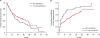
The abilities of plasma miRNA-21, -26a, and -29a-3p levels in predicting LT-free survival and overall TACE refractoriness were assessed using an IAUC plot. The base model included age, sex, tumor number, tumor size, and vascular invasion, alone or in combination. AFP or miRNAs were added and compared to the base model. The discriminatory ability was fair in the base model for LT-free survival (Fig. 2A, Harrell's C-index, 0.728). Adding miRNA-26a and AFP levels to the base model showed a slight improvement (Harrell's C-index, 0.731; 95% CI, −0.02837, −0.0004). With regards to the overall TACE refractoriness, the model that utilized both AFP and miRNA-26a levels showed the best discriminatory ability compared to the base model (Fig. 2B, 0.766 vs. 0.762, respectively; 95% CI, −0.03199, −0.00007).
 | Fig. 2IAUC plots of predictive models, based on a model with or without AFP and miRNAs. (A) IAUC plot of predictive models of LT-free survival. (B) IAUC plot of predictive models of overall TACE refractoriness. The base model consisted of sex, age, tumor number, size, and vascular invasion. Models with AFP and miRNA-26a showed the best discriminatory performance.
IAUC = integrated area under the receiver operating characteristic curve, miRNA = microRNA, AFP = alpha-fetoprotein, LT = liver transplantation, TACE = transarterial chemoembolization.
|
Correlation between pretreatment miRNA-21, -26a, and -29a-3p levels and patient characteristics
The correlation between miRNA-21, -26a, and -29a-3p levels and baseline characteristics are summarized in Supplementary Table 1. miRNA-21 levels showed a positive correlation with tumor size (r = 0.157; P = 0.027). miRNA-26a levels varied significantly with tumor number, and did not show a linear correlation. Plasma miRNA-29a-3p showed a positive correlation with alanine aminotransferase level (r = 0.152; P = 0.032), depending on the Child-Pugh class, but did not show a linear relationship.
DISCUSSION
To the best of our knowledge, this is the first study to report the association between plasma miRNA-21, -26a, and -29a-3p levels and outcomes following TACE treatment in patients with unresectable HCC. A combination of pretreatment plasma miRNA-21, -26a, and -29a-3p levels predicted early TACE refractoriness in patients with HCC who were subsequently treated with TACE.
Several studies have reported the diagnostic value of miRNA-21, a typical “onco-miRNA,” and its association with a wide variety of cancers, including HCC.1415 However, few studies have evaluated the prognostic value of miRNA-21 for HCC, especially after TACE. A recent study reported that higher levels of miRNA-21 were associated with shorter post-operative survival in patients with HCC.25 In contrast, other researchers found serum miRNA-21 level was not significantly associated with survival after TACE.26 In the present study, high pretreatment plasma miRNA-21 levels were associated with early TACE refractoriness, as part of a panel with miRNA-26a and -29a-3p, but not with LT-free overall survival.
The miRNA-26 family genetic markers are emerging as key regulators in carcinogenesis and tumor progression. miRNA-26a induces cell cycle arrest at the G1 phase in human HCC cells, in part through direct downregulation of cyclin D2 and cyclin E2.16 Furthermore, therapeutic miRNA-26a delivery using an adeno-associated virus inhibited liver cancer cell formation while also inducing tumor-specific apoptosis and providing considerable protection from disease progression without toxicity.16 In the present study, a high miRNA-26a level (≥ 1.5) was associated with overall TACE refractoriness in univariate analysis and with early TACE refractoriness, as part of a panel, in multivariate analysis. This result contradicts previous reports that patients with HCC and low miRNA-26a expression had shorter overall survival than those with high expression.17 Subsequent studies by the same researchers also demonstrated that miRNA-26a could suppress tumor growth and metastasis of HCC through interleukin-6-signal transduction and activation of transcription 3 signaling, and exerting an anti-angiogenesis function by inhibiting hepatocyte growth factor-cMet.2728 The reason for this difference is not clear; however, it might be partly due to the different cellular contexts of the tumors. For example, miRNA-26 is consistently downregulated in HCC, nasopharyngeal carcinoma, lung cancer, and breast cancer,17293031 but is overexpressed in high-grade glioma and cholangiocarcinoma.3233 This has ramifications for the present study, as HCC was mainly diagnosed based on imaging studies without pathologic confirmation. Therefore, patients with combined HCC-cholangiocarcinoma may have been accidentally included in the study population. This difference could also be explained by the difference in study populations. The previous study only included patients whose tumors had been resected, using a median value ≈ 0.8 as the cut-off level for differentiating high versus low level of miRNA-26a. This cut-off value is much lower than that of the current study (1.5), in which the median value was 1.655.
miRNA-29a may act as either an oncogene or a tumor suppressor gene. For example, miRNA-29a is known to be under-expressed in lung cancers,34 but upregulated in ovarian and colorectal cancers.3536 There are conflicting reports about the association between miRNA-29a and HCC pathology. High levels of miRNA-29a-5p in HCC tissue are correlated with early tumor recurrence after HCC surgery, especially in patients with BCLC stage 0/A.37 However, the same study found miRNA-29a-3p levels in HCC tissues were not different depending on early recurrence. A recent study proposed that a high concentration of serum miRNA-29a-3p is associated with poor overall survival and progression-free survival in patients with HCC treated with resection or local ablation.38 However, a contrasting study found miRNA-29 genetic (miRNA-29a-3p/b/c) expression was downregulated in HCC tissue suggesting a potential tumor suppressive role.39 The reason for discrepancy in these results is not clear, but a deeper investigation into different miRNA-29 family members would likely explain the varied reports. miRNA-29a-3p is the counterpart of miRNA-29a-5p in the development and progression of cancers. Consequently, in the present study, pretreatment low miRNA-29a-3p levels tended to be associated with both early and overall TACE refractoriness. This is consistent with a recent study that demonstrated downregulation of miRNA-29a-3p in AFP-positive HCCs. AFP inhibited miRNA-29a expression and induced DNA methyltransferase 3A through c-MYC binding to the transcript of miRNA-29a/b-1. Low levels of miRNA-29a-3p in tumor tissues have been associated with poor overall survival in patients with HCC.40 In the present study, however, miRNA-29a-3p levels did not support a linear correlation with AFP levels (r = −0.081; P = 0.259).
In the present study, individual levels of miRNA-21, -26a, and -29a-3p failed to show a statistically significant predictive power for early TACE refractoriness. However, combining miRNA-21, -26a, and -29a-3p together, the panel proved to be a predictive factor for early TACE refractoriness. This result suggests that multi-biomarker panels may have stronger diagnostic and prognostic power in panels.
The present study has a few limitations. First, the international community lacks a consensus definition of TACE refractoriness. The lack of a precise objective definition has led to conflicting conclusions regarding the benefit of repetitive TACE therapy. The present study borrowed the Japanese definition of TACE refractoriness, which seems to be easily applicable in clinical settings.21 Second, tumor stages of patients with HCC vary, and include all 4 BCLC stages (A, B, C, and D). Because this study was not a planned prospective clinical trial but a retrospective cohort study based on real clinical practice, patients with HCC with heterogeneous tumor stages underwent TACE as the first-line therapy for different reasons. In the present study, BCLC stage A was the most frequently observed.
In conclusion, a panel consisting of pretreatment miRNA-21, -26a, and -29a-3p can predict early TACE refractoriness in patients with TACE-treated HCC. Pretreatment circulating miRNA-21, -26a, and -29a-3p levels may also serve as potential candidate biomarkers for the prediction of treatment outcomes in patients with TACE-treated HCC.
 XML Download