Abstract
Purpose
To compare the success rates between silicone tube intubation using a lacrimal endoscope and using a conventional nasal endoscope alone in adult patients suffering from epiphora.
Methods
We conducted a retrospective chart review of 80 eyes of 55 patients who underwent silicone tube intubation from January 2014 to June 2017. Patients were preoperatively diagnosed with syringing and dacryocystography. The silicone tube was removed 3 months after surgery and success rates were evaluated at 4 and 12 months. Success rates were analyzed by dividing the patients into two groups, according to lacrimal endoscope use.
Results
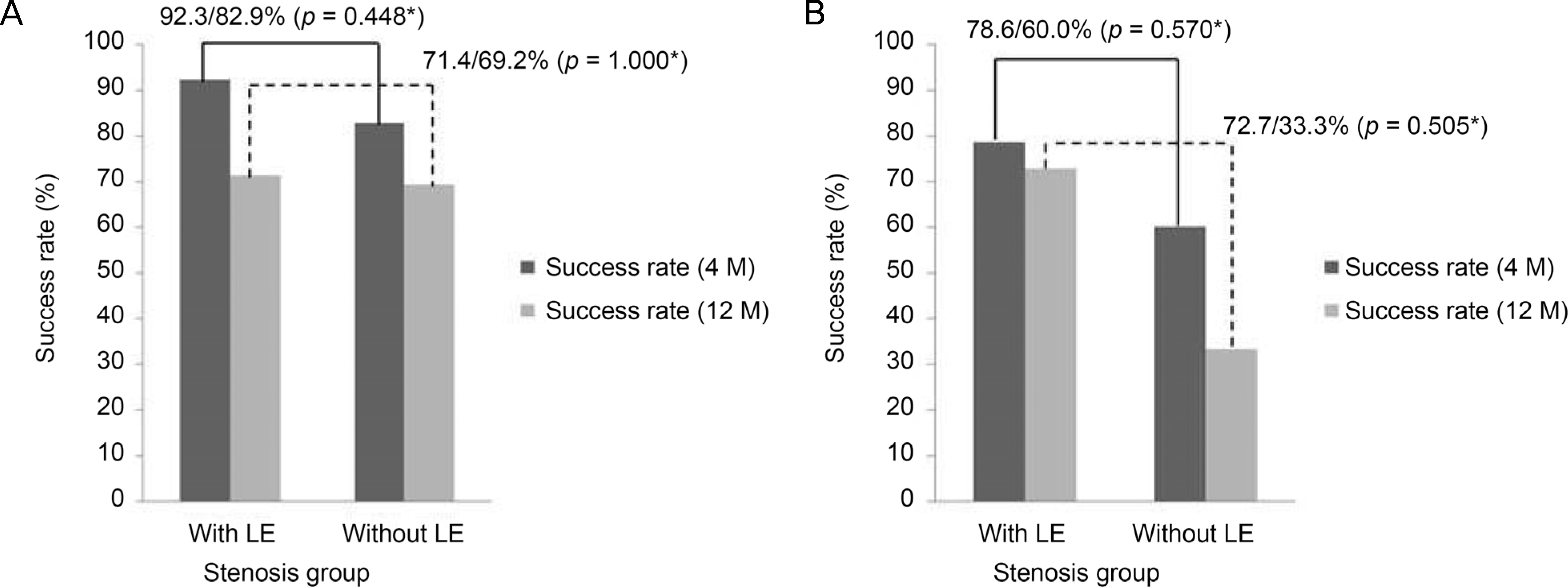
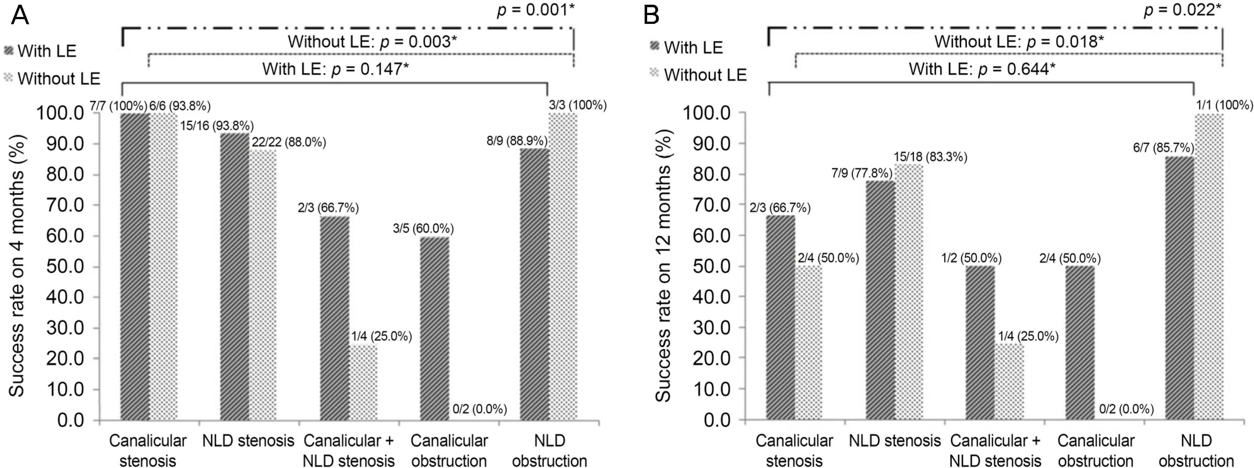
A lacrimal endoscope was used in 40 eyes. In the group using a lacrimal endoscope, preoperative diagnoses were partial obstruction in 26 eyes and complete obstruction in 14 eyes. In the group without lacrimal endoscope use, preoperative diagnoses were partial obstruction in 35 eyes and complete obstruction in 5 eyes (p = 0.018). The success rates at 4 and 12 months after surgery in the two groups (with and without lacrimal endoscope use) were 87.5% and 80.0% and 72.0% and 62.1% (p = 0.546 and p = 0.565), respectively. The success rates of patients with partial obstruction in the two groups were 92.3% and 82.9% at 4 months and 71.4% and 69.2% at 12 months (p = 0.448 and p = 1.000), respectively. The success rates of patients with complete obstruction in the two groups were 78.6% and 60.0% at 4 months and 72.7% and 33.3% at 12 months (p = 0.570 and p = 0.505), respectively. Site differences, the degree of obstruction, and lacrimal endoscope use had a significant impact on the success rate at 4 and 12 months (p = 0.001 and p = 0.022, respectively).
Conclusions
Although silicone tube intubation using a lacrimal endoscope cannot guarantee a significant success rate, it is possible to observe the anatomical structure of the nasolacrimal pathway in real time, such that the appropriate diagnosis and treatment can be performed simultaneously. Because patients diagnosed as having a complete obstruction had a good success rate, we can extend indication of silicone tube intubation as a less invasive approach.
Go to : 
References
1. Oum JS, Park JW, Choi YK, et al. Result of partial nasolacrimal duct obstruction after silicone tube intubation. J Korean Ophthalmol Soc. 2004; 45:1777–82.
2. Jones LT. An anatomical approach to problems of the eyelids and lacrimal apparatus. Arch Ophthalmol. 1961; 66:111–24.

3. Lee SM, Chung SJ, Lew H. Clinical efficacy of lacrimal endoscopy assisted silicone tube intubation in patients with nasolacrimal duct obstruction. J Korean Ophthalmol Soc. 2018; 59:582–8.

5. Kwon YH, Lee YJ. abdominal results of silicone tube intubation in incomplete nasolacrimal duct obstruction (NLDO). J Korean Ophthalmol Soc. 2008; 49:190–4.
6. Peterson NJ, Weaver RG, Yeatts RP. Effect of short-duration abdominal intubation in congenital nasolacrimal duct obstruction. Ophthal Plast Reconstr Surg. 2008; 24:167–71.
7. Salari AM, Tokhmehchi MR. A simplified method for abdominal silicone intubation. Acta Ophthalmol. 2008; 86:230.
8. Cohen SW, Prescott R, Sherman M, et al. Dacryoscopy. Ophthalmic Surg. 1979; 10:57–63.
9. al-Hussain H, Nasr AM. Silastic intubation in congenital abdominal duct obstruction: a study of 129 eyes. Ophthal Plast Reconstr Surg. 1993; 9:32–7.
10. Boyrivent V, Ruban JM, Ravault MP. Role of nasolacrimal abdominal in the treatment of lacrimation caused by congenital abdominal duct obstruction in infants. J Fr Ophtalmol. 1993; 16:532–7.
11. Kim YR, Ahn M. Long term effect of double silicone tube abdominal for acquired nasolacrimal duct obstruction. J Korean Ophthalmol Soc. 2012; 53:1554–8.
12. Jung JJ, Jang SY, Jang JW, In JH. Comparison results of silicone tube intubation according to syringing and dacryocystography. J Korean Ophthalmol Soc. 2014; 55:1584–8.

13. Connell PP, Fulcher TP, Chacko E, et al. Long term follow up of nasolacrimal intubation in adults. Br J Ophthalmol. 2006; 90:435–6.

14. Kim JH, You IC, Ahn M. The clinical outcome of endoscopic abdominal silicone tube intubation according to nasolacrimal duct resistance. J Korean Ophthalmol Soc. 2016; 57:1–5.
15. Sasaki T, Nagata Y, Sugiyama K. Nasolacrimal duct obstruction classified by dacryoendoscopy and treated with inferior meatal dacryorhinotomy. Part I: Positional diagnosis of primary abdominal duct obstruction with dacryoendoscope. Am J Ophthalmol. 2005; 140:1065–9.
16. Lim SW, Sung YJ, Lew H. Clinical efficacy of lacrimal endoscopy in patients with epiphora. J Korean Ophthalmol Soc. 2017; 58:495–502.

17. Park HJ, Hwang WS. Clinical results of silicone intubation for abdominal patients. J Korean Ophthalmol Soc. 2000; 41:2327–31.
Go to : 
 | Figure 1.Intraoperative photographs of silicone tube intubation using lacrimal endoscope with nasal endoscope. (A) The schematic diagram of lacrimal endoscope (RUIDO fiberscope, Fibertechco., Tokyo, Japan): probe, bent type 0.9 mm diameter and peripheral apparatus including monitor, recording and imaging system. (B, C) Insertion. (D) Examination of nasolacrimal pathway while as-sisting irrigation thorough water channel. |
 | Figure 2.Bar graphs showing the success rates of silicone tube intubation with or without lacrimal endoscope on 4 and 12 months since intubation. (A) Patients preoperatively diagnosed as nasolacrimal duct stenosis (or partial obstruction). There were no statistically significant differences between the two groups (p = 0.448, p = 1.000, respectively). (B) Patients preoperatively diagnosed as nasolacrimal duct obstruction. There were no statistically significant differences between the two groups (p = 0.570, p = 0.505, respectively). LE = lacrimal endoscope; M = months. * Fisher's exact test. |
 | Figure 3.Bar graphs showing the success rates on (A) 4 months and (B) 12 months after silicone tube intubation with or without lacrimal endoscope according to the site of stenosis or obstruction in dacryocystography. In group with lacrimal endoscope use (black hatch pattern), there were no significant differences in the success rate (p = 0.147, p = 0.644, respectively). However, there were significant differences in group without lacrimal endoscope use (white dot pattern) (p = 0.003, p = 0.018). When considering both lacrimal endoscope use and the site of stenosis or obstruction, there were significant differences in the success rate (p = 0.001, p = 0.022). LE = lacrimal endoscope; NLD = nasolacrimal duct. * Fisher's exact test. |
Table 1.
Baseline characteristics of the patients (80 eyes)
| With lacrimal endoscope | Without lacrimal endoscope | p-value | |
|---|---|---|---|
| Sex | 0.586* | ||
| Male | 7/40 (17.5) | 10/40 (25.0) | |
| Female | 33/40 (82.5) | 30/40 (75.0) | |
| Age (years) | 64.6 ± 13.6 | 59.0 ± 14.1 | 0.073† |
| Duration of symptoms (months) | 36.1 ± 45.6 | 34.2 ± 54.5 | 0.864† |
| Duration of intubation (months) | 3.5 ± 1.4 | 3.2 ± 1.2 | 0.311† |
| Follow up periods (months) | 12.7 ± 5.9 | 11.4 ± 8.9 | 0.461† |
Table 2.
Comparison of preoperative diagnosis
| Preoperative diagnosis | With lacrimal endoscope | Without lacrimal endoscope | Total |
|---|---|---|---|
| Stenosis | 26 (65.0) | 35 (87.5) | |
| Canalicular | 7 (17.5) | 6 (15.0) | 13 (16.2) |
| Nasolacrimal duct | 16 (40.0) | 25 (62.5) | 41 (51.2) |
| Canalicular + Nasolacrimal duct | 3 (7.5) | 4 (10.0) | 7 (8.8) |
| Obstruction | 14 (35.0) | 5 (12.5) | |
| Canalicular | 5 (12.5) | 2 (5.0) | 7 (8.8) |
| Nasolacrimal duct | 9 (22.5) | 3 (7.5) | 12 (15.0) |
| Total p-value | 40 | 40 | 800.018* |
Table 3.
Success rates with or without lacrimal endoscope use on 4 and 12 months after intubation
| With lacrimal endoscope | Without lacrimal endoscope | p-value* | |
|---|---|---|---|
| Success rate (4 months after intubation) | 35/40 (87.5) | 32/40 (80.0) | 0.546 |
| Success rate (12 months after intubation) | 18/25 (72.0) | 18/29 (62.1) | 0.565 |


 PDF
PDF ePub
ePub Citation
 Print
Print


 XML Download