

 PDF
PDF Citation
 Print
Print


INTRODUCTION
Dental caries is a highly prevalent disease [1]. The conservative intervention technique, which has been introduced in the past few years for extensive caries-attacked teeth, involves elimination of the affected tissues and replacement of the defect with restorative materials [2]. It has been recommended to remove the outer carious infected layer of dentin, while conserving the inner-caries affected dentin [3]. Caries-affected dentin (CAD) differs from normal dentin in its intrinsic properties, for example, it exhibits reduced permeability due to the formation of whitlockites inside dentinal tubules and partially demineralized intertubular dentin [2]. Recent management strategies involve the remineralization of CAD to avoid its unnecessary removal, as current tissue-engineering technologies are not capable of synthesizing artificial dentin [4]. This minimally invasive conservative approach has led to an increasing demand for remineralizing agents and adhesive restorative materials that bond to the remaining CAD.
Changes in both the structural and biochemical compositions of dentin during the caries process make CAD a challenging substrate for bonding with resin adhesives [5]. Carious dentin consists of two distinct layers: a bacterially infected dentin (outer) layer and an affected dentin (inner) layer [6]. Caries-infected dentin has been described as highly demineralized and physiologically unremineralizable, being accompanied by irreversibly denatured collagen fibrils with a virtual loss of cross-linkages. In contrast, the inner layer of CAD is uninfected, it is partially demineralized but has a remineralizing ability due to the presence of intact collagen fibers, and it is protected by hydroxyapatite crystals; therefore, it should be conserved during clinical treatment. Consequently, during cavity preparation for an adhesive restoration and after removal of caries-infected dentin, CAD forms large areas of the cavity floor. It differs from normal dentin in many aspects, including physical, chemical, and biomechanical properties, which significantly affect the outcomes of resin-dentin bonding. It has around half the hardness of normal dentin and displays excessive porosity due to mineral loss. Moreover, its intrinsic properties are different, for example, its permeability is reduced due to the formation of whitlockites inside the dentinal tubules and partially demineralized intertubular dentin. This obliteration of dentinal tubules by acid-resistant mineral crystals explains the lower bond strength to CAD than normal dentin [789].
Several trials have been conducted to improve bonding to CAD, including treatments with fluoride, tricalcium phosphate agents, and casein phosphopeptide-amorphous calcium phosphate (CPP-ACP) [710]. These are new remineralization approaches in preventive dentistry which that aim to reduce the lesion depth and to increase both the remineralization and mineralized content of the tooth structure, by improving the ratios and saturation of both calcium and phosphate. Recent studies have reported that the application of CPP-CAP containing remineralizing agents can decrease demineralization and enhance remineralization of either bovine or human dentin [1112]. Furthermore, CPP-ACP has a direct anti-cariogenic effect on oral biofilms [9].
Glass ionomer cements (GICs) are commonly used as dental restorative materials, particularly in patients at high risk of caries [13]. They have many desirable advantages such as chemical bonding to the tooth structures [514], promotion of remineralization due to gradual fluoride release and cariostatic effects [15]. They are superb choices as fluoride-releasing materials, since they have a higher quality than other restorative materials such as giomers and compomers in terms of continuous fluoride release and recharge. Bonding of GIC is based mainly on an ionic chemical bond formed between the liquid component of the cement and the calcium of the hydroxyapatite of the tooth structure [16]. Several improvements have been introduced to overcome the drawbacks of conventional GICs. One such modification is the development of resin-modified glass ionomer cement (RMGIC) which has favorable properties, including bonding to the tooth structure through chemical and micromechanical mechanisms, increased working time, decreased setting time, ease of handling, and improved physical properties, fluoride release and aesthetics [17].
A previous study investigated CPP-ACP modified GICs and reported very promising outcomes, such as increased compressive strength and micro-shear bond strength to dentin. Moreover, it showed an ability to increase enamel resistance to acid demineralization, without an unfavorable influence on the GICs' mechanical properties [18]. CPP-ACP-modified GIC could counteract the incidence of secondary root caries when exposed to cariogenic biofilms of many species in an in vitro study [19]. As well, it was reported that CPP-ACP-modified GIC exhibited higher shear bond strength to enamel than conventional GIC [20].
In the light of the recently introduced approaches for conservative treatment of dentin caries and the unique features of GICs, this study is aimed to evaluate the impact of CPP-ACP-remineralized CAD on fluoride release and the micro-shear bond strength of RMGIC bonded to it. The research hypothesis was that a CPP-ACP remineralizing agent would remineralize CAD and influence its depletion of fluoride released from RMGIC into the solution. Furthermore, this agent would increase the micro-shear bond strength of RMGIC bonded to dentin.
MATERIALS AND METHODS
Teeth selection, specimens preparation and grouping of the specimens
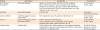
The commercial product Tooth Mousse (Recaldent, GC International, Tokyo, Japan) and light-activated GIC (Fuji II LC, GC Corporation, Tokyo, Japan) were used. A description of the materials utilized in the study is presented in Table 1. Thirty extracted human sound third molar teeth (obtained from the out-patient clinic of the Faculty of Dentistry, Mansoura University, Egypt, and surgically extracted due to impaction; ethics committee approval No. 16080518) were collected, thoroughly washed under distilled water, scaled using a sharp hand sickle scaler to remove soft tissues, and stored in a solution of 1% chloramine-T at 4°C to inhibit microbial growth.
Table 1
Materials used in the study
![]()
Fifteen teeth were sliced horizontally in the bucco-lingual direction beneath the dentino-enamel junction using a hard tissue microtome (Isomet, Buehler, Lake Bluff, IL, USA) at 2,500 rpm under copious water spray coolant to expose a flat dentin surface. Another parallel section 2 mm away from the first one was made to produce a flat dentin disc (used for evaluating fluoride release). The other fifteen teeth had their occlusal surfaces removed below the dentino-enamel junction to expose a flat dentin surface, and their roots were mounted in self-cure acrylic resin blocks (used for testing the micro-shear bond strength of RMGIC to dentin). The 15 specimens designated for each test were divided into 3 groups (5 specimens each) as follows: 1) control (sound dentin); 2) artificially demineralized dentinal specimens (demineralized CAD specimens); 3) demineralized then CPP-ACP remineralized dentinal specimens (remineralized CAD specimens).
Artificial dentinal caries induction
Artificial carious dentinal lesions were introduced in the specimens of groups 2 and 3. The specimens were immersed into an acidified gel of 5% gelatin solution with pH adjusted to 4.5 using lactic acid for 14 days, following the Silverstone-recommended protocol. At room temperature, each specimen was immersed in 10 mL of the acidified gel. This gel was replaced once after 7 days. The specimens were washed in hot running water (50°C–60°C) to remove the gel remnants and gently air-dried [9].
Application of casein phosphopeptide-amorphous calcium phosphate remineralizing agent
The remineralizing agent was applied only to the specimens of groups 3 immediately after demineralization. A thin coat of CPP-ACP was painted on the flat exposed dentinal surface using a micro-brush for 15 min/day and left undisturbed. After remineralization, specimens were rinsed with distilled water for 20 seconds and stored in artificial saliva until the next application; the process was repeated for 5 consecutive days [13].All specimens were stored in artificial saliva that was prepared and maintained at a pH of 7.4 to 8.1 in an incubator at 37°C [21].
Evaluation of fluoride release by ion chromatography
Fifteen disc-shaped dentinal specimens were classified into the 3 prescribed groups and prepared as previously described. The specimens were bonded to RMGIC (to study the influence of CAD remineralization on its consumption of fluoride released from RMGIC). The discs were treated with polyacrylic acid conditioner, which was left undisturbed for 10 seconds, water-rinsed for 10 seconds and then gently air-dried for 5 seconds to leave a moist surface, as recommended by the manufacturer.
The specimens were bonded with RMGIC using a split Teflon mold (4 mm in diameter and 2 mm in thickness) [22]. The RMGIC capsules were triturated for 10 seconds in an amalgamator (Fushion SyG-200, TRIUP International Corporation, Shanghai, China). The mold was placed on the conditioned dentin surface, filled with RMGIC, and covered with a celluloid strip. For the specimen to be easily suspended into the solution, a nylon thread was placed in each specimen, and excess material was removed by placing a glass slide over the filled mold and gently pressing to extrude the excess material. The specimens were cured using a Demetron LC curing light (Kerr Corporation, Orange, CA, USA) with a minimum output of 600 mW/cm2, removed gently from the mold, and the whole assembly was stored in 5 mL of distilled water at 35°C in a plastic container.
At intervals of 24 hours, 3, 5, and 7 days from the beginning of immersion, the amount of fluoride released from specimens was determined. At each measurement interval, the specimens were extracted from the solution, filter paper-dried, and then, immersed in another 5 mL of distilled water. For free fluoride ion determination in distilled water-collected specimens, an Ion Chromatograph (DX 500, Dionex, Camberley, UK) with suppressed conductivity was used. The fluoride ion concentration was determined to an accuracy of 0.001 ppm [14].
Measurement of micro-shear bond strength of resin-modified glass ionomer cement to dentin
Fifteen specimens were prepared, classified into 3 groups, and bonded with RMGIC as described above. A Tygon tube (TYGON Medical Tubing, Saint Gobain, Akron, OH, USA) of 2 mm in height and 1 mm in diameter was fixed onto the conditioned flat dentinal surface, filled with RMGIC, and light cured. After light curing, the specimens were stored in distilled water at 37°C for 24 hours prior to testing.
Each specimen was secured with tightening screws to the lower fixed compartment of a universal testing machine (Model 3345, Instron Corporation, Canton, MA, USA). An orthodontic wire loop of 0.14 mm diameter was wrapped around the bonded micro-cylinder assembly as close as possible to its base and aligned parallel to the loading axis of the upper movable compartment. A shearing load was applied at a crosshead speed of 0.5 mm/min with a load cell of 500 N until debonding. Data were recorded using the software Bluehill 3 (Instron Corporation) included with the machine. Micro-shear bond strength was calculated through the following equation [23]:
Where, δ = bond strength (MPa), P = load at failure (N), and r = radius of the micro-cylinder.
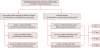
The experimental design of this study is summarized in Figure 1. All data were analyzed using 1- and 2-way analysis of variance (for micro-shear bond strength and fluoride release, respectively) and the post hoc least significant difference test for pairwise comparisons with a significance level of α = 0.05 in SPSS software version 20 (IBM Corp., Armonk, NY, USA).
RESULTS
Fluoride release
Table 2 presents the level of fluoride release in each studied group at different intervals; 24 hours, 3, 5, and 7 days and shows the influence of time on fluoride release in each group (p < 0.05). Groups 1 and 3 both showed the same behavior of significantly higher initial fluoride release (8.38 ± 0.33 ppm and 4.48 ± 0.28 ppm, respectively) on the 1st day which decreased gradually on the 3rd and 5th days, and then was the lowest (1.11 ± 0.13 ppm and 1.88 ± 0.08 ppm, respectively) on the 7th day (p < 0.05). As well, group 2 had significantly higher initial fluoride release (1.25 ± 0.09 ppm) on the 1st day than on the other days (p < 0.05). However, in group 2, fluoride release on the 7th day was not significantly different from those on the 3rd and 5th days, while a significant difference was detected between the results of the 3rd and 5th days.
Table 2
Fluoride release (ppm) of different groups at different intervals

The values are presented as means ± standard deviations. The values with different superscript letters within the same column are significantly different at p < 0.05.
Group 1, control (RMGIC-sound dentin assembly specimens); Group 2, RMGIC-artificially demineralized dentin assembly specimens (CAD specimens); Group 3, RMGIC to demineralized and then CPP-ACP remineralized dentin assembly specimens (remineralized CAD specimens); LSD, least significant difference; RMGIC, resin-modified glass ionomer cement; CAD, caries-affected dentin; CPP-ACP, casein phosphopeptide-amorphous calcium phosphate.
![]()
The mean amount of fluoride release varied with groups at each interval (Table 2). The control groups had significantly higher fluoride release on the 1st, 3rd, and 5th days (8.38 ± 0.33 ppm, 4.68 ± 0.27 ppm, and 3.39 ± 0.30 ppm, respectively) than the other groups (groups 2 and 3, p < 0.05). On the 7th day, the highest fluoride release was recorded for the remineralized specimens (1.88 ± 0.08 ppm, p < 0.05).
Micro-shear bond strength
The micro-shear bond strengths (MPa) of the studied groups are shown in Table 3. The micro-shear bond strength of the control group (25.03 ± 2.73 MPa) was significantly higher than that of both the demineralized and remineralized groups (p < 0.05). Additionally, remineralization significantly enhanced the micro-shear bond strength of the demineralized specimens (5.70 ± 1.71 MPa) by 10 MPa to 15.16 ± 1.53 MPa (p < 0.05).
Table 3
Micro-shear bond strength(SBS) of the groups tested in this study

| Group | SBS (MPa) | F value | p value |
|---|---|---|---|
| Group 1 | 25.03 ± 2.73a | - | - |
| Group 2 | 25.03 ± 2.73a | 681.48 | 0.0001 |
| Group 3 | 15.16 ± 1.53b | - | - |
| LSD | 2.84 | - | - |
The values are presented as means ± standard deviations. The values with different superscript letters are significantly different at p < 0.05.
Group 1, control (RMGIC-sound dentin assembly specimens); Group 2, RMGIC-artificially demineralized dentin assembly specimens (CAD specimens); Group 3, RMGIC to demineralized then CPP-ACP remineralized dentin assembly specimens (remineralized CAD specimens); LSD, least significant difference; RMGIC, resin-modified glass ionomer cement; CAD, caries-affected dentin; CPP-ACP, casein phosphopeptide-amorphous calcium phosphate.
![]()
DISCUSSION
A state of oral health exists when net mineral loss (demineralization) and net mineral gain (remineralization) remain in equilibrium [24]. The primary objective of deep caries management is to conserve the tooth structure, as well as to maintain the integrity of pulp health. CAD is characterized by intact collagen framework but with loss of minerals [25]. This layer is able to remineralize and retain such dentin during pulp capping procedures providing a chance to heal and repair stressed pulp without invasive procedures such as root canal treatment [26].
CPP-amorphous calcium fluoride phosphate (CPP-ACFP) and CPP-ACP compounds are natural remineralizing agents derived from milk, to promote the general trend found in nature. A range of laboratory, animal, and short and long-term human clinical trials have confirmed the capability of these compounds to prevent demineralization and to enhance remineralization by replacing both calcium and phosphate ions lost due to caries, making the enamel more resistant to further acid attacks [27]. Therefore, the application of CPP-ACP may improve the bonding ability of different types of dental restorations to CAD.
Calcium phosphate is ordinarily insoluble, (i.e., it forms a crystalline structure at neutral pH). CPP-based products, such as CPP-ACP, which contains 18% calcium ion and 30% phosphate ion in weight, are the basis for the anticariogenicity of milk derivatives [28]. The action of CPP derivatives is partially based on the regulation of bioavailable calcium phosphate levels. CPP can adhere to 25 calcium ions, 15 phosphate ions, and 5 fluoride ions per molecule and can stabilize calcium and phosphate ‘nanoclusters’ of ions in solution. Moreover, CPP can free its weight in calcium and phosphate to form a colloid compound that prevents calcium phosphate crystals from growing to the size required for precipitation [29]. ACP is formed at neutral or alkaline pH. When CPP adheres to ACP aggregates, their growth is controlled, preventing them from reaching the critical size for precipitation [20]. These characteristics result in maintained ion phosphate and calcium supersaturation with increased remineralization and reduced hydroxyapatite demineralization [30].
To determine the amount of fluoride release from GICs into aqueous solutions, ion chromatography was chosen, both because this technique is appropriate for the measurement of free fluoride ions and because it senses low concentrations of fluoride ions that may not be detected by the ion-selective electrode method [31]. Fluoride release from GIC is a complex process influenced by several intrinsic and extrinsic factors. Intrinsic factors may include the formulation, the powder/liquid ratio, the geometry of the specimen, mixing time, material solubility or porosity, temperature, surface finishing, and treatment. Extrinsic factors encompass the type and pH of the storage medium, the environmental temperature, experimental design, and analytical methods. Three mechanisms are responsible for fluoride release from GIC: surface loss, diffusion through pores and cracks, and bulk diffusion [3233343536]. Our present findings showed that RMGIC bonded to the specimens of the three groups continued to release fluoride in distilled water over the 7 days test period. The maximum release took place on the first day, soon after, the amount steadily diminished until reaching a reasonably constant rate. This may be related to the initial ‘burst’ of fluoride release from the glass particles. The burst release is attributed to the reaction between polyalkenoic acid and fluoride-containing glass particles on setting, and also to the rapid dissolution of fluoride from the outer surface into the solution [3233]. The gradually diminished rate of fluoride release during the subsequent days until the seventh day may have been due to the earlier burst of fluoride released from glass particles dissolved in the polyacid during setting [37]. This is in agreement with Forsten [38], who concluded that the highest burst of fluoride release was on the first day and decreased over the first week.
Additionally, the CAD-RMGIC group exhibited the lowest level of fluoride release in the solution, which may have been related to higher fluoride deposition into CAD than into the mineralized dentin forming complexes with the remaining acid-resistant mineral crystals [39]. This is in agreement with another study [40] that reported an immediate high fluoride deposition onto demineralized dentin immersed into fluoride calcium phosphoric acid complex solution, which had the ability to remineralize it. That complex was considered to be a source of fluoride as was the RMGIC presented in this study. Since CPP-ACP partially remineralized CAD, there was less fluoride uptake and deposition within the specimens of group 3 than within those of group 2, while group 3 showed higher fluoride content within its solution than did group 2. As well, Mazzaoui et al. [41], explained that CPP-ACP remains as a contaminant on the treated CAD surface which promotes the release of fluoride ions from RMGIC to form CPP-ACFP nano-complexes.
The results revealed that the micro-shear bond strength of RMGIC to artificial CAD was significantly lower than that to sound dentin. The total strength of normal dentin is primarily dependent on the mineral phase (71.3%) while the remaining strength is correlated to the collagen matrix (28.7%) [42]. This indicates that the minerals lost in the inter-tubular matrix impair the mechanical properties of the CAD concerning bond strength [8]. Taking into account the chemical bonding mechanism between the carboxylic groups of the polyacrylic acid and the calcium ions of hydroxyapatite, the lower bond strength of RMGIC to carious dentin is reasonable, because the loss of calcium ions through demineralization gives less opportunity for bonding [43].
In contrast, demineralized dentin treated with CPP-ACP showed a significant improvement in micro-shear bond strength. A feasible explanation may be the ability of this agent to increase the deposition of hydroxyapatite crystals on the dentin surface which in turn increases bond strength [44]. This occurred through the enhanced ionic bond to more hydroxyapatite crystals around collagen fibers and the micromechanical interlocking of RMGIC in contrast to the demineralized tooth structure [13]. These findings are consistent with those reported by Choi et al. [45], who concluded that the micro-tensile bond strengths of both conventional GIC and RMGIC to demineralized dentin were significantly lower than the corresponding micro-tensile bond strengths to mineralized dentin.
According to Sattabanasuk et al. [46], the application of CPP-ACP on CAD surface might have an impact on the bond strength of some adhesive systems. It can adhere intraorally to plaque pellicles, hydroxyapatite and soft tissues, supplying bioactive calcium and phosphate to the tooth structure thereby, facilitating the remineralization process. These findings correspond with the results of this study. Another supporting study by Rahiotis and Vougiouklakis [5], indicated that CPP-ACP improved remineralization of CAD compared to specimens without pretreatment. It was explicated that CPP-ACP technology, which has been introduced recently, can remineralize CAD. Calcium and phosphate ions easily diffuse through porous lesions and are deposited in partly demineralized crystals, reforming apatite crystals. These crystals are the fundamental component of the chemical bond of GIC to dentin.
CONCLUSIONS
Within the limitations of this study, CPP-ACP treatment can be suggested for use in vivo to motivate CAD remineralization, subsequently arresting the progress of carious lesions. Regarding fluoride release, CAD treated with CPP-ACP uses less fluoride discharged from RMGIC into solution for further remineralization. These findings are of interest, as they are evidence of the ability of CPP-ACP to partially mineralize CAD. As well, CPP-ACP enhances the bonding stability of RMGIC to CAD. CPP-ACP is a promising conservative approach that encourages minimal intervention and leads to dramatic improvements in dentistry.
 XML Download