

 PDF
PDF ePub
ePub Citation
 Print
Print


Dear Editor,
Autoantibodies to centromere protein-F (CENP-F), which is closely associated with cell proliferation, are known to be a specific marker for malignant tumors [123]. However, a few cases of autoantibodies to CENP-F have been reported in autoimmune diseases. Despite some studies indicating no association between autoimmune diseases and CENP-F [23], the association still remains unclear. Particularly, there are no reports on autoantibodies to CENP-F or CENP-F-like pattern by antinuclear antibody (ANA) test in rheumatoid arthritis (RA). We observed the first case of a CENP-F-like pattern in RA without any underlying diseases.
A 37-year-old female visited Hanyang University Medical Center, Seoul, Korea, with 3-month history of pain in both knees and right ankle. The laboratory test results were as follows: erythrocyte sedimentation rate (ESR), 74 mm/hr; C-reactive protein (CRP), 4.4 mg/dL; rheumatoid factor (RF), 89 IU/mL; and anti-cyclic citrullinated peptide (anti-CCP), 176 U/mL. X-ray imaging of the hands and feet revealed soft tissue swelling, and whole-body bone scanning revealed abnormally increased bone uptake in the right foot, both hands, and both knees. The ANA test showed a CENP-F-like pattern and the titer of 1:640 (Fig. 1). All other findings were negative, with no underlying disease, family history, or drug history. She was diagnosed as having RA on the basis of the 2010 American College of Rheumatology/European League Against Rheumatism RA classification criteria [4] with a total score of 7: two small and two large joints involved (score 2), high positive RF >42 IU/mL and anti-CCP >30 U/mL (score 3), abnormal ESR >20 mm/hr in females and CRP >0.8 mg/dL (score 1), and >6 weeks of symptoms (score 1).
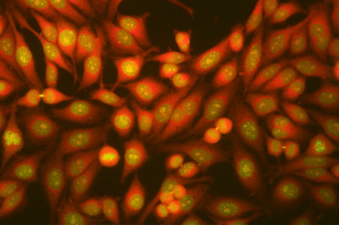 | Fig. 1The antinuclear antibody test of HEp-2 cells using the indirect immunofluorescence method. The image shows the centromere protein-F-like pattern. It comprises the nuclear speckled pattern, multiple bright paired foci inside the nucleus at the early G2 phase; smooth nuclear envelope at the late G2 phase; centromere pattern, multiple aligned small and faint dots at the prophase and metaphase; and midbody pattern, an intense staining dot located at the midzone at the late anaphase and telophase.
|
CENP-F is a 367 kDa nuclear protein first reported in 1993 [56]. It is distributed in the nuclear matrix at the early G2 phase, forms a kinetochore at the late G2 phase to promote activation of the centromere and cell division, and disappears after the completion of M phase. Perhaps, it promotes cell proliferation, an increase in the number of mitotic cells as the cell cycle gets faster, because it is specific to malignant tumors, breast cancer, lung cancer, ovarian cancer, cervical cancer, non-Hodgkin lymphoma, and esophageal squamous cell carcinoma [1236]. Particularly, the high expression level of CENP-F in primary breast cancer is considered a molecular background of the rapid proliferation and high aggressiveness of cancer cells [7].
The ANA test is the best method to detect fluorescence of a CENP-F-like pattern, which is expressed during the cell cycle (G2-M phase). It appears similar to the nuclear speckled pattern at the early G2 phase, multiple bright paired foci inside the nucleus and a smooth nuclear envelope similar to the anti-proliferating cell nuclear antigen (anti-PCNA) antibody pattern at the late G2 phase. The centromere pattern, multiple aligned small dots, appears from prophase to metaphase; the midbody pattern, an intense dot located at the midzone, appears from late anaphase to telophase [256].
Several studies have reported autoantibodies to CENP-F; however, none have explained their correlation with RA. In previous studies, patients with malignant tumors and autoantibodies to CENP-F did not exhibit any relationship with RA [23]. Moreover, six patients with autoantibodies to CENP-F did not have RA (undefined connective tissue disease, 2; primary antiphospholipid syndrome, 1; colorectal carcinoma, 1; hepatitis C virus, 1; and fever of unknown origin, 1) [8]. Another six patients with both RA and malignant tumors were reported without any correlation with CENP-F [9].
As our patient's symptoms and laboratory tests did not suggest malignant tumors, the likelihood of malignancy was low, and the CENP-F-like pattern was related to RA rather than malignancy. However, no case having a CENP-F-like pattern in RA without any underlying diseases has been reported thus far, and the correlation between CENP-F and RA is not clear. Temporary acute inflammatory reaction to RA might account for an increase in autoantibodies to CENP-F. Therefore, periodic ANA tests to continuously detect a CENP-F-like pattern are imperative.
Several patterns of the ANA test, including speckled, homogeneous, and centromere patterns, are commonly reported in RA [10]. The CENP-F-like pattern might be misinterpreted as the speckled, centromere, or anti-PCNA antibody pattern. We think that CENP-F-like patterns in RA have been incorrectly reported and dismissed. To date, the importance of autoantibodies to CENP-F in RA remains understated. Therefore, if more cases of CENP-F-like patterns in RA are reported, further studies on its clinical relevance in RA are warranted.
 XML Download