Journal List > Ann Lab Med > v.39(2) > 1107512

Kim, Kim, Chang, Kong, Lim, Kong, Cho, Lee, Shin, Park, Eom, and Lee: T-Cell Receptor Rearrangements Determined Using Fragment Analysis in Patients With T-Acute Lymphoblastic Leukemia

Abstract

Background

Chromosomal abnormalities and common genetic rearrangements related to T-acute lymphoblastic leukemia (T-ALL) are not clear. We investigated T-cell receptor (TCR) rearrangement in Korean T-ALL patients by fragment analysis, examining frequency, association between clinicopathologic characteristics and TCR clonality, and feasibility for detecting minimal residual disease (MRD).

Methods

In 51 Korean patients diagnosed as having T-ALL, TCR rearrangement was analyzed using the IdentiClone TCR gene clonality assay (InVivoScribe Technologies, San Diego, CA, USA) from archived bone marrow specimens. Limit of detection (LOD) and clonal stability at relapse were evaluated. The association between clinical prognosis and TCR clonality was examind by age and immunophenotypic classification.

Results

Thirty-eight patients (74.5%) had 62 clonal products of TCRβ, TCRγ, and/or TCRδ rearrangements at diagnosis. Children with T-ALL (<12 years) showed a higher frequency of clonality (93.8%) than adolescents/adults (65.7%; ≥12 years). Patients with a mature immunophenotype (84.4%) showed a relatively higher frequency of clonality than those with the immature immunophenotype (57.9%). Survival and event-free survival were not influenced by immunophenotype or TCR clonality. The LOD was 1%. Clonal evolution at the relapse period was noted.

Conclusions

The overall detection rate of TCR clonality was 74.5%. Survival did not differ by TCR clonality or immunophenotype and age group. Fragment analysis of TCR rearrangement cannot be used to assess MRD due to low sensitivity. Further research on the relationship between prognosis and frequency of TCR rearrangements is needed, using more sensitive methods to detect clonality and monitor MRD.

INTRODUCTION

T-acute lymphoblastic leukemia (T-ALL) is characterized by the proliferation of T-lineage lymphoid blasts but represents a heterogeneous group of neoplastic cells. Although 80–95% of adults and 95% of pediatric patients with ALL achieve complete remission (CR) at the end of induction therapy, a substantial proportion of T-ALL patients are inadequately treated and experience disease relapse, while others may be over-treated because of the inability to individualize clinical treatment [123].
There are several classifications of T-ALL according to immunophenotypic analysis and T-cell receptor (TCR) genetic analysis [456]. The European Group for the Immunological Characterization of Leukemias (EGIL) defined four developmental stages in T-ALL [5]: prothymocytic (pro-T), prethymocytic (pre-T), cortical, and medullary T-ALL. Pro-T and pre-T-ALLs represent immature immunophenotypes, and cortical and medullary T-ALLs represent mature immunophenotypes. Patients with cortical or medullary T-ALL immunophenotypes have higher CR rates than those with pro-T or pre-T-ALL immunophenotypes [4]. Until recently, it has been difficult to make an accurate diagnosis or classify T-ALL into precise subgroups [2]. Nevertheless, the differences in maturation arrest during T-cell development may be of prognostic significance to T-ALL patients [47]. Alternatively, T-ALL can be classified according to TCR rearrangements, since the TCRβ, TCRγ, and TCRδ configurations show distinct gene expression signatures related to maturation arrest at different stages of T-cell differentiation, which may better reflect T-cell development [8].
Chromosomal abnormalities and common genetic rearrangements related to T-ALL have not been clearly identified [910]. TCR clonality detection rates in European pediatric and adult T-ALL patients have been found to be 95% [111] and 84% [12], respectively. In particular, a TCR gene can be identified in most pediatric cases of T-ALL, and the TCR gene rearrangement can be considered a marker for minimal residual disease (MRD). Among the several methods to detect TCR clonality status, the multiplex PCR-based fragment analysis technique is currently available and provides information on the TCR repertoire at a previously inaccessible level of analysis [13].
We evaluated the frequency of TCR rearrangements in the Korean T-ALL patients using fragment analysis. After clonality testing, we investigated the association between the clinicopathologic findings and clonality status, with age and immunophenotype as the core variables. Overall survival and event-free survival (EFS) were also investigated, and the correlation between survival and TCR clonality was assessed. Finally, we examined the feasibility of TCR rearrangement as an MRD marker using follow-up specimens. The limit of detection (LOD) and clonal stability at relapse were evaluated to determine the MRD sensitivity and stability during disease progression.

METHODS

Patients and specimens

A total of 51 adult and pediatric patients diagnosed as having T-ALL between 2002 and 2012 from three tertiary hospitals, Pusan National University Hospital (N=20, Busan), Pusan National University Yangsan Hospital (N=12, Yangsan), and the National Cancer Center (N=19, Goyang), in Korea were included in this retrospective study. The study was approved by the Institutional Review Board of the Pusan National University Yangsan Hospital (No. 05-2014-061). Informed consent was obtained from all the patients.
Among the archival specimens, bone marrow (BM) aspirates obtained at initial diagnosis and at the end of induction chemotherapy (days 23–45) from each patient were used for this study. Baseline and clinicopathologic characteristics, including age, immunophenotype, cytogenetics, and clinical outcomes, were obtained from reviews of electronic medical records at the three hospitals in December 2016.
The patients were arbitrarily divided into two age groups: children (N=16, <12 years) and adolescents/adults (N=35; ≥12 years) [14]. EFS was defined as survival with no objective evidence of disease progression or relapse. Immunophenotypic analysis was performed using fresh specimens in the clinical laboratories of each participating hospital. Patients were classified into immunophenotypic subgroups based on the EGIL criteria [5]: pro-T (CD7+, CD2−, and/or CD34+, but CD1− or sCD3−), pre-T (CD7+, CD2+, and/or CD34+, but CD1− or sCD3−), cortical T (CD34− and CD4+CD8+ double positivity), or medullary T (sCD3+, CD34−, and either CD4+ or CD8+) subgroups.
The BM specimens and baseline and clinicopathologic information were sent to Pusan National University Yangsan Hospital. Genomic DNA was extracted from the archival BM specimens using a QIAmp DNA Mini kit (Qiagen, Hilden, Germany), according to the manufacturer's protocol, at this hospital. The purity and concentration of DNA were verified using a NanoDrop ND-1000 spectrophotometer (Thermo Fisher Scientific, Wilmington, DE, USA).

TCR rearrangements using fragment analysis

Fragment analysis was performed using IdentiClone TCR Gene Clonality Assay (InVivoScribe Technologies, San Diego, CA, USA) at Pusan National University Hospital for two years from October 2015 to March 2016. Analysis of TCR arrangements included three reactions that targeted TCRβ (Vβ-Jβ1/2.2/2.6/2.7 for TCRβA, Vβ-Jβ2.1/2.3/2.4/2.5 for TCRβB, and Dβ-Jβ for TCRβC), two reactions that targeted TCRγ (Vγ1-8/Vγ10-Jγ1/2/P1/P2 for TCRγA and Vγ9/Vγ11-Jγ1/2/P1/P2 for TCRγB), and one reaction that targeted TCRδ (Vδ–Jδ). The fragments as PCR products were analyzed on an automated capillary electrophoresis system (ABI 3,130, Applied Biosystems, Foster City, CA, USA), and the sizes of the PCR products were determined using GeneMapper v4.0 (Applied Biosystems). All electropherograms were examined by at least two experts in fragment analysis.
For the initial diagnostic specimens, fragment analysis for TCRβ, TCRγ, and TCRδ rearrangements was simultaneously conducted. Rearrangements were interpreted as clonotypes according to the EuroClonality/BIOMED-2 guidelines [613]. The criteria for defining a positive peak were the display of discrete bands by the products within the expected size range and the largest peak being at least three times higher than the third largest peak in the polyclonal background. To evaluate its feasibility as an MRD marker, TCR rearrangements were also examined using fragment analysis at the end of induction chemotherapy. MRD is detected based on an initially identified leukemia-specific clonotype, which is the same fragment with a unique length on an electropherogram. Because of BM hypoplasia at the end of induction chemotherapy, the DNA content obtained was not sufficient for analysis in 14 specimens. In addition, three patients without TCR clonality at initial diagnosis were excluded from this assay; therefore, the TCR rearrangements were investigated in BM specimens from only 34 patients at the end of induction chemotherapy.
To investigate the possibility for MRD monitoring, LOD and clonal stability at relapse were also evaluated. Initial diagnostic specimens from three T-ALL patients were used for assessing the LOD of this technology. The blasts in the BM of the three patients were 98.8%, 82.7%, and 55.9%, respectively. Serial dilutions of the three selected specimens that were positive for TCR rearrangement were prepared, ranging from 10-1 to 10-4 of normal peripheral blood mononuclear cells. All assays were performed in duplicate. We performed fragment analysis for four randomly selected patients from Pusan National University Hospital who relapsed within two years of the initial diagnosis, and compared the clonotypes at the initial diagnosis with those at relapse to investigate clonal stability.

Statistical analysis

Categorical data were summarized as counts and percentages, and continuous data were summarized as medians with interquartile range (IQR). Categorical data were analyzed using the chi-square test, and continuous data were analyzed using the Student's t-test or Mann-Whitney U test as appropriate. Frequencies of TCR rearrangement and the distribution of immunophenotypes based on the EGIL classification of TCR clonality subgroups were compared between children and adolescent/adult T-ALL patients using Student's t-test, Pearson's chi-squared test, Fisher's exact test (when expected cell counts were <5), one-way ANOVA, or Mann-Whitney U test. Kaplan-Meier and log-rank tests were used to compare overall survival or EFS in different groups. P<0.05 was considered statistically significant. All statistical analyses were performed using SPSS version 22.0 (SPSS Inc., Chicago, IL, USA).

RESULTS

Frequency of TCR rearrangements

A total of 62 clonal PCR products of different TCR rearrangements were identified through fragment analysis in 38 (74.5%) of the 51 T-ALL patients at diagnosis. Fragment analysis identified at least one clonal rearrangement in the 38 specimens, using the three-target (TCRβ, TCRγ, and TCRδ) sets. Among the 51 patients, monoclonal rearrangements were found in 18 patients for TCRβ, 31 patients for TCRγ, and 14 patients for TCRδ. Two or more TCR rearrangements occurring in a single specimen were identified for 22 patients (43.1%). Among the 38 patients, the median and IQR of rearrangements per patient were 2.0 and 1.0, respectively. Depending on the serial dilution for assessing LOD, this method detected TCR rearrangements at an LOD above 10-2 (sensitivity 1%).

Association of TCR clonality with patient characteristics

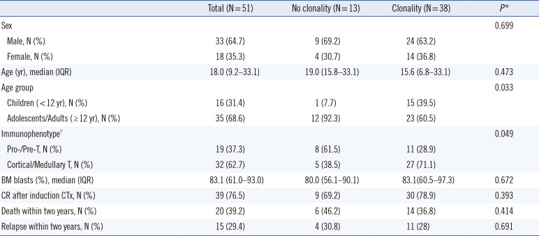
The clinical characteristics of patients with or without TCR clonality at diagnosis are presented in Table 1. TCR clonality was significantly related to age and immunophenotype but not to sex, BM blast counts, CR, or survival. Children with T-ALL (15/16, 93.8%) showed a higher frequency of clonality than adolescents/adults (23/35, 65.7%). Clonality was associated with age (P=0.033). Further analyses according to the three age groups, children, adolescents, and adults, are described in Supplemental Data Tables S1, S2, S3.
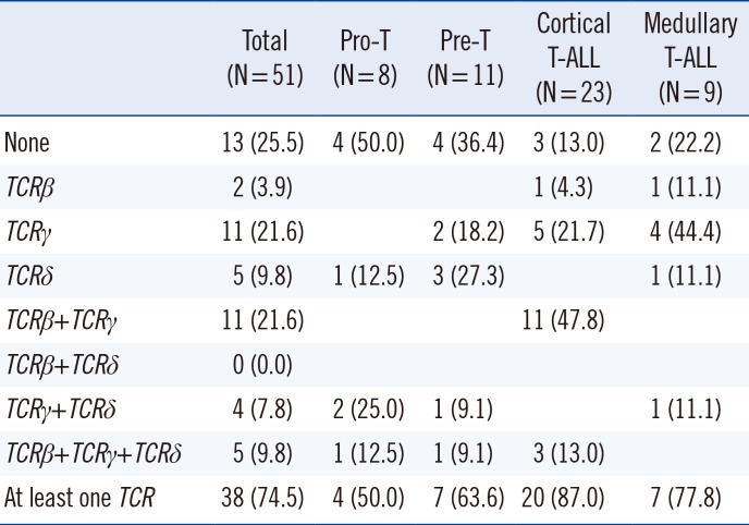
To investigate the association between clonal TCR rearrangement and T-cell development stage, patients were assigned to the following T-cell development subgroups based on the EGIL criteria 2 (Table 2): the pro-/pre-T subgroup (N=19) as an immature subtype and the cortical/medullary T-cell subgroup (N=32) as the mature subtype. Since the number of patients in each of the four subgroups was relatively small, the immature and mature subtypes were clustered.
Overall, TCR clonality was associated with the immunophenotypic subgroups (P=0.049, Table 2). Among the patients with TCR clonality (N=38), 11 had the immature phenotype, whereas among the patients without TCR clonality (N=13), three patients had cortical-T and two had medullary T-ALL mature phenotypes. Although the association between immunophenotype and TCR clonality was significant, neither immunophenotype nor TCR clonality influenced overall survival or EFS (P=0.561 and P=0.449, respectively).

Clinical significance of TCR clonality at the end of induction chemotherapy

Among the 34 tested patients, 30 were interpreted to be in morphological remission (<5% blasts on the all nucleated cells) and cytogenetic remission; however, four patients showed persistent leukemic blasts. A total of 15 clonotypes (five TCRβ, eight TCRγ, and two TCRδ rearrangements) were detected in 14 (41.2%) patients. Among these 14 patients with TCR clonality, three showed persistent leukemic blasts on BM aspirates, and 11 had no residual leukemic cells. One persistent patient did not exhibit clonality and thus was among the 20 patients whose TCR rearrangements were not detected. The 11 patients showing morphological remission were regarded as having MRD based on the presence of TCR rearrangements.
After excluding the four patients with morphological persistence, the association of clinical characteristics with TCR clonality was assessed for a total of 30 patients. As shown in Table 3, no significant difference was observed in BM blast count, BM cellularity, and relapse or death within two yrs between the two groups. Patients with MRD detected at the end of induction chemotherapy had relatively lower survival; however, this finding was not significant (P=0.416). Among the eight relapsed patients, only three showed early positive MRD results (Table 3).

Stability of TCR clonality at relapse

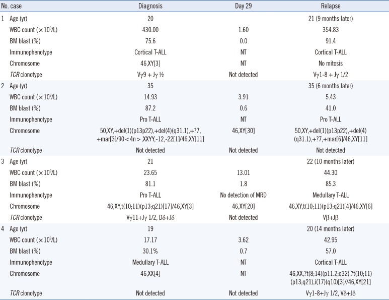
To investigate the stability of TCR clonality, clinicopathologic characteristics at initial diagnosis and relapse were compared in four randomly selected relapsed patients at Pusan National University Hospital. Three of the four relapsed patients were positive for TCR clonality at relapse and had received allogenic stem cell transplantation before relapse; all three of these relapsed patients had clonotypes different from those detected at initial diagnosis. Patient 3 relapsed within four months after transplantation; however, the immunophenotype and TCR clonality were different from those at the initial diagnosis, although the initial chromosomal abnormality was retained. Patient 4 relapsed at seven months after allogenic stem cell transplantation (Table 4).

DISCUSSION

The overall frequency of TCRβ, TCRγ, and TCRδ rearrangements was 74.5% in Korean patients with T-ALL at initial diagnosis. Korean children with T-ALL showed a higher frequency of clonality (93.8%) than adolescents/adults (65.7%). The frequency of TCR clonality in European pediatric T-ALL patients (95%) was similar to that of the Korean children T-ALL group [1]; however, the frequency of TCR clonality in European adult T-ALL patients is 84% [12] and higher than that of Korean adolescent/adult T-ALL patients. Age distribution might be a main factor contributing to the difference in frequencies in Korean and European patients. Two previous studies conducted in Taiwan and Korea included both children and adult patients, and detected TCR clonality in 59–67% of the patients [1315]. Compared with that of European adult T-ALL patients, this study showed relatively low detection rates of patients suggesting that ethnic diversity may also contribute to the low frequency of TCR rearrangements in Asians.
Using the EGIL and TCR classification systems, Bene et al. [5] demonstrated that approximately a half of the T-ALL patients could be assigned to relatively comparable developmental stages. However, neither the EGIL nor the TCR subgroups predicted clinical outcomes in our study. Nevertheless, the TCR classification system may be more applicable to pediatric T-ALL cases than the EGIL classification system. We used complementary immunophenotypic and TCR genotypic analysis to confirm and extend these data. In a previous study [4], 4%, 4%, 39%, and 10% of adult T-ALL patients were classified with the pro-T, pre-T, cortical T, and medullary immunophenotypes, respectively (age range: 14–52 years; median: 26.6 years). Similar results were observed in the present study. Furthermore, the association between immunophenotype and TCR clonality was confirmed, wherein immature groups had lower frequencies of TCR rearrangement (P=0.049).
The overall proportion of patients exhibiting TCR clonality was similar to those previously reported for T-ALL patients through real-time quantitative PCR (RQ-PCR) or sequence analysis [1617]. Using RQ-PCR, Flohr et al. [16] detected two or more instances of clonality in 88% of 422 children with T-ALL and at least one clonal TCR rearrangement in 93% of the patients. Therefore, either technique appears to be suitable for detecting T-ALL clonality at diagnosis.
The LOD of this method in our study was 1%, indicating that this assay is not sufficiently sensitive to detect MRD. Research has suggested that detection of the post-therapy MRD burden (>10-4) in BM aspirates or detection of TCR clonality can be a more powerful prognostic marker for subsequent relapse than typically used markers, such as age, white blood cell count at diagnosis, and cytogenetic alterations [1718]. However, in our study, TCR clonality at diagnosis or at the end of induction chemotherapy was not useful for predicting overall survival and EFS, such as death within two yrs and relapse within two years.
From the perspective of stability, three of the four relapsed patients showed clonal evolution. Although fragment analysis was not suitable for assessing MRD due to the low LOD, it can be used to sensitively detect newly proliferating leukemic clones when using all three clonality combinations. The immunophenotypes and TCR clonotypes of Patient 3 (Table 4) were different at diagnosis and relapse, but the karyotype was identical. The discrepancy between the same cytogenetic findings and clonal evolutions by TCR rearrangement and immunophenotyping in this relapsed patient might be due to the low detection sensitivity of conventional karyotyping. Indeed, translocations, such as t(1;14)(p32;q11), cryptic interstitial deletion at 1p32 (TAL1), and deletion of 9p, are often not detected by karyotyping in T-ALL but are identified through only molecular genetic analysis [1920]. Therefore, the TCR rearrangement assay is useful for surveillance, but its ability to predict early relapse in the CR state is limited due to clonal evolution.
The current clinical strategies to assess MRD rely on next-generation sequencing (NGS) and RQ-PCR-based methods using patient-specific primers [91721]. In our study, the relapsed patients showed different clonotypes, suggesting clonal evolution compared with the state at initial diagnosis. Compared with RQ-PCR, NGS has the advantage of detecting clonal evolution during the course of disease, thus abrogating the risk of obtaining false-negative results. Therefore, NGS-based TCR rearrangement will likely become a part of routine practice for the diagnosis and follow-up of patients with hematological neoplasms [2223].
The low frequency of TCR rearrangement in Korean adolescent/adult patients might also be due to the quality of archived specimens. Because this study was a retrospective study, specimen quality was the most important limitation of the study. The qualty of archived specimens depends on the storage period and conditions. Therefore, low specimen quality could have contributed to the low detection rate. Conducting fragment analysis at diagnosis with fresh, good-quality specimens could reveal the exact frequency of TCR rearrangement in Korean T-ALL patients. Other major limitations of this study were the very small number of patients, prolonged storage of specimens, and low sensitivity of the detection method. Further studies, including a larger number of patients and highly sensitive methods may help improve the clinical diagnosis and subsequent MRD monitoring of T-ALL patients.
In summary, the TCR clonal frequencies in children (<12 years) and adolescent/adult patients (≥12 years) with T-ALL in Korea were found to be 93.8% and 65.7%, respectively. Although a higher frequency of TCR rearrangements was noted in children and in patients with a cortical or medullary immunophenotype, no significant association was found between TCR clonality and clinical outcomes such as EFS. Moreover, clonal evolution in TCR rearrangement was noted between diagnosis and relapse. This phenomenon should be explored in future studies involving a large and diverse set of patients, using a high-throughput technique.

Acknowledgment

This study was supported by the Research Institute for Convergence of Biomedical Science and Technology (30-2015-030), Pusan National University Yangsan Hospital, and Pusan National University Research Grant, 2018.

Notes

Authors' Disclosures of Potential Conflicts of Interest: There are no conflicts of interest to declare.

References

1. Thörn I, Forestier E, Botling J, Thuresson B, Wasslavik C, Björklund E, et al. Minimal residual disease assessment in childhood acute lymphoblastic leukaemia: a Swedish multi-centre study comparing real-time polymerase chain reaction and multicolour flow cytometry. Br J Haematol. 2011; 152:743–753. PMID: 21250970.
2. Chiaretti S, Foà R. T-cell acute lymphoblastic leukemia. Haematologica. 2009; 94:160–162. PMID: 19181788.
3. Brüggemann M, Gökbuget N, Kneba M. Acute lymphoblastic leukemia: monitoring minimal residual disease as a therapeutic principle. Semin Oncol. 2012; 39:47–57. PMID: 22289491.
4. Vitale A, Guarini A, Ariola C, Mancini M, Mecucci C, Cuneo A, et al. Adult T-cell acute lymphoblastic leukemia: biologic profile at presentation and correlation with response to induction treatment in patients enrolled in the GIMEMA LAL 0496 protocol. Blood. 2006; 107:473–479. PMID: 16179376.
5. Bene MC, Castoldi G, Knapp W, Ludwig WD, Matutes E, Orfao A, et al. Proposals for the immunological classification of acute leukemias. European Group for the Immunological Characterization of Leukemias (EGIL). Leukemia. 1995; 9:1783–1786. PMID: 7564526.
6. Groenen PJ, Langerak AW, van Dongen JJ, van Krieken JH. Pitfalls in TCR gene clonality testing: teaching cases. J Hematop. 2008; 1:97–109. PMID: 19669208.
7. Thiel E, Kranz BR, Raghavachar A, Bartram CR, Löffler H, Messerer D, et al. Prethymic phenotype and genotype of pre-T (CD7+/ER−)-cell leukemia and its clinical significance within adult acute lymphoblastic leukemia. Blood. 1989; 73:1247–1258. PMID: 2467704.
8. Asnafi V, Beldjord K, Boulanger E, Comba B, Le Tutour P, Estienne MH, et al. Analysis of TCR, pT alpha, and RAG-1 in T-acute lymphoblastic leukemias improves understanding of early human T-lymphoid lineage commitment. Blood. 2003; 101:2693–2703. PMID: 12446444.
9. Brüggemann M, Raff T, Kneba M. Has MRD monitoring superseded other prognostic factors in adult ALL? Blood. 2012; 120:4470–4481. PMID: 23033265.
10. Brüggemann M, Schrauder A, Raff T, Pfeifer H, Dworzak M, Ottmann OG, et al. Standardized MRD quantification in European ALL trials: proceedings of the Second International Symposium on MRD assessment in Kiel, Germany, 18–20 September 2008. Procs. Second Int Symp. on MRD Assessment in Kiel, Germany, 18–20 September 2008. Leukemia. 2010; 24:521–535. PMID: 20033054.
11. Thörn I, Forestier E, Thuresson B, Wasslavik C, Malec M, Li A, et al. Applicability of IG/TCR gene rearrangements as targets for minimal residual disease assessment in a population-based cohort of Swedish childhood acute lymphoblastic leukaemia diagnosed 2002–2006. Eur J Haematol. 2010; 84:117–127. PMID: 19895569.
12. Gameiro P, Mortuza FY, Hoffbrand AV, Foroni L. Minimal residual disease monitoring in adult T-cell acute lymphoblastic leukemia: a molecular based approach using T-cell receptor G and D gene rearrangements. Haematologica. 2002; 87:1126–1134. PMID: 12414341.
13. Chen YL, Su IJ, Cheng HY, Chang KC, Lu CC, Chow NH, et al. BIOMED-2 protocols to detect clonal immunoglobulin and T-cell receptor gene rearrangements in B- and T-cell lymphomas in southern Taiwan. Leuk Lymphoma. 2010; 51:650–655. PMID: 20233058.
14. van Grotel M, Meijerink JP, van Wering ER, Langerak AW, Beverloo HB, Buijs-Gladdines JG, et al. Prognostic significance of molecular-cytogenetic abnormalities in pediatric T-ALL is not explained by immunophenotypic differences. Leukemia. 2008; 22:124–131. PMID: 17928886.
15. Kim Y, Choi YD, Choi C, Nam JH. Diagnostic utility of a clonality test for lymphoproliferative diseases in koreans using the BIOMED-2 PCR assay. Korean J Pathol. 2013; 47:458–465. PMID: 24255634.
16. Flohr T, Schrauder A, Cazzaniga G, Panzer-Grümayer R, van der Velden V, Fischer S, et al. Minimal residual disease-directed risk stratification using real-time quantitative PCR analysis of immunoglobulin and T-cell receptor gene rearrangements in the international multicenter trial AIEOP-BFM ALL 2000 for childhood acute lymphoblastic leukemia. Leukemia. 2008; 22:771–782. PMID: 18239620.
17. Wu D, Sherwood A, Fromm JR, Winter SS, Dunsmore KP, Loh ML, et al. High-throughput sequencing detects minimal residual disease in acute T lymphoblastic leukemia. Sci Transl Med. 2012; 4:134ra63.
18. van der Velden VH, Panzer-Grümayer ER, Cazzaniga G, Flohr T, Sutton R, Schrauder A, et al. Optimization of PCR-based minimal residual disease diagnostics for childhood acute lymphoblastic leukemia in a multi-center setting. Leukemia. 2007; 21:706–713. PMID: 17287857.
19. Kikuchi A, Hayashi Y, Kobayashi S, Hanada R, Moriwaki K, Yamamoto K, et al. Clinical significance of TAL1 gene alteration in childhood T-cell acute lymphoblastic leukemia and lymphoma. Leukemia. 1993; 7:933–938. PMID: 8321044.
20. Okuda T, Shurtleff SA, Valentine MB, Raimondi SC, Head DR, Behm F, et al. Frequent deletion of p16INK4a/MTS1 and p15INK4b/MTS2 in pediatric acute lymphoblastic leukemia. Blood. 1995; 85:2321–2330. PMID: 7727766.
21. Campana D. Progress of minimal residual disease studies in childhood acute leukemia. Curr Hematol Malig Rep. 2010; 5:169–176. PMID: 20467922.
22. de Haas V, Verhagen OJ, von dem Borne AE, Kroes W, van den Berg H, van der Schoot CE. Quantification of minimal residual disease in children with oligoclonal B-precursor acute lymphoblastic leukemia indicates that the clones that grow out during relapse already have the slowest rate of reduction during induction therapy. Leukemia. 2001; 15:134–140. PMID: 11243381.
23. Sekiya Y, Xu Y, Muramatsu H, Okuno Y, Narita A, Suzuki K, et al. Clinical utility of next-generation sequencing-based minimal residual disease in paediatric B-cell acute lymphoblastic leukaemia. Br J Haematol. 2017; 176:248–257. PMID: 27861730.
SUPPLEMENTARY MATERIALS

Supplemental Data Table S1

Baseline characteristics of T-ALL patients according to three age groups
alm-39-125-s001.pdf

Supplemental Data Table S2

Frequency of TCR clonality detected using fragment analysis at initial diagnosis according to three age groups of T-ALL patients
alm-39-125-s002.pdf

Supplemental Data Table S3

Characteristics of T-ALL patients with morphological remission according to age groups at the end of induction chemotherapy
alm-39-125-s003.pdf
Table 1

Baseline characteristics of T-ALL patients according to TCR clonality

alm-39-125-i001
Total (N=51) No clonality (N=13) Clonality (N=38) P*
Sex 0.699
 Male, N (%) 33 (64.7) 9 (69.2) 24 (63.2)
 Female, N (%) 18 (35.3) 4 (30.7) 14 (36.8)
Age (yr), median (IQR) 18.0 (9.2–33.1) 19.0 (15.8–33.1) 15.6 (6.8–33.1) 0.473
Age group 0.033
 Children (< 12 yr), N (%) 16 (31.4) 1 (7.7) 15 (39.5)
 Adolescents/Adults (≥ 12 yr), N (%) 35 (68.6) 12 (92.3) 23 (60.5)
Immunophenotype 0.049
 Pro-/Pre-T, N (%) 19 (37.3) 8 (61.5) 11 (28.9)
 Cortical/Medullary T, N (%) 32 (62.7) 5 (38.5) 27 (71.1)
BM blasts (%), median (IQR) 83.1 (61.0–93.0) 80.0 (56.1–90.1) 83.1(60.5–97.3) 0.672
CR after induction CTx, N (%) 39 (76.5) 9 (69.2) 30 (78.9) 0.393
Death within two years, N (%) 20 (39.2) 6 (46.2) 14 (36.8) 0.414
Relapse within two years, N (%) 15 (29.4) 4 (30.8) 11 (28) 0.691

*Calculated using Student's t-test, Pearson's chi-squared test, or Mann-Whitney U test; Patients were classified into immunophenotypic subgroups based on the criteria of the European Group for the Immunological Characterization of Leukemias and assigned to the pro-/pre-T subgroup as an immature subtype and the cortical/medullary T-cell subgroup as a mature subtype.

Abbreviations: T-ALL, T-acute lymphoblastic leukemia; TCR, T-cell receptor; IQR, interquartile range; Pro-T, prothymocytic; Pre-T, prethymocytic; BM, bone marrow; CR, complete remission; CTx, chemotherapy.

Table 2

Frequency of TCR clonality using fragment analysis at initial diagnosis according to the immunophenotype subgroups* of T-ALL patients

alm-39-125-i002
Total (N = 51) Pro-T (N = 8) Pre-T (N = 11) Cortical T-ALL (N = 23) Medullary T-ALL (N = 9)
None 13 (25.5) 4 (50.0) 4 (36.4) 3 (13.0) 2 (22.2)
TCRβ 2 (3.9) 1 (4.3) 1 (11.1)
TCRγ 11 (21.6) 2 (18.2) 5 (21.7) 4 (44.4)
TCRδ 5 (9.8) 1 (12.5) 3 (27.3) 1 (11.1)
TCRβ+TCRγ 11 (21.6) 11 (47.8)
TCRβ+TCRδ 0 (0.0)
TCRγ+TCRδ 4 (7.8) 2 (25.0) 1 (9.1) 1 (11.1)
TCRβ+TCRγ+TCRδ 5 (9.8) 1 (12.5) 1 (9.1) 3 (13.0)
At least one TCR 38 (74.5) 4 (50.0) 7 (63.6) 20 (87.0) 7 (77.8)

Values are presented as N (%).

*T-ALL patients were stratified into different stages of intrathymic differentiation according to the antigens expressed: pro-T (CD7+, CD2−, and/or CD34+, but CD1− or sCD3−), pre-T (CD7+, CD2+, and/or CD34+, but CD1− or sCD3−), cortical T (CD34− and CD4+CD8+ double positivity), or medullary T (sCD3+, CD34−, and either CD4+ or CD8+) subgroups based on the criteria of the European Group for the Immunological Characterization of Leukemias. The pro-thymocyte and pre-thymocyte stages are double-negative for CD4 and CD8, and the cortical thymocyte stage shows a double-positive (CD4+CD8+) phenotype. The medullary T stage expresses only either CD4 or CD8.

Abbreviations: TCR, T-cell receptor; T-ALL, T-acute lymphoblastic leukemia; Pro-T, prothymocyte.

Table 3

Characteristics of T-ALL patients with morphological remission according to TCR clonality at the end of induction chemotherapy

alm-39-125-i003
Total (N = 30)* No clonality (N = 19) Clonality (N = 11) P
Age (yr), median (IQR) 15.1 (5.7–22.5) 13.2 (6.2–33.0) 16.5 (4.0–22.0) 0.523
Age group 0.442
 Children (<12 yr), N (%) 13 (43.3) 9 (47.4) 4 (36.4)
 Adolescents/Adults (≥ 12 yr), N (%) 17 (56.7) 10 (52.6) 7 (63.6)
BM blasts (%), median (IQR) 0.4 (0.2–0.6) 0.4 (0.2–0.5) 0.2 (0.2–1.2) 0.689
Death within two years, N (%) 11 (35.5) 6 (31.6) 5 (45.5) 0.643
Relapse within two years, N (%) 6 (19.4) 4 (21.1) 2 (18.2) 0.724

*Among 34 patients who were tested with fragment analysis at the end of induction chemotherapy, four persistently leukemic patients were excluded; Student's t-test, Pearson's chi-squared test, or Mann-Whitney U test was used for statistical test.

Abbreviations: TCR, T-cell receptor; T-ALL, T-acute lymphoblastic leukemia; IQR, interquartile range; BM, bone marrow; T-ALL, T-acute lymphoblastic leukemia.

Table 4

Clinicopathologic characteristics of the four relapsed patients at Pusan National University Hospital, Busan, Korea

alm-39-125-i004
No. case Diagnosis Day 29 Relapse
1 Age (yr) 20 21 (9 months later)
WBC count (×109/L) 430.00 1.60 354.83
BM blast (%) 75.6 0.0 91.4
Immunophenotype Cortical T-ALL NT Cortical T-ALL
Chromosome 46,XY[3] NT No mitosis
TCR clonotype Vγ9 + Jγ ½ Not detected Vγ1-8 + Jγ 1/2
2 Age (yr) 35 35 (6 months later)
WBC count (×109/L) 14.93 3.91 5.43
BM blast (%) 87.2 0.6 41.0
Immunophenotype Pro T-ALL NT Pro T-ALL
Chromosome 50,XY,+del(1)(p13p22),+del(4)(q31.1),+?7, +mar[3]/90<4n≤,XXYY,-12,-22[1]/46,XY[11] 46,XY[30] 50,XY,+del(1)(p13p22),+del(4)(q31.1),+?7,+mar[6]/46,XY[11]
TCR clonotype Not detected Not detected Not detected
3 Age (yr) 21 22 (10 months later)
WBC count (×109/L) 23.65 13.01 44.30
BM blast (%) 81.1 1.8 85.3
Immunophenotype Pro T-ALL No detection of MRD Medullary T-ALL
Chromosome 46,XY,t(10;11)(p13;q21)[17]/46,XY[3] 46,XY[20] 46,XY,t(10;11)(p13;q21)[4]/46,XY[6]
TCR clonotype Vγ11+Jγ 1/2, Dδ+Jδ Not detected Vβ+Jβ
4 Age (yr) 19 20 (14 months later)
WBC count (×109/L) 17.17 3.62 42.95
BM blast (%) 0.301 0.7 57.0
Immunophenotype Medullary T-ALL NT Cortical T-ALL
Chromosome 46,XX[4] NT 46,XX,?t(8;14)(p11.2;q32),?t(10;11)(p13;q21),i(17)(q10)[3]//46,XY[21]
TCR clonotype Not detected Not detected Vγ1-8+Jγ 1/2, Vδ+Jδ

Abbreviations: BM, bone marrow; WBC, white blood cell; TCR, T-cell receptor; NT, not tested; T-ALL, T-acute lymphoblastic leukemia; MRD: minimal residual disease.

TOOLS
Similar articles