

 PDF
PDF ePub
ePub Citation
 Print
Print


Introduction
Cervical cancer is the leading cause of cancer-related mortality in women worldwide, and 80% of cases occur in developing countries [1]. Although the incidence rates of cervical cancer have consistently declined in Koreans, it was identified as the third most common cancer in women aged between 15 and 34 years [23]. There are 3,500 newly diagnosed cervical cancer cases with 960 deaths in Korea [3]. Human papillomavirus (HPV) infection has been definitively implicated in the pathogenesis of cervical intraepithelial neoplasia (CIN) and cervical cancer [45]. Most HPV infections are cleared by the immune system within one to two years [6]. However, persistent infection with high-risk HPV can trigger the development of high-grade squamous intraepithelial lesions (HSIL) and cervical cancer [7]. Among the 170 known HPV types, a few have been classified as a high-risk variety because of their high rate of detection in women with cervical cancers [8]. HPV 16 and 18 are the most prevalent high-risk types that account for more than 70% of all invasive cervical cancers [9]. Additionally, the ten-year cumulative incidence of HSIL or cancer in women with HPV 16 infection (17%) is significantly higher than that in women infected by other high-risk genotypes (3%) [10]. Thus, the detection of HPV 16 infection in cervical samples is useful to identify women at high risk of developing HSIL or cancer.
Although women with both HPV infection and abnormal Pap results are commonly observed in clinical practice, most of them do not develop cervical cancer. Previous studies have reported that the risk of development of subsequent HSIL or cancer following low-grade lesions such as atypical squamous cells of undetermined significance (ASC-US) and low-grade squamous intraepithelial lesions (LSIL) of the cervix ranges between only 1.9% and 13% [111213]. The clinical significance of low-grade cervical lesions remains unclear. An interaction between demographic, immunological, and environmental factors can affect the persistence of HPV infection and the development of high-grade lesions [14]. Therefore, a population-based study is warranted to evaluate the risk factors determining the progression of low-grade cervical lesions and the subsequent management of these women. We evaluated the cytological prognosis and epidemiological risk factors for cytological progression in HPV 16-positive women with ASC-US or LSIL.
Materials and methods
We analyzed data obtained as part of the Korean HPV cohort study, a large, prospective, multicenter study including 5 university medical centers nationwide, funded by the Korea Centers for Disease Control and Prevention [15]. The Korean HPV cohort study is an ongoing research project with long-term follow-up that aims to establish the infrastructure for the optimal management of HPV infection. This study enrolled a total of 1,158 women between April 2010 and October 2014 and included women aged 20–60 years who were HPV-positive (ASC-US or LSIL on cytological examination), regardless of previous HPV infection. Exclusion criteria were pregnancy, malignant disease including cervical cancer diagnosed at the time of enrollment, actively treated psychological disease, and a history of hysterectomy or CIN treated within six months of enrollment. Institutional Review Board approval (CGH-IRB-2010-13) was obtained at each study center, and all women provided written informed consent.
Upon enrollment, all women completed a self-administered questionnaire comprising the following categories of data: sociodemographic status, health-related lifestyle, women-specific conditions, and sexual habits. Additionally, women were followed-up with cervical cytology and HPV DNA testing at six-month intervals. For cervical cytological examination, both conventional and liquid-based Pap smears were acceptable as baseline evaluation tests; however, only the liquid-based Pap smear using the Cervex-Brush (Rovers Medical Devices, Oss, Netherlands) was used during follow-up examinations. The cytological results were reported at Cheil General Hospital. HPV genotyping was performed using a DNA microarray technique based on a polymerase chain reaction method using a Cheil HPV DNA chip kit (Cheil General Hospital, Seoul, Korea). Samples were tested for the presence of 19 high-risk HPV types (16, 18, 31, 33, 35, 39, 45, 51, 52, 53, 56, 58, 59, 66, 67, 68a, 68b, 69, and 82) and 17 low-risk HPV types (6, 11, 30, 32, 40, 42, 43, 44, 54, 55, 62, 70, 72, 81, 84, 90, and 91).
We categorized the cytological results obtained at follow-up as progression, regression, or no change. Progression was defined as atypical squamous cells, and the inability to exclude a high-grade squamous intraepithelial lesion (ASC-H), HSIL or LSIL from ASC-US, and HSIL from LSIL. Regression was defined as a change to normal cytological features, to ASC-US from LSIL, or to normal cytological features from ASC-US. HPV test results observed during follow-up were classified into three groups as follows: persistence, incidence, and clearance. Persistence was defined as positivity for both the same HPV type as that observed at the time of enrollment or the same HPV type concomitant with other types. Incidence was defined as positivity only for types different from HPV types at enrollment. Clearance was defined as a negative HPV test.
1. Statistical analysis
Baseline data are presented as mean±standard deviation for continuous variables or frequency (%) for categorical variables. Patients' general and clinical characteristics were compared using the Pearson's χ2 test. The Kruskal-Wallis test was used to analyze prognosis based on the HPV status. A logistic regression model was used to estimate relative risk (RR) and 95% confidence intervals (CIs) to characterize the associations between HPV infection and epidemiological characteristics. All P-values <0.05 were considered statistically significant. The IBM SPSS Statistics software, version 20.0 (SPSS Inc., Chicago, IL, USA) was used for all data analyses.
Results
The Korean HPV cohort enrolled 654 (56.5%) and 504 (43.5%) women with ASC-US and LSIL, respectively. However, 359 women did not undergo any six-month follow-up tests. All 799 women included in the study were followed-up at least once. Six-month follow-up examination showed that ASC-US regressed to normal cytological features in 46.3% of the women, persisted in 31.0% women, and progressed to LSIL, ASC-H, or HSIL in 7.2%, 6.8%, and 8.7% women, respectively. Similarly, LSIL showed regression to normal cytology in 42.5% or to ASC-US in 29.9% and persisted as LSIL in 14.4%. Progression to HSIL occurred in only 45 women (13.2%).
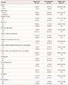
Among the 1,158 enrolled patients, the 10 most commonly identified HPV types were all high-risk types. In the order of decreasing prevalence these were HPV 16, 58, 56, 53, 52, 39, 18, 51, 68, and 66. HPV 16 showed the highest prevalence (12.3%). Among the 799 women who underwent six-month follow-up tests, 72 women infected with unknown HPV types at the time of enrollment were excluded. Of the remaining women, 345 (47.5%), 184 (25.3%), and 198 (27.2%) showed persistent, incidental, and cleared infection, respectively. The most common type was HPV 16 (n=143, 12.3%) presenting as a single infection (n=85, 59.4%) and multiple infections (n=58, 40.6%). Among those showing cytological progression at the six-month follow-up, 27.0% were infected with HPV 16, which represented a significantly higher progression rate than that observed with other HPV types (RR, 1.75; 95% CI, 1.08–2.84; P=0.028) (Table 1).
Table 1
Changes in cervical cytology and human papillomavirus infection status in women initially diagnosed with atypical squamous cells of undetermined significance/low-grade squamous intraepithelial lesion

![]()
HPV 16 infection status at follow-up testing was associated with cervical cytology prognosis (Table 2). Persistent HPV 16 infection was significantly associated with cytological progression over one year (P<0.001). At the six-month follow-up, the rate of persistent infection was 85.2% in the cytological progression and only 32.0% in the cytological regression group. Only 3.7% of the women in the cytological progression group had a negative HPV test at follow-up, and HPV 16 infection was cleared in 38.0% of the women in the cytological regression group. The rate of cytological progression was 42.6% (23 of 54) in the HPV 16 persistence group and only 4.8% (1 of 21) in the clearance group. The rate of cytological regression was only 29.6% (16 of 54) in the HPV 16 persistence group and as high as 90.5% (19 of 21) in the clearance group. The results were similar at the 12-month follow-up. The cytological progression rate in women with multiple HPV 16 infections (27.9%) was similar to that in women with HPV 16 infection alone (26.3%).
Table 2
The association between human papillomavirus status and cervical cytology in human papillomavirus 16 infected women

![]()
We performed logistic regression analysis to evaluate the epidemiological risk factors affecting cytological progression in HPV 16-positive women (Table 3). Of the 91 women who underwent six-month follow-up, 53 women had an HPV 16 single infection and 38 had HPV 16 multiple infections at the time of enrollment. The RR of progression in cigarette smokers (including former smokers) was higher than that in non-smokers (RR, 4.15; 95% CI, 1.01–17.0). We performed multivariate logistic regression analysis to assess the compound effects of variables on the risk of progression. Smoking was the only significant independent predictor of cytological progression (odds ratio, 4.15; 95% CI, 1.01‒17.00; P=0.048). Young age, alcohol use, obesity, oral contraceptive use, change in sexual partners within a year prior to enrollment in the study, gravidity, menopause, and HPV vaccination were not significantly associated with a high risk of cytological progression.
Table 3
Analysis of epidemiologic risk factors for cytological progression in human papillomavirus 16 infected women (n=91)
![]()
Discussion
The results of this study suggest that HPV 16 infection is a significant risk factor for the development of cervical cancer. A meta-analysis including 14,595 patients reported that most invasive cervical cancers worldwide are associated with HPV 16 infection [16]. Current guidelines recommend additional evaluation as a component of cervical cancer screening in women with HPV 16 infection [17]. We evaluated the persistent HPV 16 infection rates and the epidemiological factors associated with cytological progression in HPV 16-positive women. This study showed that the rate of development of high-grade lesions in women with HPV 16 infection was higher than that in women with other types of HPV infection (RR, 1.75; 95% CI, 1.08–2.84; P=0.028). Among HPV 16-positive women, persistent infection was observed in 54% at the six-month follow-up and in 39.1% at the 12-month follow-up. Both rates were higher than the persistence rates of 47.5% and 34.1%, respectively in the overall HPV cohort, suggesting that HPV 16 infection is more likely to persist and is associated with progression to high-grade cervical lesions.
Risk factors for cervical cancer include age, cigarette smoking, multiple sexual partners, gravidity, an immunocompromised status, and long-term use of oral contraceptives [18]. Among the HPV 16-positive women evaluated in this study, cigarette smokers showed a significantly higher risk for progression to abnormal cervical cytology. Smoking has been significantly associated with an increased risk of cervical cancer [192021]. Smoking interferes with HPV clearance by impairing the immune response that decreases the number of CD4 lymphocytes and Langerhans cells, as well as reducing the activity of natural killer cells [222324252627]. A higher risk of cervical cancer was observed in women with HPV 16 infection with a history of smoking (not necessarily current smokers). Moreover, a dose-dependent relationship with the number of cigarettes smoked has been reported [28]. Additionally, young age, alcohol use, obesity, oral contraceptive use, change of sexual partners within a year prior to enrollment in the study, gravidity, and menopause were associated with a higher risk of cytological progression. However, several other factors correlated with cytological progression were statistically non-significant owing to the short-term follow-up and small sample size of this study.
The strengths of this study include the long-term prospective study design, the fact that we included women from five university medical centers across four metropolitan cities in Korea, and that we minimized interobserver variability in result interpretation by performing all cytological evaluations at a single center (Cheil General Hospital). Limitations of our study are as follows: 1) The Korean HPV cohort was designed such that follow-up biopsies were not mandatory, and only cytology and HPV testing were performed at each follow-up visit. Although cytology and HPV testing are effective screening tools for precancerous lesions, colposcopy guided biopsies remain the diagnostic standard for cervical lesions. 2) An incomplete understanding of other potential risk factors (previous HPV infection status, age at which HPV vaccination was administered) may limit the interpretation of results. 3) The 12-month follow-up used in this study is relatively short. A previous study has shown that cytological regression from ASC-US to normal and from LSIL to ASC-US or normal cytology occurred in 16.8 and 13.8 months, respectively in women with oncogenic HPV types [29]. In this study, among women with cytological progression at the six-month follow-up, the progression rate in HPV 16-positive women was significantly higher than that in women infected with other HPV types (27.0% vs. 17.5%; RR, 1.75; 95% CI, 1.08–2.84; P=0.028). However, the 12-month follow-up results did not show a statistically significant difference. Thus, long-term follow-up studies are needed to draw more accurate inferences. Although this study included a relatively short follow-up period, the Korean HPV cohort is an ongoing study. Owing to the relatively large sample size and the systematic study design, the results of that cohort study are expected to be more representative of Korean women as a whole.
In conclusion, the cytological progression rate in HPV 16-positive women with ASC-US or LSIL is higher than that in women infected with other HPV types. Furthermore, smoking among women with HPV 16 infection is a major risk factor for the progression of cervical lesions. This study would provide a better understanding of the epidemiological risk factors that determine the prognosis of low-grade cervical abnormalities in Korean women.
 XML Download