

 PDF
PDF ePub
ePub Citation
 Print
Print


Abstract
Purpose
The aims of this study were to evaluate risk factors for knee stiffness after the fixation of distal femoral fractures, and to analyze the clinical and radiologic outcomes.
Materials and Methods
This is a retrospective case control study of 104 consecutive patients who have a distal femoral fracture and were treated with a submuscular locking plate. The case group comprised of patients with 12-month postoperative range of motion (ROM) ≤90° or a history of manipulation under anesthesia. The case group was compared with the control group of patients with a 12-month postoperative ROM >90°. The possible risk factors were evaluated by univariate and logistic regression analysis. The postoperative ROM and Knee Society clinical rating system was evaluated for the clinical assessment and the distal femoral angle on a whole-extremity scanogram was measured for radiologic assessments.
Results
Fifty-four patients were included in the study (14 in the case group, 40 in the control group). Univariate analysis showed that comminuted fracture, intra-articular fracture, open fracture, temporary external fixation, severe osteoarthritis, and prolonged immobilization placed patients at an increased risk for knee stiffness. On the other hand, multivariate logistic regression showed that an extensor mechanism injury was the only significant predictor (p=0.001; odds ratio, 42.0; 95% confidence interval, 5.0–350.7). The ROM and Knee Society score were significantly lower in the case group; however, the coronal alignment was similar in the case and control group.
Conclusion
Various factors that delay postoperative knee motion place patients at increased risk of knee stiffness. Understanding these risk factors may help surgeons prevent postoperative knee stiffness after distal femoral fractures. In particular, extensor mechanism injury, such as patella fracture or open quadriceps injury, was found to be an independent predictable factor associated with knee stiffness.
Go to : 
References
1. Court-Brown CM, Caesar B. Epidemiology of adult fractures: a review. Injury. 37:691–697. 2006.

2. Martinet O, Cordey J, Harder Y, Maier A, Bühler M, Barraud GE. The epidemiology of fractures of the distal femur. Injury. 31(Suppl 3):C62–63. 2000.
3. Arneson TJ, Melton LJ 3rd, Lewallen DG, O'Fallon WM. Epidemiology of diaphyseal and distal femoral fractures in Rochester, Minnesota, 1965–1984. Clin Orthop Relat Res. 234:188–194. 1988.
4. Sanders R, Swiontkowski M, Rosen H, Helfet D. Double-plating of comminuted, unstable fractures of the distal part of the femur. J Bone Joint Surg Am. 73:341–346. 1991.
5. Seinsheimer F 3rd. Fractures of the distal femur. Clin Orthop Relat Res. 153:169–179. 1980.
6. Prevosti LG, Subramainian VA, Rothaus KO, Dineen P. A comparison of the open and closed methods in the initial treatment of sternal wound infections. J Cardiovasc Surg (Torino). 30:757–763. 1989.
7. Halpenny J, Rorabeck CH. Supracondylar fractures of the femur: results of treatment of 61 patients. Can J Surg. 27:606–609. 1984.
8. Schandelmaier P, Partenheimer A, Koenemann B, Grün OA, Krettek C. Distal femoral fractures and LISS stabilization. Injury. 32(Suppl 3):SC55–63. 2001.
9. Bolhofner BR, Carmen B, Clifford P. The results of open reduction and internal fixation of distal femur fractures using a biologic (indirect) reduction technique. J Orthop Trauma. 10:372–377. 1996.
10. Schütz M, Müller M, Krettek C, et al. Minimally invasive fracture stabilization of distal femoral fractures with the LISS: a prospective multicenter study. Results of a clinical study with special emphasis on difficult cases. Injury. 32(Suppl 3):SC48–54. 2001.
11. Ostrum RF, Geel C. Indirect reduction and internal fixation of supracondylar femur fractures without bone graft. J Orthop Trauma. 9:278–284. 1995.
12. Haller JM, Holt DC, McFadden ML, Higgins TF, Kubiak EN. Arthrofibrosis of the knee following a fracture of the tibial plateau. Bone Joint J. 97:109–114. 2015.
13. Bishop J, Agel J, Dunbar R. Predictive factors for knee stiffness after periarticular fracture: a case-control study. J Bone Joint Surg Am. 94:1833–1838. 2012.
14. Schiavone Panni A, Cerciello S, Vasso M, Tartarone M. Stiffness in total knee arthroplasty. J Orthop Traumatol. 10:111–118. 2009.
15. Robertson GA, Coleman SG, Keating JF. Knee stiffness following anterior cruciate ligament reconstruction: the incidence and associated factors of knee stiffness following anterior cruciate ligament reconstruction. Knee. 16:245–247. 2009.
16. Ehlinger M, Dujardin F, Pidhorz L, Bonnevialle P, Pietu G, Vandenbussche E. SoFCOT. Locked plating for internal fixation of the adult distal femur: influence of the type of construct and hardware on the clinical and radiological outcomes. Orthop Traumatol Surg Res. 100:549–554. 2014.
17. Kregor PJ, Stannard JA, Zlowodzki M, Cole PA. Treatment of distal femur fractures using the less invasive stabilization system: surgical experience and early clinical results in 103 fractures. J Orthop Trauma. 18:509–520. 2004.
18. Kolb W, Guhlmann H, Windisch C, Marx F, Kolb K, Koller H. Fixation of distal femoral fractures with the less invasive stabilization system: a minimally invasive treatment with locked fixed-angle screws. J Trauma. 65:1425–1434. 2008.
19. Insall JN, Dorr LD, Scott RD, Scott WN. Rationale of the Knee Society clinical rating system. Clin Orthop Relat Res. 248:13–14. 1989.
20. Bong MR, Di Cesare PE. Stiffness after total knee arthroplasty. J Am Acad Orthop Surg. 12:164–171. 2004.
21. Desai AS, Karmegam A, Dramis A, Board TN, Raut V. Manipulation for stiffness following total knee arthroplasty: when and how often to do it? Eur J Orthop Surg Traumatol. 24:1291–1295. 2014.
22. Parvizi J, Tarity TD, Steinbeck MJ, et al. Management of stiffness following total knee arthroplasty. J Bone Joint Surg Am, 88 Suppl. 4:175–181. 2006.
23. Pivec R, Issa K, Kester M, Harwin SF, Mont MA. Long-term outcomes of MUA for stiffness in primary TKA. J Knee Surg. 26:405–410. 2013.
24. Shelbourne KD, Patel DV. Treatment of limited motion after anterior cruciate ligament reconstruction. Knee Surg Sports Traumatol Arthrosc. 7:85–92. 1999.
25. Magit D, Wolff A, Sutton K, Medvecky MJ. Arthrofibrosis of the knee. J Am Acad Orthop Surg. 15:682–694. 2007.
26. Laubenthal KN, Smidt GL, Kettelkamp DB. A quantitative analysis of knee motion during activities of daily living. Phys Ther. 52:34–43. 1972.
27. Rowe PJ, Myles CM, Walker C, Nutton R. Knee joint kinematics in gait and other functional activities measured using flexible electrogoniometry: how much knee motion is sufficient for normal daily life? Gait Posture. 12:143–155. 2000.
28. Massè A, Biasibetti A, Demangos J, Dutto E, Pazzano S, Gal-linaro P. The judet quadricepsplasty: longterm outcome of 21 cases. J Trauma. 61:358–362. 2006.
29. Lee DH, Kim TH, Jung SJ, Cha EJ, Bin SI. Modified judet quadricepsplasty and Ilizarov frame application for stiff knee after femur fractures. J Orthop Trauma. 24:709–715. 2010.
Go to :
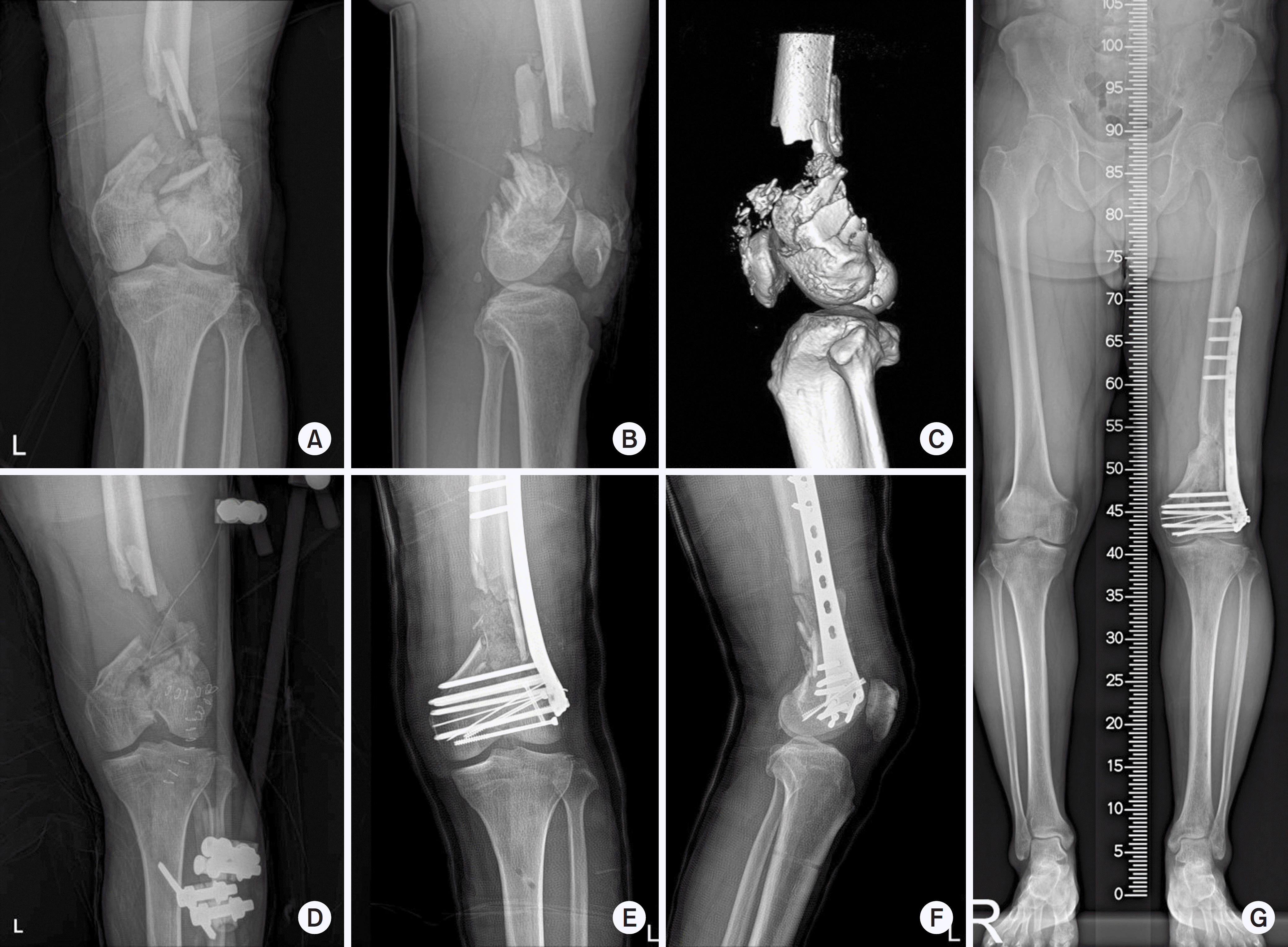 | Fig. 1.(A, B) A 58-year-old man with an open distal femoral fracture. He had a severe open comminuted distal femoral fracture. (C) The distal femoral segment was not large enough to allow rigid fixation. (D) Spanning temporary external fixation was used for severe soft tissue injury, which was maintained for 12 days. (E, F) Because fracture site instability was observed intraoperatively, cast immobilization was applied for 6 weeks. (G) Although bone union was observed after postoperative 9 months, the range of motion of the knee was not recovered completely. |
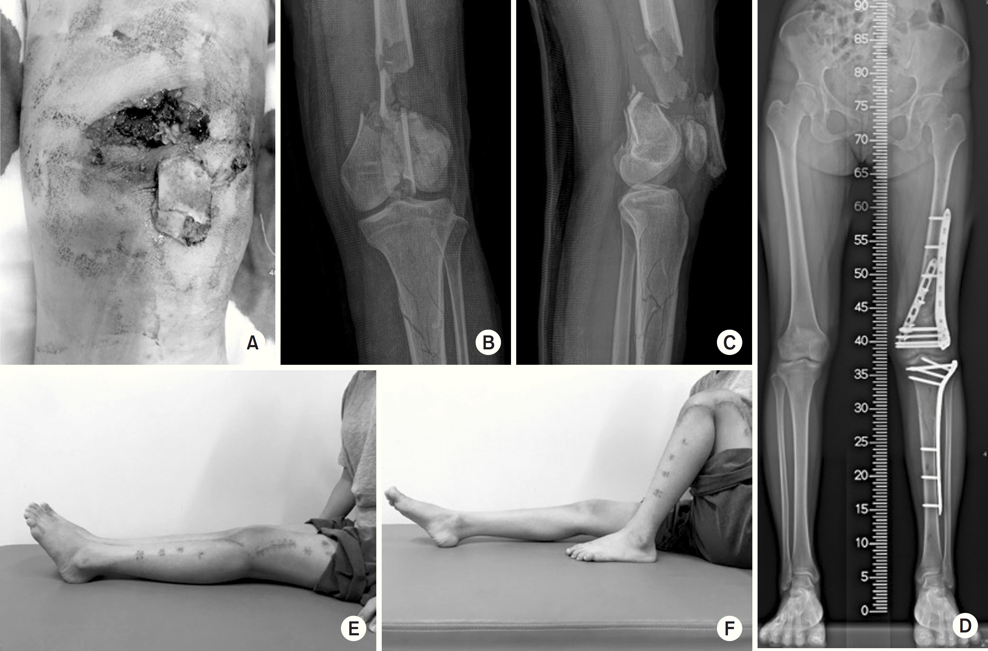 | Fig. 2.(A) A 26-year-old woman with open distal femoral fracture. She had an open fracture and quadriceps muscle injury. A bony fragment penetrated through the skin and the wound was contaminated. (B, C) Preoperative radiographs demonstrating comminuted and intra-articular fractures in the distal femur. After complete debridement and a weak spanning external fixation, the patient underwent auto- and allo-bone graft and firm-plate fixation. Two days later, she started knee exercise. (D) Follow-up radiograph after 3 months demonstrating fracture union. (E, F) The range of motion of the knee was recovered completely through early exercise and firm internal fixation. |
Table 1.
Demographic Characteristics of the Case and Control Groups
Table 2.
Comparisons of the Frequency of Variables Known to be Risk Factors of Knee Joint Stiffness according to the Range of Motion
| Variable | Case (n=14) | Control (n=40) | p-value* |
|---|---|---|---|
| High-energy injury | 14 | 23 | <0.001 |
| Comminuted fracture | 12 | 16 | 0.005 |
| Intra-articular fracture | 14 | 15 | <0.001 |
| Extensor injury | 10 | 2 | <0.001 |
| Open fracture | 10 | 4 | <0.001 |
| Temporary external fixator | 8 | 2 | <0.001 |
| Osteoarthritis K-L grade >4† | 4 | 2 | 0.033 |
| Immobilization >3 weeks | 8 | 6 | 0.004 |
Table 3.
Clinical and radiologic outcomes of the case and control groups
| Variable | Case (n=14) | Control (n=40) | p-value |
|---|---|---|---|
| 3-month postoperative ROM (o) | 82±8 (70–90) | 125±14 (95–145) | <0.001* |
| 12-month postoperative ROM (o) | 88±17 (70–120) | 125±15 (90–145) | <0.001* |
| Knee Society score | 79±12 (65–95) | 89±6 (72–97) | <0.001* |
| Bone union time (wk) | 22±4 (16–28) | 14±3 (9–21) | <0.001* |
| Mechanical LDFA (o) | 86±1 (85–87) | 87±4 (79–93) | 0.215 |
| Anatomical LDFA (o) | 80±1 (79–82) | 80±4 (70–86) | 0.155 |
 XML Download