Journal List > Korean Circ J > v.48(12) > 1106766

Kolla, Larsen, Click, Chaliki, and Maltais: Pivotal Role of Intraoperative Transesophageal Echocardiography for Detecting Iatrogenic Aortic Regurgitation due to Cardiac Catheterization
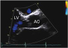
A 67-year-old man with a history of fatigue and dyspnea on exertion was diagnosed with atrial fibrillation and severe mitral regurgitation. Preoperative transthoracic echocardiography (TTE) showed a normal aortic valve without aortic regurgitation (Figure 1 and Supplementary Video 1) and bileaflet mitral valve prolapse with severe mitral regurgitation. As part of the preoperative evaluation, cardiac catheterization was performed, which showed normal coronary arteries. The patient was referred for mitral valve repair.
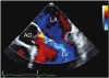
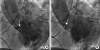
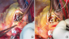
Intraoperative transesophageal echocardiography (TEE) before sternotomy showed a moderate, unexpected eccentric jet of aortic regurgitation (Figure 2 and Supplementary Video 2). The preoperative left ventriculogram showed the multipurpose guiding catheter intermittently entering the aortic sinus (Figure 3 and Supplementary Video 3). Because aortic regurgitation was not present before catheterization, the catheter likely caused trauma to the aortic leaflet, resulting in aortic regurgitation. Two areas with defects were found during surgery: an apparent cleft in the right coronary cusp and a tear in the central right coronary cusp (Figure 4A). Aortic valve repair consisted of commissuroplasty between the right and noncoronary cusps, repair of the tear in the right coronary cusp, and resuspension of all 3 commissures (Figure 4B). Mitral valve repair, ligation of the left atrial appendage, and surgical CryoMaze of the left atrium were performed as planned. Follow-up TTE showed insignificant aortic regurgitation, trivial mitral regurgitation, and normal left ventricular ejection fraction.
Iatrogenic aortic regurgitation due to cardiac catheterization is a rare complication most often associated with aortic dissection.1) Few cases have been reported of iatrogenic aortic regurgitation due to aortic leaflet trauma during cardiac catheterization.2)3)4) This case highlights the importance of intraoperative TEE in identifying unexpected findings such as aortic regurgitation secondary to iatrogenic aortic valve cusp trauma.

Figures and Tables

Figure 1

Transthoracic echocardiogram, showing a normal aortic valve without aortic regurgitation.

Ao = aorta; LV = left ventricle.
kcj-48-1160-g001
Figure 2

Intraoperative transesophageal echocardiogram before surgery, showing aortic regurgitation.

Ao = aorta; LA = left atrium; LV = left ventricle.
kcj-48-1160-g002
Figure 3

Cineangiogram during contrast injection of the left ventriculogram. (A) Left ventricular catheter (arrow) just below the right coronary cusp. (B) Left ventricular catheter (arrow) just above the right coronary cusp.

kcj-48-1160-g003
Figure 4

Intraoperative image. (A) Tear in the right coronary cusp (arrows). (B) Aortic valve repair (arrows).

kcj-48-1160-g004

Notes

Conflict of Interest The authors have no financial conflicts of interest.

Author Contributions

  • Conceptualization: Chaliki HP, Maltais S.

  • Data curation: Kolla KR, Larsen CM, Click RL.

  • Writing - original draft: Kolla KR, Chaliki HP.

  • Writing - review & editing: Kolla KR, Click RL, Chaliki HP, Maltais S.

References

1. Wyss CA, Steffel J, Lüscher TF. Isolated acute iatrogenic aortic dissection during percutaneous coronary intervention without involvement of the coronary arteries. J Invasive Cardiol. 2008; 20:380–382.
2. Denyer MH, Elliott CM, Robicsek F. Pericardial patch repair of aortic cusp perforation caused by cardiac catheterization. J Card Surg. 1988; 3:155–157.
crossref
3. Fundarò P, Di Mattia DG, Cialfi A, Santoli C. Repair of aortic cusp traumatic laceration caused by cardiac catheterization. J Heart Valve Dis. 1996; 5:281–282.
4. Rolf T, Tozzi P, Roumy A, Iglesias JF, von Segesser LK. Aortic valve lesion after coronary angiography. Interact Cardiovasc Thorac Surg. 2011; 12:205–206.
crossref

SUPPLEMENTARY MATERIALS

Supplementary Video 1

Preoperative transthoracic echocardiogram showing the presence of mitral regurgitation and absence of aortic regurgitation.

Supplementary Video 2

Intraoperative transesophageal echocardiogram showing an eccentric jet of aortic regurgitation.

Supplementary Video 3

Left ventricular cineangiogram showing the multipurpose guiding catheter briefly entering the aortic sinus from the left ventricle during the contrast injection.
TOOLS
ORCID iDs

Kantha R. Kolla
https://orcid.org/0000-0003-1959-928X

Carolyn M. Larsen
https://orcid.org/0000-0001-6530-3545

Hari P. Chaliki
https://orcid.org/0000-0002-9470-9605

Similar articles