

 PDF
PDF ePub
ePub Citation
 Print
Print


INTRODUCTION
Cleft lip and/or cleft palate (CLP) occur in 1 in 700 infants [1]. Oronasal fistulae (ONF) can occur during the treatment of CLP [2]. Although the phenotype of this malformation varies, all defects ultimately require transplantation therapy to block the defect. The most commonly used donor site for such transplantation is the tongue of the patient, and all surgical procedures are performed in the oral cavity, thereby requiring caution about peri-operative airway obstruction [3].
In particular, tongue flap surgery is performed in two steps to repair ONF, and the first surgery involves pulling and attaching the dorsal part of the tongue [4]. A part of the tongue dorsum is lifted up to form a long anteroposterior flap; one part is attached to the tongue, and the other end is sutured to the defective area of the palate and transplanted. The second surgery is performed approximately 2 weeks after the first surgery, and the flap, which becomes a part of the palate with stable circulation from collateral vessels in palate, is cut off and separated from the tongue. A common complication is detachment or bleeding of the palate portion of the flap [5].
In the second surgery, anesthesiologists face a great challenge in airway management because the patient's tongue is attached to the palate [6]. According to a report, a safe flap division was performed on a patient willing to undergo sedation and adequate local anesthesia [7]. However, the use of this method for children has limitations. Thus, we report a case of oral intubation using a flexible fiberoptic bronchoscope in general anesthesia for tongue flap division after inhalational anesthesia induction in a child.
CASE
A 6-year-old boy (American Society of Anesthesiologists Class I, weight 17 kg, height 115 cm) had undergone cheiloplasty at 100 days of age and palatorrhaphy at 19 months of age due to cleft lip and palate. However, oronasal fistulous opening had remained, and thus, oronasal fistula closure using a vomer flap and tongue flap had been performed 20 days prior to presentation. Then, the flap had been placed on the recipient site, i.e, the palate, without any signs of necrosis. Subsequently, the patient visited our hospital for tongue flap division surgery.
Preoperative laboratory test results, electrocardiographic findings, and chest radiographic results were all within normal ranges. Although there was a flap in the airway, mouth opening was 3 finger breadths, and there was no limitation of neck extension.
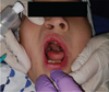
However, the tongue flap was attached to the palate from the root of the tongue with the base near the tongue tip. In addition, as the palate portion of the tongue flap was close to the soft palate (Fig. 1), intubation using a laryngoscope was difficult. Therefore, as reported in a previous study [7], we planned to perform surgery using local anesthesia and sedation. However, the patient seemed to be very anxious in the operation room, and thus, performing surgery under local anesthesia seemed difficult. After explaining and obtaining consent from the patient's parents, we determined to perform the surgery under general anesthesia. To make the patient feel relaxed, the caregiver was brought to the operation room, and subsequently, facemask ventilation was performed. After preoxygenation with 100% O2 for 5 mins, inhalation anesthesia induction was performed using 8% sevoflurane. An intravenous line was then placed, and 8 mg of rocuronium was injected as a neuromuscular blocker.
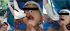
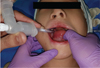
The use of a direct laryngoscope could damage the lingual artery or intrinsic muscle of the tongue, thereby causing a risk for complications such as bleeding when the flap connected to the palate is stretched excessively or the blade of the laryngoscope exerts excessive force on the flap [8]. Therefore, oral intubation was immediately carried out using a flexible fiberoptic bronchoscope (FOB) (LF-DP, Olympus Co., Japan). In preparation for the situation in which visual field is not clear and the use of the laryngoscope is required when the FOB is used, we approached from the right side of the tongue flap. The pediatric FOB passed the space of the flap and buccal cavity without any difficulty, and a 5.0-mm cuffed Ring-Adair-Elwin (RAE) tube was also stably positioned (Fig. 2, 3). There was no decrease in oxygen saturation and bleeding from the toungue flap during intubation, and the surgery took approximately 45 minutes.
After confirming hemostasis in the surgical site, an antagonist of the neuromuscular blocker was administered, and extubation was performed in the operation room after the respiration and consciousness of the patient were fully restored. No complications associated with the general anesthesia and operation were noted.
DISCUSSION
Cleft lip and/or cleft palate (CLP) occurs at a ratio of 1 in 700 infants, possibly due to fusion failure of the maxillary and medial nasal processes at 4–9 weeks of gestation, but no clear etiology has been identified [1].
Infants with CLP are not adequately nourished because negative pressure with proper oronasal saealing does not occur in their oral cavity, which leads to inadequate nursing including breastfeeding. Thus, they are more vulnerable to infection and gastrointestinal disorders as compared to healthy babies [9]. In particular, in patients with cleft palate, delayed treatment may result in dysfunctions in respiration, swallowing, mastication, and language acquisition due to abnormal dental arches and consequent deformations of the facial bone [10]. Various methods can be selected depending on the degree of the pediatric CLP.
The tongue flap for repairing large anterior palatal fistulas [4] is a 2-stage operation. In the first operation, there is no structure in the oral cavity acting as an obstacle during general anesthesia. However, in the second operation, the flap connected to the palate causes difficulty in airway management. In this case, the rigid laryngoscope was hard to apply, thereby requiring sufficient planning before anesthesia induction [11]. In particular, children feel very anxious about the second operation, and thus, it is impossible to perform awake endotracheal intubation even though it involves a difficult airway. Thus, sedation and intubation should be carried out using a method such as inhalation anesthesia. However, if a flap is present, the supra-glottic airway cannot be used, which may lead to a fatal outcome when the intubation fails [12].
In our case, to avoid the condition of “cannot Intubate, cannot ventilate” [12], the surgical site and other facial abnormalities in the patient were repaired. Mask ventilation was confirmed while the patient was conscious, and inhalation anesthesia induction was performed.
In the tongue flap surgery, nasotracheal intubation is performed in the first surgery, while oral intubation is used in the second surgery because of the possibility of injury of the flap that has been lifted in the root of the tongue [13].
There are two intubation methods available for tongue flap division surgery: intubation using a Miller-type laryngoscope and intubation using an FOB [1415]. In both cases, the deeper and larger the flap, the more difficult it may be to obtain a clear view. Oral cavity damage due to the laryngoscope has also been reported in adult patients. If there is a flap, it may be difficult to have a clear view due to soft tissue damage, and there is also a risk for blood aspiration [8]. In the case of using only the FOB, it may be difficult to proceed with intubation owing to the presence of a structure in the larynx when the tip of the FOB is inserted into the trachea and the FOB is guided to advance the endotracheal tube. When the endotracheal tube is advanced, it may sweep through the soft tissue of the root of the tongue in a somewhat deformed position and cause bleeding, or the flap itself may be damaged due to stretching force acting on the tongue. If the size of the flap is large, it may be helpful to pack the flap with gauze soaked with epinephrine in advance [16].
Rapid sequence intubation can be performed in some emergency situations. In this case, 1.0 mg/kg rocuronium or 1.0 mg/kg succinylcholine can be used. The condition that enables easy intubation is the same for both [17], but rocuronium yields a faster reversal than succinylcholine because of the presence of sugammadex in the case of intubation failure [18].
In conclusion, repairing the oronasal fistula of patients with CLP is essential, but a difficulty in anesthesia on children has been an issue especially in the second surgery. And airway surgery is associated with a risk before and after surgery. It is necessary to establish an airway management plan after thorough consultation with surgeons, and intubation using an FOB, as in this case, can be an effective method to achieve airway management in such patients.
 XML Download