

 PDF
PDF ePub
ePub Citation
 Print
Print


INTRODUCTION
Sudden headache onset may rarely be caused by spontaneous intracranial hypotension (SIH). SIH-induced headache typically, but not always, shows a postural or exertional pattern, with relief achieved by lying in a supine or recumbent position [1]. Symptoms that commonly accompany SIH are nausea, vomiting, vertigo, and hearing alteration [23]. SIH is also frequently associated with visual disturbances that are caused by cranial nerve palsies of innervated oculomotor muscles [45].
SIH is typically characterized by a benign and self-limiting course, and in most cases, resolves spontaneously without any specific treatment [1]. Although conservative therapies, including bed rest, hydration, simple analgesics, caffeine intake, steroid administration, and the use of an abdominal binder are available, the injection of autologous blood into the epidural space (epidural blood patch [EBP]) may be the treatment of choice to relieve symptoms [678].
We describe the case of a patient diagnosed with SIH who developed diplopia following resolution of sudden headache. This case report shows that an EBP may successfully treat visual symptoms, which were not effectively treated with conservative management, after relief of headache in SIH.
CASE
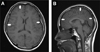
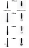
A 43-year-old female patient visited the emergency department 4 days after the abrupt onset of a headache (numeric rating scale 8), nausea, and tinnitus, which began following a session of heavy lifting. She complained that the headache extended from the left parietal to the frontal areas. Her symptoms were not affected by posture and she had no notable past medical history. Laboratory analysis and a brain computed tomogram showed no remarkable findings. She was discharged with a diagnosis of migraine after symptom improvement following simple analgesic administration. She returned to the emergency room 6 days after discharge due to horizontal diplopia on right gaze that developed with improvement of the headache. Intraocular examination was normal. She was admitted to the neurology department for further tests. The physical examination revealed limited abduction in the right eye. Other cranial nerves and systemic examinations were unremarkable. A diagnostic spinal puncture showed the cerebrospinal fluid (CSF) opening pressure was 6 cmH2O and CSF analysis was normal. In general, the normal range of CSF opening pressure measured at lumbar puncture is 10–18 cmH2O, with the patient lying on the side. Brain magnetic resonance imaging (MRI) without contrast enhancement showed diffuse pachymeningeal enhancement, with thickening and venous sinus engorgement (Fig. 1). Mild obliteration of the basal, prepontine, and suprasellar cistern was also observed. There was no other abnormality on the MRI. Furthermore, radionuclide cisternography revealed multiple CSF leakages in mid to lower thoracic spine levels, notably T9 and T10 (Fig. 2). The patient was diagnosed with SIH and secondary right sixth cranial nerve palsy.
Although conservative treatment management, including bed rest and hydration, relieved the headache, double vision in the right gaze was not fully relieved. The department of anesthesiology and pain medicine was consulted for consideration of EBP treatment. Fourteen days after developing diplopia, an EBP was applied. At this time, the headache had completely resolved. With the patient lying in prone position, 14 ml of autologous blood was injected aseptically into the epidural space at the T9-T10 level using an 18G Tuohy needle under fluoroscopic guidance. Because the patient complained of back pain, only 14 ml of epidural blood was administered. After EBP application, the patient maintained the supine position overnight. Decreased diplopia was reported the day after EBP treatment.
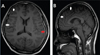
The patient was discharged 3 days after the procedure. One month after discharge, symptoms of diplopia and headache were resolved. Furthermore, follow-up examination revealed complete resolution of sixth cranial nerve palsy without any neurologic deficit. A follow-up MRI also showed decreased extent of the diffuse pachymeningeal enhancement with thickening and venous sinus engorgement (Fig. 3). In addition, although it was not clinically significant, a small subacute subdural hematoma was observed.
DISCUSSION
The most common manifestation of SIH is an orthostatic headache, which increases in the upright position. Headache relief by postural change (when lying down) is well-described [1]. However, in the present case, the headache was not affected by posture. A previous report has described patients with SIH who experienced severe headache with no posture-related component [9]. Additional symptoms, such as hearing disturbance, tinnitus, nausea, and vomiting have been documented in patients diagnosed with SIH [1]. Although they are less common, visual problems are associated with SIH, and include blurred vision, visual field defects, and diplopia. Among ocular manifestations in SIH, approximately 80% of patients have been reported to have ophthalmoplegia, which resulted from abducens nerve palsies [4]. Initially, our patient experienced not only headache, but also nausea and tinnitus. Although her initial symptoms were decreased by conservative management, delayed onset diplopia developed and the patient was diagnosed with secondary right sixth cranial nerve palsy. New diagnostic criteria for spinal spontaneous CSF leakage and SIH have proposed based on both radiographic and clinical manifestations [10]. The MRI and cisternography of the present patient showed typical radiologic findings observed in SIH (Fig. 1 and 2).
It has been established that SIH may result from spontaneous CSF leakage. The precise mechanism and pathophysiology of spontaneous CSF leakage remain unknown. However, it is generally accepted that mechanical factors may play an important role in CSF leakage [11]. For example, trivial traumatic events often precede the onset of SIH symptoms. In addition, heritable connective tissue disorders may contribute to the development of spontaneous CSF leakage, including Marfan and Ehlers-Danlos syndromes [12]. Our patient denied any underlying disorder or notable medical history. Therefore, mechanical factors, such as heavy lifting, may be responsible for the development of SIH in our patient.
Although SIH is considered a benign and self-limiting disorder, conservative management, including bed rest, hydration, simple analgesics, caffeine intake, steroid administration, and the use of an abdominal binder are available [1]. In addition to conservative therapies, it is well established that the mainstay of treatment for SIH is autologous EBP [67]. When CSF volume decreases, the burden on the vessels, nerves, and other dural pain sensitive structures, which are subject to traction and distortion, may cause headache or cranial nerve dysfunction. Therefore, the blood patch is considered a definitive treatment since it can prevent CSF leakage. In our SIH patient, while conservative treatments effectively resolved the headache, it was ineffective in the development of diplopia after headache relief. The application of EBP successfully treated diplopia, even 14 days after the onset of visual symptoms. The exact timing and volume of the blood patch have not been disclosed. Ten to 20 ml of blood is typically injected into the epidural space and is effective in relieving symptoms [67]. Béchard et al. [13] have reported that EBP, performed within 24 hours of diplopia onset, may lead to improvement and early resolution of symptoms in patients with sixth cranial nerve palsy after dural puncture. Dural puncture, such as spinal analgesia and spinal tapping, can cause iatrogenic intracranial hypotension, which leads to abrupt leakage of CSF and a rapid decrease of CSF pressure. Therefore, early treatment can be important. However, SIH may result from spontaneous CSF leakage, which progress slowly. Some reports have documented that EBP completely resolved visual symptoms in patients with sixth [14] and third [15] cranial nerve palsies, which was secondary to SIH, several weeks after symptom onset. Our patient also received EBP 14 days after the onset of diplopia as well, suggesting that relatively late application of EBP can resolve cranial nerve dysfunction due to SIH. This discrepancy in timing may be explained by different underlying mechanisms causing intracranial hypotension (spontaneous vs. iatrogenic).
To our knowledge, this is the first report of successful application of EBP for the treatment of diplopia that developed after resolution of a headache, which was relieved with conservative treatment, in a patient with SIH. This case also illustrates the important role of EBP in the treatment of diplopia after resolution of headache in SIH.
 XML Download