Abstract
Objective
Placement of a ventriculoperitoneal (VP) shunt is a common neurosurgical procedure for cerebrospinal fluid diversion. A rare complication is delayed intracranial hemorrhage (ICH) secondary to VP shunting, and only a few patients with this complication have been reported. We investigate the incidence and risk factors of delayed ICH development following VP shunt placement.
Methods
Over an 11-year period, 167 patients received a VP shunt for hydrocephalus, and of these, 138 patients were eligible for this study. All medical records and computed tomography scans obtained within 48 h after the operation and at postoperative day 7 were reviewed. The risk factors of developing delayed ICH (≥48 hr after VP shunt placement) were analyzed according to the demographic data, including sex and age, original intracranial lesions, co-morbid diseases, and laboratory findings.
Results
Delayed ICH following VP shunt placement developed in 34 (24.6%) of the 138 patients. Risk factors for developing delayed ICH were age (p=0.037) and the partial thromboplastin time (PTT) (p=0.032). Intraventricular hemorrhage after VP shunting was the most common complication, occurring in 16 cases. Hemorrhagic volume was <1 mL in 28 cases and >1 mL in 6 cases.
Conclusion
This study suggests that old age and delayed PTT are major risk factors for developing delayed ICH following VP shunting. Additionally, delayed ICH after VP shunting commonly occurs even when most patients are asymptomatic. Therefore, extra care should be taken to observe and follow-up with patients who have undergone VP shunt placement.
Hydrocephalus is a common condition accounting for 69,000 hospital discharges each year in the United States.3) Cerebrospinal fluid (CSF) shunt procedures have dramatically reduced the morbidity and mortality of hydrocephalus. Among CSF diversion techniques, the ventriculoperitoneal (VP) shunt is the routine, first choice, due to easy placement and functional reliability as compared to the other alternatives. However, there are potential complications that may require multiple surgical procedures during the lifetime of a patient. Postoperative complations after VP shunt placement include shunt obstruction, subdural hematoma, infection, seizures, catheter migration, and shunt malfunction. 26)
Among these complications, delayed intraparenchymal hemorrhage (IPH) or intraventricular hemorrhage (IVH) secondary to VP shunt placement is not common. Although the mechanism of delayed intracranial hemorrhage (ICH) after VP shunt has been reported, the risk factors for the development of delayed ICH are still not understood. Therefore, we evaluate the risk factors and incidence of delayed ICH following VP shunt with a review of the relevant literature.
Clinical data were investigated in patients who underwent VP Shunt between March 2003 and December 2013. Initially, 167 patients were identified as having a VP shunt using a Codman Hakim programmable valve (Codman, Johnson & Johnson, Raynham, MA, USA). Twenty-nine patients were excluded from this study because they had not undergone brain computed tomography (CT) scanning within 48 hours after operation and within postoperative 7 days, and early hemorrhage developed within 48 hours after VP shunt.
The inclusion criteria were as follows: 1) VP shunt was performed in our single center; 2) Post-operation brain CT scanning was done within 48 hours after operation; 3) Follow-up brain CT scan was done within post-operative 7 days. The exclusion criteria were as follows: 1) VP shunt in another institution; 2) a revision of the VP shunt; 3) less than 15 years old. The patients were categorized into two groups according to delayed ICH. We defined delayed ICH as subsequent occurrence of ICH which was not seen in CT scans within post-operative 48 hours.
Non-enhanced brain CT scans were obtained using a standard technique with thickness of 4.5 mm supratentorial slices. Brain CT scans were obtained immediately after operation, within 48 hours after VP shunt, and postoperative day 7. ICH was defined as hyperdense lesion in the cerebral parenchyme, ventricle, or along the catheter track. When neurologic deficits or any symptomatic change of mentation were developed in patients, brain CT scans were performed immediately. Delayed ICH associated with a VP shunt was classified according to the volume of the hematoma: 1) <1 mL; 2) 1 to 15 mL; 3) ≥15 mL. The volumetric measurement of hematoma was calculated by ellipsoid method (a x b x c)/2 in CT images.11)
All medical records including emergency department records, admission notes, progress notes, outpatient notes, and serial brain CT scans were reviewed in all patients. The following baseline patient characteristics were collected: demographic data including sex and age; predisposing intracranial lesions including trauma, brain tumor, spontaneous intracerebral hemorrhage, normal hydrocephalus, ischemic cerebral infarction and subarachnoid hemorrhage (SAH), co-morbid conditions including hypertension (HT), diabetes mellitus (DM), laboratory assessment including international normalized ratio (INR), partial thromboplastin time (PTT), platelet counts, and blood glucose level.
SPSS version 18.0 (SPSS Inc., Chicago, IL, USA) was used to analyze all data. Pearson's χ2 test, Fisher's exact test, and the Student's t-test were used depending on the characteristics of the variables being compared. Statistical significance was accepted for p values of <0.05.
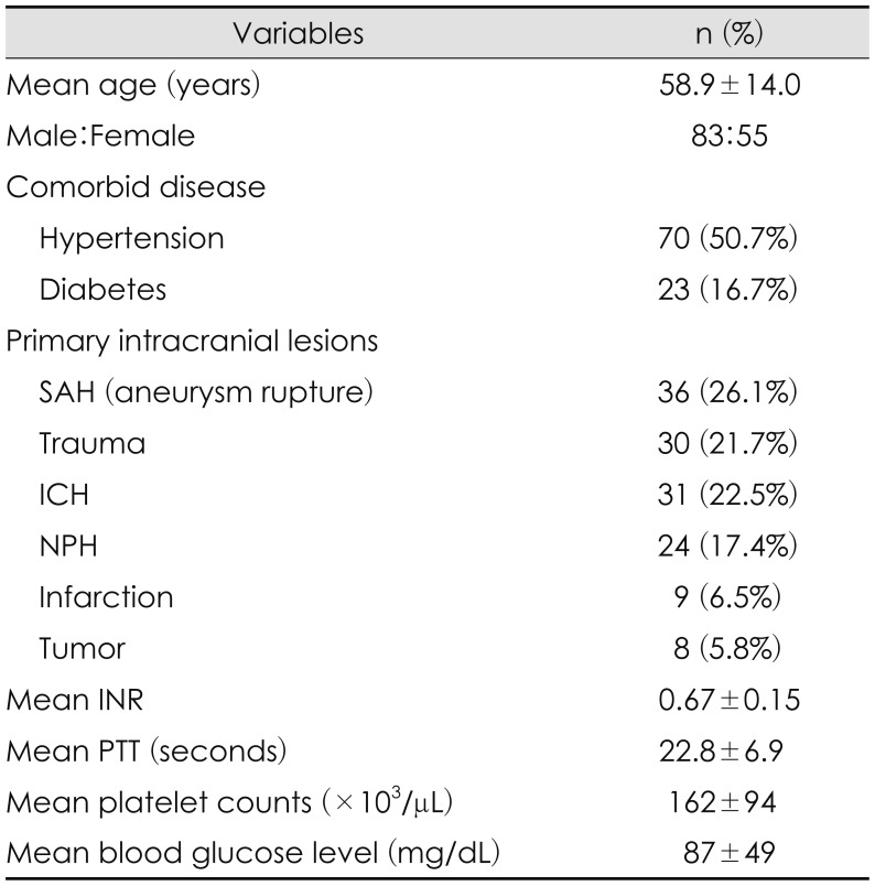
ICH following VP shunt was detected on CT scan in 40 (27.9%) of the 143 patients. The early ICH (within 48 hours after VP shunt) were 6 (4.2%) and delayed ICH were 34 (23.7%) among 143 patients. Therefore, a total 138 patients were involved in this study, excluding 6 patients who developed early hemorrhage (Table 1). The mean age of the 138 patients was 58.9±14.0 years (range, 17–88 years), and 83 (60.1%) patients were male. Original intracranial lesions were aneurysmal SAH in 36 (26.1%), trauma in 30 (21.7%), ICH in 31 (22.5%), normal pressure hydrocephalus (NPH) in 24 (17.4%), cerebral infarction in 9 (6.5%), and brain tumor in 8 (5.8%) patients. Regarding co-morbid diseases, 70 patients had HT and 23 patients had DM. In laboratory assessment, the mean INR was 0.67±0.15, mean PTT was 22.8±6.9 seconds, mean platelet count was 162±94×103/µL, and mean glucose level was 87±49 mg/dL.
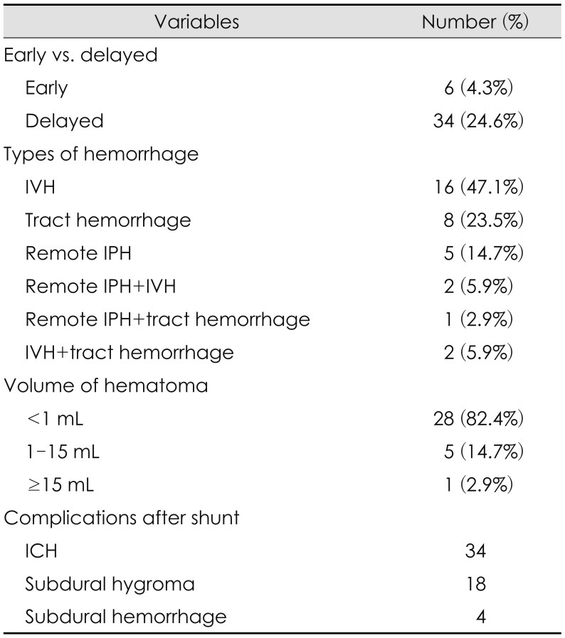
IVH and hemorrhage along the track were the most common, and were present in 16 (47.1%) and 8 (23.5%) patients, respectively. Five patients (14.7%) had IPH, 2 patients (5.9%) had concomitant IPH with IVH and two had IVH with hemorrhage along the track, and 1 patient (2.9%) had IPH and hemorrhage along the track (Table 2).
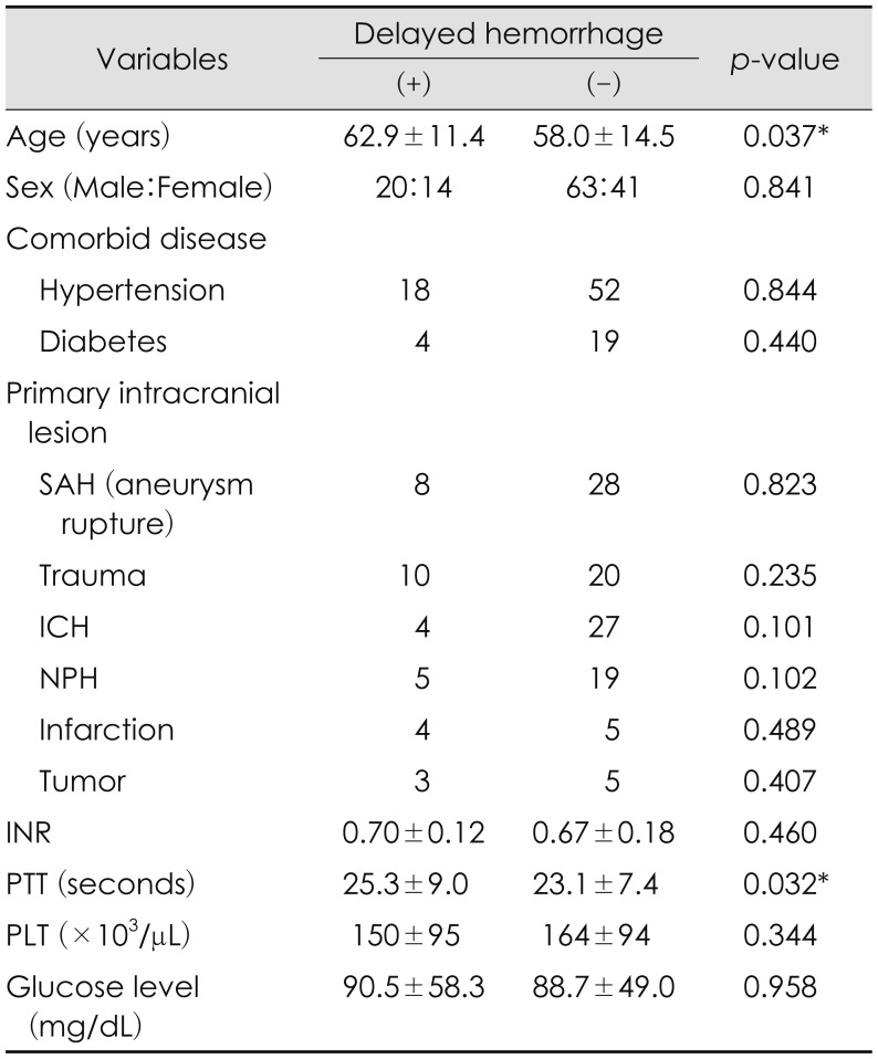
No statistically significance was found for sex ratio, comorbid diseases (HT, DM), or preferred intracranial lesions (SAH, trauma, ICH, NPH), INR, or the platelet count in delayed hemorrhage after VP shunt (Pearson's χ2 test) (Table 3). Previous co-morbid disease like cerebral infarction and tumor history did not have significant correlation in delayed ICH after VP shunt (Fisher's exact test). Although mean INR, blood glucose level tended to be greater in the delayed ICH group, it was not statistically significant (p=0.460 and p=0.958, Student's t-test). However, the old age and the prolonged PTT were found to be significantly associated with developing delayed ICH (p=0.037 and p=0.032, Student's t-test).
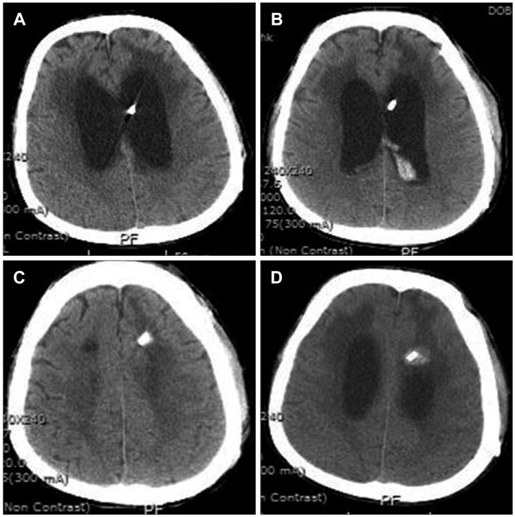
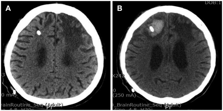
A 57-year-old woman presented with declined mentation and difficulty voiding. Initial brain CT scans revealed that ventricular size was enlarged, and communicating hydrocephalus was confirmed with radioisotope cisternography. She was suffering from cerebral infarction, but there were no co-morbid disease. In laboratory examination, INR/PTT, and platelet counts were within normal range, but the blood glucose level was slightly increased on 131 mg/dL. VP shunt was performed and ICH did not occur postoperatively 1 day after VP shunt (Figure 1A). However, delayed ICH along the path of the catheter developed on postoperative day 7 (Figure 1B). Fortunately, ICH did not increase, nor did neurologic deficits develop. As a result, ICH was treated conservatively.
A 56-year-old man presented with voiding difficulty and gait disturbance. He had a history of burr hole trephination for chronic subdural hemorrhage. INR and PTT were within normal ranges, but the platelet counts were decreased to 109×103/µL. A VP shunt was performed and no hemorrhage occurred postoperative a day after VP shunt (Figure 2A). But the delayed IVH developed on postoperative day 3 (Figure 2B). No intracerebral hemorrhage was seen along the path of the catheter on postoperative day 3 (Figure 2C). In addition, the other hemorrhage in catheter path occurred on postoperative day 7 (Figure 2D).
VP shunt is one of common procedure for neurosurgeons to control the communicating hydrocephalus.24) As is well-known, complications after insertion of a CSF shunt, such as infection, obstruction, subdural hematoma, malfunction, seizures, migrating or kinked catheter; obstruction or blockage of the ventricular and abdominal ends of the catheter, and infection of the shunt apparatus are the major problems of CSF shunting procedures.1925) In this study, radiological complications of VP shunt developed ICH in 34 patients, hygroma in 18 patients, and subdural hematoma in 4 patients.
IPH or IVH secondary to VP shunt placement is not common. Savitz and Bobroff18) reported that the incidence of delayed ICH was 4% following VP shunt. Fukamachi et al.8) performed CT scans on 242 patients within the first week of ventricular shunting procedures and found that ten of them had small ICH (3 cm in the maximal diameter). Postoperative IPH or IVH usually develops at the timing of ventricular puncture.8) Sayers19) reported six cases with ICH among 1,390 case with VP shunt. The CT findings of IVH with small amounts were observed in 10% of the cases in the Palmieri's series;16) less frequently, small pools of blood were noted under the ependyma next to the catheter tip or along its path in the parenchyma.12) ICH has been reported in 0.4% to 4% of patients undergoing shunt procedures.425) In this study, delayed ICH of less than 1 mL was observed in 20% patients with VP shunt. Only 6 (4%) patients showed the delayed ICH with more than 1 mL. This is comparable to the incidence of delayed ICH of Savitz and Bobroff's report.18) However, the overall incidence of delayed ICH was 24%, which is a higher incidence than other studies. The reason may be that it included a small ICH with less than 1 mL.
Several mechanisms leading to ICH after insertion of the shunt catheter have been proposed: 1) co-existence of coagulopathy; 2) shunt-induced disseminated intravascular coagulation (DIC); 3) mechanical disruption of intravascular vessel by catheter; 4) hemorrhage into intraparenchymal tumor; 5) hemorrhage from an occult intravascular malformation; and 6) head trauma occurring shortly after VP shunt placement.22) DIC has been reported after head trauma, brain tumor surgery, or VP shunting.8101320) Bleeding along the path of the ventricular catheter and into the ventricular system has been reported previously following VP shunt placement; it may be caused by puncture of the choroid plexus, repeated attempts at perforation of the ventricles or inadequate placement of the catheters in the parenchyma of the brain.58) Savitz and Bobroff18) reported the mechanism was more likely disruption of a cerebral blood vessel by the catheter; the normal pulsations of the CSF transmitted to the catheter might have caused the catheter to erode through a blood vessel with subsequent ICH. Fujioka et al.7) presented ICH in the same location after a ventricular puncture. He hypothesized that the bleeding might have occurred by the same mechanism as traumatic delayed ICH. Hypoxia, hypercapnia, and venous congestion also produce cerebral hyperemia, which encourages gradual haematoma formation particularly at the sites of injury.1621) Misaki et al.14) suggested that early hemorrhage was attributable to venous occlusion due to intraoperative manipulation whereas delayed bleeding has occurred in fragile brain tissue compromised by co-existence of brain disorders. However, previous intracranial diseases were not affected the development of delayed ICH in our study.
A previous study demonstrated the mechanisms of delayed intracerebral hemorrhage after a VP shunt. However, there have been few studies of factors influencing the actual occurrence of delayed ICH. In this study, the risk factors of delayed ICH were old age and PTT. Zia et al.27) found that only age was an independent risk factors for recurrence of ICH. Pennlert et al.17) suggested that age and diabetes mellitus were associated with recurrent ICH in their study. DIC is commonly a manifestation of a severe systemic disorder, including infection, malignancies, trauma or surgery, shock, or obstetric complications.912) The laboratory diagnosis of DIC is made in the presence of thrombocytopenia, elevated prothrombin time (PT) and activated PTT values, low fibrinogen, and elevated fibrin degradation products. Potential risk factors for progressive intracerebral hemorrhage include abnormalities in coagulation parameters such as PTT, PT, and platelet count, which have been associated with delayed and postoperative ICH.2324) Oertel et al.15) determined that male, time to 1st CT scan, older age, PTT was correlated with progressive hemorrhage and PT, but platelet did not show a similar correlation. Even if a small amount of ICH or IVH developed, it can induce a mechanical obstruction of the VP shunt requiring shunt revision.
This study suggests that old age and prolonged PTT are major risk factors for development of delayed ICH following VP shunt. In addition, delayed ICH after VP shunt frequently develops, even though most patients are asymptomatic even when there develops a small amount of IPH or IVH that does not require surgical evacuation. The findings of this study indicate that patients should be carefully observed after VP shunt and followed-up with a neuroimaging study.
References
1. Baratham G, Dennyson WG. Delayed traumatic intracerebral haemorrhage. J Neurol Neurosurg Psychiatry. 1972; 35:698–706. PMID: 5084138.
2. Blount JP, Campbell JA, Haines SJ. Complications in ventricular cerebrospinal fluid shunting. Neurosurg Clin N Am. 1993; 4:633–656. PMID: 8241787.
3. Bondurant CP, Jimenez DF. Epidemiology of cerebrospinal fluid shunting. Pediatr Neurosurg. 1995; 23:254–258. PMID: 8688350.
4. Choux M, Genitori L, Lang D, Lena G. Shunt implantation: reducing the incidence of shunt infection. J Neurosurg. 1992; 77:875–880. PMID: 1432129.
5. Derdeyn CP, Delashaw JB, Broaddus WC, Jane JA. Detection of shunt-induced intracerebral hemorrhage by postoperative skull films: a report of two cases. Neurosurgery. 1988; 22:755–757. PMID: 3374789.
6. Evans JP, Scheinker IM. Histologic studies of the brain following head trauma; post-traumatic petechial and massive intracerebral hemorrhage. J Neurosurg. 1946; 3:101–113. PMID: 21018500.
7. Fujioka S, Matsukado Y, Kaku M, Yano T, Yoshioka S. Delayed apoplexy following ventricular puncture-a case report. No Shinkei Geka. 1982; 10:955–958. PMID: 6983661.
8. Fukamachi A, Koizumi H, Nukui H. Postoperative intracerebral hemorrhages: a survey of computed tomographic findings after 1074 intracranial operations. Surg Neurol. 1985; 23:575–580. PMID: 3992457.
9. Goodnight SH Jr. Bleeding and intravascular clotting in malignancy: a review. Ann N Y Acad Sci. 1974; 230:271–288. PMID: 4522874.
10. Keimowitz RM, Annis BL. Disseminated intravascular coagulation associated with massive brain injury. J Neurosurg. 1973; 39:178–180. PMID: 4719695.
11. Kothari RU, Brott T, Broderick JP, Barsan WG, Sauerbeck LR, Zuccarello M, et al. The ABCs of measuring intracerebral hemorrhage volumes. Stroke. 1996; 27:1304–1305. PMID: 8711791.
12. Mant MJ, King EG. Severe, acute disseminated intravascular coagulation. A reappraisal of its pathophysiology, clinical significance and therapy based on 47 patients. Am J Med. 1979; 67:557–563. PMID: 495626.
13. McGauley JL, Miller CA, Penner JA. Diagnosis and treatment of diffuse intravascular coagulation following cerebral trauma. Case report. J Neurosurg. 1975; 43:374–376. PMID: 1151476.
14. Misaki K, Uchiyama N, Hayashi Y, Hamada J. Intracerebral hemorrhage secondary to ventriculoperitoneal shunt insertion--four case reports. Neurol Med Chir (Tokyo). 2010; 50:76–79. PMID: 20098034.
15. Oertel M, Kelly DF, McArthur D, Boscardin WJ, Glenn TC, Lee JH, et al. Progressive hemorrhage after head trauma: predictors and consequences of the evolving injury. J Neurosurg. 2002; 96:109–116. PMID: 11794591.
16. Palmieri A, Pasquini U, Menichelli F, Salvolini U. Cerebral damage following ventricular shunt for infantile hydrocephalus evaluated by computed tomography. Neuroradiology. 1981; 21:33–35. PMID: 7219699.
17. Pennlert J, Eriksson M, Carlberg B, Wiklund PG. Long-term risk and predictors of recurrent stroke beyond the acute phase. Stroke. 2014; 45:1839–1841. PMID: 24788972.
18. Savitz MH, Bobroff LM. Low incidence of delayed intracerebral hemorrhage secondary to ventriculoperitoneal shunt insertion. J Neurosurg. 1999; 91:32–34. PMID: 10389877.
20. Shurin S, Rekate H. Disseminated intravascular coagulation as a complication of ventricular catheter placement. Case report. J Neurosurg. 1981; 54:264–267. PMID: 7452342.
21. Smith DR, Ducker TB, Kempe LG. Experimental in vivo microcirculatory dynamics in brain trauma. J Neurosurg. 1969; 30:664–672. PMID: 4977840.
22. Snow RB, Zimmerman RD, Devinsky O. Delayed intracerebral hemorrhage after ventriculoperitoneal shunting. Neurosurgery. 1986; 19:305–307. PMID: 3489202.
23. Stein SC, Young GS, Talucci RC, Greenbaum BH, Ross SE. Delayed brain injury after head trauma: significance of coagulopathy. Neurosurgery. 1992; 30:160–165. PMID: 1545882.
24. Touho H, Hirakawa K, Hino A, Karasawa J, Ohno Y. Relationship between abnormalities of coagulation and fibrinolysis and postoperative intracranial hemorrhage in head injury. Neurosurgery. 1986; 19:523–531. PMID: 3785592.
25. Udvarhelyi GB, Wood JH, James AE Jr, Bartelt D. Results and complications in 55 shunted patients with normal pressure hydrocephalus. Surg Neurol. 1975; 3:271–275. PMID: 1154252.
26. Wu Y, Green NL, Wrensch MR, Zhao S, Gupta N. Ventriculoperitoneal shunt complications in California: 1990 to 2000. Neurosurgery. 2007; 61:557–562. PMID: 17881969.
27. Zia E, Engström G, Svensson PJ, Norrving B, Pessah-Rasmussen H. Three-year survival and stroke recurrence rates in patients with primary intracerebral hemorrhage. Stroke. 2009; 40:3567–3573. PMID: 19729603.