

 PDF
PDF ePub
ePub Citation
 Print
Print


The most common cause of extensor pollicis longus (EPL) tendon rupture is a distal radius fracture; however, spontaneous EPL tendon rupture also shows a high frequency of occurrence, which cannot be ignored. A retrospective study of 27 EPL ruptures suggested that trauma or iatrogenic causes account for more than half of the cases, but spontaneous rupture is also considered an important etiological factor1. The etiology of spontaneous EPL tendon rupture includes systemic or local steroid injections, tenosynovitis, synovitis, rheumatoid arthritis, and repetitive wrist motions2.
This report presents a case of idiopathic EPL tendon rupture in a patient who was a daily laborer with repetitive wrist motion. No cases of EPL rupture caused by mechanical attrition due to an osteophyte caused by progression of carpometacarpal (CMC) arthritis, rather than the generally known etiology involving repetitive wrist motions, have been reported yet. Herein, we provide the case report and a review of the literature.
CASE REPORT
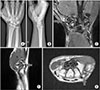
A 42-year-old man visited our hospital complaining of limited thumb extension for six months, without trauma history. The patient had not received any injection since the onset of symptoms. As a daily laborer, he was a worker on a construction site for 8 hours a day, 5 to 6 days a week for a total of 15 years. Physical examination was performed on the right wrist and a 1×1 cm mass was palpated around the first CMC joint of the right wrist, with tenderness. Plain radiography was performed, from which the osteophyte was suspected around the dorsum of the first CMC joint (Fig. 1A, B). Magnetic resonance imaging of the right wrist revealed an osteoarthritic change around the first CMC joint and a subcortical cyst on the first metacarpal base (Fig. 1C) and the osteophyte in the dorsum of the first CMC joint (Fig. 1D, E). A chronic rupture of the EPL tendon around the dorsum of the first CMC joint was observed.
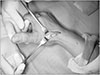
Surgery was performed by placing a tourniquet around the upper arm under regional anesthesia. An incision was made in the anatomical snuff box of the hand following the running of the EPL tendon (Fig. 2A). We found both ends of the ruptured EPL tendon (Fig. 2B), as well as the osteophyte in the dorsum of the first CMC joint (Fig. 2C), which was removed with an osteotome and rongeur. After removal, osteophyte was confirmed by histologic examination.
Tendon transfer using extensor indicis proprius (EIP) tendon was performed because the injured tendon could not be repaired end-to-end. A short incision was made over the index metacarpophalangeal joint, and the EIP tendon was divided immediately proximal to the extensor hood. The EIP tendon was delivered into the incision made in the anatomical snuff box, and along with its muscle, it was freed of soft tissue attachments. The tendon was then transferred to the distal stump of the EPL tendon by using Pulvertaft's technique (Fig. 3).
Postoperative short arm thumb spica splint was applied for 4 weeks with the wrist in near full extension and the thumb extended and abducted. Then the thumb was gradually permitted to move with the wrist splinted in extension for another 4 weeks. Independent flexion and extension of the thumb was possible compared with the other side at the third postoperative month (Fig. 4A) and there was no recurrence of the osteophyte at the CMC joint of the thumb on computed tomography of the right wrist (Fig. 4B). It is scheduled to be followed regularly in the outpatient department in the future.
DISCUSSION
Spontaneous rupture of the EPL tendon is relatively common. Although it is mostly an inflammatory response, systemic or local steroid injections are considered one of the major causative factors. The most closely associated inflammatory disease is rheumatoid arthritis. Spontaneous rupture of the EPL tendon can also be caused by tophaceous gout infiltration, ankylosing spondylitis, wrist fractures, bone spurs developing after metastatic distal radius or scaphoid fractures, misplaced external fixators or metal plates, non-metastatic distal radius or scaphoid fractures, prolonged nonunion of the scaphoid, dorsal subluxation of the distal ulna after trauma, and Madelung's deformity1234.
Moreover, the nutrition impairment theory is the main theory describing the cause of spontaneous tendon ruptures, and increased pressure on Lister's tubercle has been reported to lead to ischemia and subsequent spontaneous rupture of the EPL tendon5. The other mechanisms are nutrition impairment, pressure caused by necrosis, a sharp fragment of bone that erodes the tendon, crushing injury, non-union of Lister's tubercle, a roughened area of the radius caused by attrition of the tendon, repetitive activities that cause tenosynovitis, or a combination of these mechanisms6.
In contrast, there are a few reports concerning EPL tendon ruptures caused by repeated abnormal movements of the wrist joint. A case of spontaneous rupture of EPL due to repetitive movements in a housewife was reported2. Another study reported a case of a spontaneous rupture of EPL due to repetitive movements in a tailor4.
A case of spontaneous rupture of the EPL tendon in a patient without severe trauma or inflammatory diseases, Lister's tubercle abnormalities, synovitis, or tenosynovitis was reported and the author concluded that EPL tendon rupture could occur without trauma due to attrition of the tendon around Lister's tubercle, regardless of age7. This conclusion was supported by other studies on EPL tendon ruptures in sports athletes who were engaged in excessive use of the wrist8.
CMC arthritis is the second most common arthritis afflicted by osteoarthritis9. It is widely known that use of the basal joint in work-related mechanical loading is one of the risk factors for CMC arthritis. Typical patients, especially men, have history of repetitive occupational joint use. Radiographic findings are considered in the diagnosis, and Eaton and Glickel's classification system is widely used to define the degree of severity of CMC arthritis. As the CMC arthritis progresses, the size of the osteophyte or joint fragment also increases10.
In the present study, a solid mass on the right wrist was identified on physical examination, which was later found to be an osteophyte of the first CMC joint, which was in the second stage of Eaton and Glickel's classification system as per radiologic evaluation. The patient, being a daily worker, had a history of repeated wrist motion, which is a risk factor for CMC arthritis and an etiology of idiopathic EPL rupture. This suggests that the EPL tendon caused mechanical attrition with the osteophyte of the CMC joint, resulting in rupture of the EPL tendon spontaneously, because the EPL tendon passes over the dorsum of the first CMC joint after a sharp turn at Lister's tubercle.
Hence, we suggest that in cases of a suspicion of EPL tendon rupture without trauma history, the patient's occupation and history of repetitive wrist motion should be checked, in addition to the patient's medication history, including local steroid injection, and systemic disease such as rheumatoid arthritis, ankylosing spondylitis, and gout. If CMC arthritis is diagnosed, accurate physical examinations and stage evaluation of the disease should be undertaken. When planning the treatment, in addition to surgery for the ruptured tendon, careful treatment of CMC arthritis may improve the patient's pain and functional satisfaction.


 XML Download