

 PDF
PDF ePub
ePub Citation
 Print
Print


Free flaps are commonly used for lower extremity reconstruction due to trauma-related injuries. There is no disagreement that one of the best tools for extremity salvage is the “free flap,” which is being used increasingly to treat soft tissue defects in cases of lower extremity trauma.
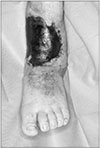
As many as 33% of flaps used to reconstruct lower limb damage require an orthopedic procedure after reconstruction1. A series of free flaps for foot and ankle defects reported by Lin et al. in 2006 found a secondary procedure rate of 86%1. They reported that re-exploration revealed necrosis in 24% of muscle flaps and 2% of fasciocutaneous flaps1. Thus, not only the microsurgery itself but postoperative flap management of the flap is important. We present a case in which, at 1 year after the initial surgery, there was total necrosis of a thoracodorsal artery perforator free flap due to inadequate U-shaped flap elevation. The flap had been placed to achieve internal fixation of an open distal tibiofibular fracture with nonunion (Fig. 1).
CASE REPORT
A 42-year-old man was referred for treatment of total flap necrosis on the left ankle caused by an almost 22-cm U-shaped incision for flap elevation. The length of the elevated flap was approximate 8 to 10 cm and the width of that was 4 cm. One year previously at another hospital, the patient had undergone placement of a thoracodorsal artery perforator free flap and hardware fixation for an open wound with a distal tibiofibular fracture caused by a conveyer belt crushing injury of the left ankle. When he was initially seen 1 day after the trauma, the circumferential defect measured 28×18 cm. The surgeons planned coverage for the defect. First, latissimus dorsi chimeric flaps were elevated, and muscle segments to be harvested were designed according to the dimensions of the dead space to be obliterated. Artery and vein anastomosis were done via an end-to-end method (thoracodorsal artery-to-anterior tibial artery, thoracodorsal vein-to-anterior tibial vein; pedicle length 7.5 cm). At that time, the trauma surgeon positioned a U-shaped flap for bone grafting and internal fixation of an open distal tibiofibular fracture with nonunion. It was that flap that had become necrotic 1 year later (Fig. 1).
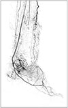
When the patient was transferred to our emergency room, we checked for vital signs, flap refill, and Doppler sound using a probe. There was no refill at the flap, and no Doppler sound was detected. Demarcation was proceeding at that time. We checked the flap angiosome with lower extremity three-dimensional angio computed tomography with contrast. The anterior tibial artery and collateral arteries were the main vessels supplying blood to the flap. The posterior tibial artery branch was intact (Fig. 2). After the margin of the flap necrosis was determined, we debrided it and covered it with an anterolateral thigh fasciocutaneous free flap using the chimeric approach. We then covered the donor site with local flap advancement and a split-thickness skin graft. There was an 11×12 cm defect on the left ankle after detaching the necrotic flap. We harvested a 12.5×13.5 cm fasciocutaneous flap from the right thigh and chose a pedicle of the former free flap as the recipient artery and its accompanying veins as recipient veins.
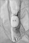
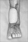
On postoperative day 1, flap refill was slow with no color change in the flap margin. Doppler sound with the probe was intact. On postoperative day 14, we removed the stitches. There were no complications during the entire postoperative management period (Fig. 3). Recently, almost 3.5 months after the surgery, the patient visited an outpatient clinic, where the flap was found to be still stable without complications (Fig. 4).
DISCUSSION
The human lower limb plays a role in balance and support, which in turn allows humans to stand and perform upright ambulation. Thus, the primary goals of reconstruction are to salvage the limb and preserve its form and function. The field of reconstruction gained a vast number of options following improvement of vascular techniques during the 1960s, opening the door to the era of microvascular reconstruction2. Although the free flap techniques has expanded the therapeutic options, it can be both medicinal and poisonous without flap related knowledge.
Flap consists of any type of tissue that is lifted from a donor site with intact blood supply. The blood supply to the tissue is crucial for flap survival. In this case, total necrosis of postoperative 1 year flap was occurred by inadequate U-shape flap elevation. Although the shape of the elevated flap was not fit into rectangular shape exactly, the length to width ratio of that was almost 2.25:1. The circulation to the tissue was not enough to flap survival, due to inadequate incision. About flap elevation in lower extremities, subdermal plexus allows elevation of a rectangular flap with a length to width ratio of approximately 2:1. As the width increases, the more large vessel will be incorporated to provide an adequate blood supply to subdermal plexus of the elevated flap.
Understanding the concept of angiosome is also essential in ensuring the success of any flap related surgical procedure. The angiosome principle was defined by Ian Taylor's landmark anatomic study and divides the body into individual angiosome: three-dimensional blocks of tissue fed by “source” arteries, especially the foot and ankle are composed of six such distinct angiosomes. Because the foot and ankle comprise an end organ, their main arteries have numerous direct arterial-arterial connections that allow alternative routes of blood flow to develop if the direct route is disrupted or compromised3. In terms of flaps requiring surgical procedure, detailed knowledge of the angiosome and adjacent vascular connections allows surgeon to design safe flap incision and tissue exposure that preserve blood supply to flap. The designed flap incision must provide both adequate exposure for the surgical procedure and adequate blood supply to preserve the viability of flap. If there is good blood flow from the source artery feeding each angiosome, the safest incisions to make are along the border between two adjacent angiosomes, because each side of the incision has maximal blood flow3. But the safest incision dose not always mean the best exposure, so compromises sometimes need to be made.
Thus, the computed tomography angiography is often required for vascular evaluation of patients who require lower extremity reconstruction, especially when planning the microsurgery. Doppler probe is also important. It is useful to map out the vascular anatomy and evaluate the direction and quality of blood flow. Triphasic sound indicates normal arterial flow; biphasic sound indicates mildly compromised flow; and monophasic sound indicates arterial compromise. A blunt, short monophasic spitting like sound indicates complete distal occlusion with no runoff4. Those examination tools involve inspecting the vascularity of the initial wound area and the postoperative flap.
We could learn a lesson from this case—that insufficient understanding of flap could interfere with the life of the flap, even in postoperative 1 year, well vascularized flap. Thus, both designing the flap elevation incision considering the circulation of tissue and evaluating flap angiosome should be undertaken with a reconstructive surgeon familiar with microsurgery. These steps are priorities for preventing flap mismanagement. Also naturally, during the surgical planning, there must be sufficient discussion about such other important factors as medical history of patient, proper timing of operation and so on. Hence, to make the most suitable decision, many factors should be assessed by a multidisciplinary team, including orthopedic, reconstructive, and vascular professionals.
 XML Download