

 PDF
PDF ePub
ePub Citation
 Print
Print


Abstract
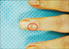
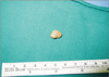
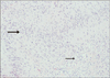
A 29-year-old male patient complained subungual palpable mass with sharp pain. Ultrasonography showed a lobulated, hypoechoic, vascularized mass that was distinct from the surrounding soft tissue of 1.2×0.9×0.6 cm in size, probably a glomus tumor. Authors performed surgical excision and excisional biopsy, and pathological study was diagnosed schwannoma. Subungual schwannoma is very rare and have been reported as a painless mass. Authors report a case of painful subungual schwannoma mimicking glomus tumor on ultrasonography.
Figures and Tables
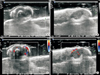 | Fig. 2Transverse (A) and longitudinal (B) ultrasonography images show 1.2×0.9×0.6-cm-sized well-defined, lobulated, round, and hypoechoic cystic mass at the radial side under the nail plate. (C, D) Color Doppler ultrasonography shows subtle flow signals around the hypoechoic area.
|
References
1. Holdsworth BJ. Nerve tumours in the upper limb. A clinical review. J Hand Surg Br. 1985; 10:236–238.

2. Rockwell GM, Thoma A, Salama S. Schwannoma of the hand and wrist. Plast Reconstr Surg. 2003; 111:1227–1232.
3. Phalen GS. Neurilemmomas of the forearm and hand. Clin Orthop Relat Res. 1976; (114):219–222.
4. Huntley JS, Davie RM, Hooper G. A subungual schwannoma. Plast Reconstr Surg. 2006; 117:712–713.
5. Moon SE, Cho YJ, Kwon OS. Subungual schwannoma: a rare location. Dermatol Surg. 2005; 31:592–594.
6. Kulkarni J, Moholkar A, Patil A. Subungual schwannoma: an uncommon location. J Hand Surg Am. 2013; 38:1258–1259.
7. Soto R, Aldunce MJ, Wortsman X, Sazunic I. Subungual schwannoma with clinical, sonographic, and histologic correlation. J Am Podiatr Med Assoc. 2014; 104:302–304.
8. Han KJ, Lee YS, Park M. Digital nerve schwannoma of the hand. J Hand Surg Eur Vol. 2012; 37:361–362.
 XML Download