

 PDF
PDF ePub
ePub Citation
 Print
Print


INTRODUCTION
Open carpal tunnel release is one of the most commonly performed procedures in patients who have idiopathic carpal tunnel syndrome12. Most hand surgeons have been trained to perform open release of the transverse carpal ligament under direct visualization with a longitudinal incision. Although excellent results have been reported, some authors have suggested that scar tenderness, pain in the thenar or hypothenar area (pillar pain), and persistent weakness may occur frequently after standard open procedures345678. Thus, minimally invasive surgical techniques, such as the limited open single incision and an approach through the flexor carpi radialis, have become widespread to treat carpal tunnel syndrome. Minimally invasive techniques include endoscopic carpal tunnel release (ECTR) and double minimal incision release. Nazzi et al.9 introduced a minimally invasive surgical technique for carpal tunnel release, which has been attributed to less surgical trauma, thereby reducing pillar pain and postoperative scar tenderness and providing better grip strength and more rapid return to work. However, surgeons considering a minimally invasive surgical technique should be aware of the complications, such as nerve injury, vascular injury, or incomplete release. Technical improvements in endoscopic techniques and subsequent reports have demonstrated fewer complications10. Many studies have compared the results of open carpal tunnel release and ECTR procedures; however, only a few studies have described the results of open carpal tunnel release against a minimally invasive technique. The purpose of the present study was to compare results of the double minimal incision technique with those of open carpal tunnel release using validated outcome questionnaires, standardized functional tests, and physical measurements.
MATERIALS AND METHODS
This was a retrospective cross-sectional clinical trial conducted from January 2010 to December 2014 at Inha University Hospital. We considered the clinical diagnosis of carpal tunnel syndrome based on the presence of three or more of the following factors: (1) history of recurrent or persistent paresthesia in the median nerve distribution; (2) worsening of symptoms (numbness, weakness, wrist pain, and stiffness); (3) nocturnal awakening with paresthesia; (4) presence of a positive Tinel's and/or Phalen's sign on physical examination; and (5) absence of a response to nonsurgical treatments (night splint, non-steroidal anti-inflammatory drugs, or steroid injections) for at least 6 months since the start of symptoms in patients >50 years old. Patients who entered the study were evaluated with electrodiagnostic studies.
In total, 111 patients with carpal tunnel syndrome were enrolled in this study. Of them, 47 had unilateral involvement and 64 were bilateral cases. Therefore, the study was comprised of 175 hands. Both methods were explained to each patient. The patients were divided into 2 groups and underwent one of the two procedures for carpal tunnel release surgery. A longitudinal incision was used in 53 patients (82 hands), designated as group A. Fifty-eight patients (93 hands) in group B were treated with the double minimal incision technique described below. Mean age, female-to-male ratio, and dominance of the operated hand were similar between the groups and electrodiagnostic studies were performed on all wrists prior to surgery (Table 1). This study protocol was approved by the Institutional Review Board of Inha University Hospital (No. 2016-08-019).
1. Assessment
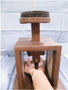
We conducted the surgery and evaluated the outcomes on an outpatient basis. Clinical assessment was made retrospectively at every outpatient visit after patients subjective symptoms consulting Boston Carpal Tunnel Questionnaire; numbness, nocturnal pain, wrist and hand pain, weakness, and stiffness were investigated. Patients were examined for subjective symptoms at the every outpatient visit and were considered positive if there was symptom. The visual analogue scale (VAS) were completed postoperatively and diagnostic tests (Tinel's and Phalen's tests) were performed. Pillar tenderness was measured with a 2.0 kg plunger for 30 seconds at the level of the distal wrist crease (Fig. 1). Hand grip strength was measured with a Jamar dynamometer (Fabrication Enterprises, Elmsford, NY, USA). Return to work was the mean time to perform normal daily life activities without subjective discomfort after surgery. We also checked nerve or vascular injury and persistent neurological symptoms as complications.
These tests and examinations were repeated 2, 4, and 8 weeks and 3, 6, and 12 months postoperatively. We also evaluated the patients' satisfaction levels with their scars 12 months postoperatively at the outpatient clinic in connection with the reduced size of the incision.
2. Statistics
We compared the outcomes of the 2 groups with analysis of covariance at each postoperative interval. The data were analyzed with SPSS ver. 17.0 (SPSS Inc., Chicago, IL, USA). A p-value less than 0.05 was considered significant. The chi-square test was used to assess numbness, nocturnal pain, wrist and hand pain, thenar atrophy, the Tinel's sign, and the Phalen's test. Linear-by-linear association was employed to assess the VAS scores, and the t-test was used to evaluate grip strength and the days required for the patient to return to work.
3. Operative technique
The operation was performed under general anesthesia or regional anesthesia (brachial plexus block) using a pneumatic tourniquet.
1) Standard open surgery technique
The transverse carpal ligament was exposed through a short longitudinal incision along the axis of the ring finger (Fig. 2). Neither the motor branch of the median nerve nor the palmar cutaneous branch was routinely identified. The carpal tunnel was decompressed from the antebrachial fascia to the distal edge of the transverse carpal ligament and any pathological features were noted and investigated. The skin was closed after releasing the tourniquet and performing meticulous hemostasis.
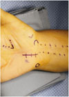
2) Double minimal incision release
Two skin incisions were used to decompress the carpal tunnel (Fig. 3); thus, leaving a 2-cm bridge of skin intact at the base of the palm. First, a 1-cm distal incision (A in Fig. 3) was made longitudinally along the proximal portion of median crease to expose the distal margin of the transverse carpal ligament, which was divided under direct vision. Secondly, a 1-cm incision was made transversely (B in Fig. 3) at the wrist crease over the palmaris longus tendon. Care was taken to avoid injury to the median palmar cutaneous nerve. We retracted the palmaris longus tendon radially using a skin hook. Blunt dissection with a freer (scalpel) was performed initially toward the distal incision site between the skin and transverse carpal ligament to make sufficient room for introducing sharp Metzenbaum scissors. The area beneath the transverse carpal ligament was probed with the freer to protect the median nerve. Then, the proximal two-thirds of the transverse carpal ligament was released by cutting with sharp Metzenbaum scissors. The remaining distal part of the transverse carpal ligament was released through the distal incision site.
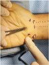
We introduced mosquito forceps along the median nerve beneath the incised transverse carpal ligament to confirm complete release and then palpated with the mosquito forceps over the skin from proximally to distally (Fig. 4). All patients were discharged home either on the same day or the following day. Wool bandages were retained for 4–7 days.
RESULTS
1. Electrodiagnostic studies
Mean sensory latency in group A was 4.7 milliseconds, and mean motor latency was 5.7 milliseconds preoperatively. Mean sensory latency in group B was 4.9 milliseconds, and mean motor latency was 5.8 milliseconds (p>0.05).
2. Outcome measurements
1) Clinical symptoms
No differences in numbness, nocturnal pain, wrist and hand pain, weakness and stiffness were observed preoperatively between groups A and B (Table 2). Group B had significantly less numbness, wrist and hand pain, and stiffness 2, 4, and 8 weeks postoperatively than those in group A (p<0.05). However, these differences disappeared ≥3 months after surgery. There was no difference in nocturnal pain and weakness between 2 groups.
2) Visual analog scale
Preoperative VAS scores were not different between groups A and B (Table 3). Both groups had a gradual decrease in VAS score. Group B showed significantly better results after 2, 4, and 8 weeks than those in group A (p<0.05). However, these differences disappeared ≥3 months after surgery.
3) Tinel's sign/Phalen's test
Positive Tinel's and Phalen's test results were observed in approximately 50% of group A and B patients 2 weeks postoperatively. No differences were observed between the 2 groups after 2 weeks.
4) Pillar tenderness/grip strength
Only 5.3% (hands, 5) of the double minimal incision release group complained of pillar tenderness with loads of 2.0 kg over the carpal tunnel 2 weeks after surgery, whereas 29.2% (hands, 24) of the minimal open group complained of pillar tenderness. Mean preoperative grip strength was 13.2 kg in the double minimal incision procedure group and 13.8 kg in the open-release group. Grip strength had decreased significantly in both groups to 10.8 kg (p<0.05) 2 weeks after surgery in the double minimal incision procedure group and to 10.8 kg in the open-release group (p>0.05).
5) Return to work and cosmetic result
The median time for patients to return to work was 38 days (range, 15–77 days) in the open-release group compared with 25 days (range, 8–60 days) in the double minimal incision release group (p<0.01). The scars in the double minimal incision release group remained significantly less tender than those in the open-release group until 2 months postoperative. Both groups had equally mild persistent scar tenderness 1 year after surgery.
Patients who underwent the double minimal incision procedure had better cosmetic satisfaction (satisfaction rate, 95%) than those in the open-release group. They were questioned at the outpatient clinic for their satisfaction/dissatisfaction with the reduced size of the incision scar.
6) Complication
No major nerve or vascular injury occurred in any patient. However, 3 patients developed complications. Symptoms consistent with reflex sympathetic dystrophy, including redness, swelling, and sweating occurred in 2 patients in the open-release group. The symptoms resolved in 1 patient after physical therapy. The symptoms remained in the other patient, and a regular therapy program was required. Two patients finally returned to regular work. One patient in the open release group required revision surgery due to long-lasting symptoms from a hematoma in the surgical lesion.
DISCUSSION
Carpal tunnel syndrome is one of the most common compressive neuropathies of the upper limbs and requires conservative treatment and occasionally carpal tunnel release surgery. Different surgical techniques can be used and both open techniques and ECTR are employed.
Most surgeons use a short, continuous, curved incision directly over the carpal tunnel for decompression and do not routinely look for the palmar cutaneous branch of the median nerve. Taleisnik11 recommended a skin incision on the ulnar side of the axis of the ring finger, and damage to the branches of the palmar cutaneous nerve can be avoided if the skin incision is made in the axis of the ring finger. However, these incisions leave a scar in the pressure-bearing area at the base of the palm and may contribute to symptoms in a heavy manual labor worker. Previous studies have suggested that open carpal tunnel release is associated with considerable morbidity, including prolonged tenderness of the scar and grip weakness for as long as 3–6 months after the operation346.
Chow12 introduced an endoscopic technique to decompress the carpal tunnel, which allows rapid recovery. Early results are satisfactory with reduced scarring and postoperative pain. Trumble et al.13 compared results from open and endoscopic surgeries techniques. In their evaluation of satisfaction level, symptomatic and functional improvement after surgery was greater during 3 months in patients who received the endoscopic technique than in those who underwent open surgery. However, this technique requires specialized, expensive equipment, and is difficult to perform. The ulnar nerve, digital nerves, superficial palmar arch, and the motor branch of the median nerve may be damaged by the endoscopic blade51012. Thus, safety of the endoscopic technique has been a major concern, but few complications occur after learning the technique13.
The double minimal incision technique has the various advantages of an endoscopic procedure but also has some disadvantages. The double-incision technique is relatively easy to perform and allows rapid postoperative recovery14. This technique did not result in a higher risk of postoperative complications, expense, or increase in surgical time compared to the single-incision technique915.
Our double minimal incision release was easy and safe for carpal tunnel release and resulted in good-to-excellent clinical outcomes. No special equipment is necessary, and no extended learning curve for the use of equipment are needed, thereby making the learning curve short. This procedure allows the distal portion of the transverse carpal ligament to be divided under direct vision to avoid any neurovascular damage. In addition, this procedure has the advantages of during endoscopic release. The pressure-bearing area is protected by leaving an intact bridge of skin at the base of the palm and it does not divide the palmaris brevis muscle or the palmaris fascia. Thus, our group B patients had less scar tenderness and greater symptom relief for the first 8 weeks following the surgery.
One of the most important functional outcomes is the interval between the operation and the patient's resumption of daily living and work activities. In a previous study, industrial employees lost an average of 54 days of work after open carpal tunnel release16. In contrast, Chow16, who retrospectively studied the results of ECTR in 456 patients, noted that 269 patients (59%) returned to normal activity and work after 2 weeks and 392 (86%) returned after 4 weeks. As in previous studies of ECTR, our patients who underwent the double minimal incision release procedure returned to work earlier (mean, 25 days after the operation) than those who underwent open release (mean, 38 days after the operation). There was no loss to follow up (111 patients, 176 hands) during the 12-month study period. Our technique led to less pain due to the minimal incision and better cosmetic satisfaction because of little scaring. Moreover, the double minimal incision procedure does not require expensive equipment and can decrease costs because the patient returns to work in a shorter period of time.
Several limitations of this study should be mentioned. We were unable to employ any self-reported questionnaires or systemic interviews to evaluate the patients' outcome preoperatively. Moreover, we did not compare the groups regarding factors that should be pair-matched, such as gender, age, or profession.


 XML Download