

 PDF
PDF ePub
ePub Citation
 Print
Print


Abstract
Primary cutaneous cryptococcosis is a fungal infection caused by Cryptococcus neoformans which is frequently occurred in the immunosuppressed host. The treatment of primary cutaneous cryptococcosis is mainly fluconazole, and the prognosis is relatively good. We report a case of primary cutaneous cryptococcosis due to intravenous line on the left forearm after lumbar stenosis surgery in a patient with rheumatoid arthritis, who finally underwent second, fourth, and fifth ray amputation.
Go to : 
References
1. Sarosi GA, Silberfarb PM, Tosh FE. Cutaneous cryptococcosis: a sentinel of disseminated disease. Arch Dermatol. 1971; 104:1–3.

2. Shin DH, Kim KS, Lee JM, Choi JS, Kim KH. Primary cutaneous cryptococcosis. Ann Dermatol. 1999; 11:27–9.
3. Neuville S, Dromer F, Morin O, et al. Primary cutaneous cryptococcosis: a distinct clinical entity. Clin Infect Dis. 2003; 36:337–47.
4. Kim YG, Kim HW, Park HC, Kim JE, Ko JY, Ro YS. Primary cutaneous cryptococcosis mimicking herpes zoster. Korean J Dermatol. 2013; 51:343–7.
5. Ko YJ, Hong MH, Park CM, Moon HW, Hur M, Yun YM. Primary cutaneous cryptococcosis in a patient with iatrogenic Cushing’s syndrome: a case report and review of the literature. Korean J Clin Microbiol. 2012; 15:70–3.
6. Revenga F, Paricio JF, Merino FJ, Nebreda T, Ramirez T, Martinez AM. Primary cutaneous cryptococcosis in an immunocompetent host: case report and review of the literature. Dermatology. 2002; 204:145–9.
7. Kim JH, Shin DJ, Byun YS, Kim SW, Kim TE. Cryptococcal tenosynovitis of the hand in a patient with rheumatiod arthritis: a case report. J Korean Soc Surg Hand. 2011; 16:251–4.
8. Kim YJ, Seo SJ, Ro BI. A case of primary cutaneous cryptococcosis misdiagnosel as skin tuberculosis. Korean J Med Mycol. 2003; 8:16–20.
9. Noble RC, Fajardo LF. Primary cutaneous cryptococcosis: review and morphologic study. Am J Clin Pathol. 1972; 57:13–22.
10. Chen SC, Sorrell TC, Chang CC, Paige EK, Bryant PA, Slavin MA. Consensus guidelines for the treatment of yeast infections in the haematology, oncology and intensive care setting, 2014. Intern Med J. 2014; 44:1315–32.
Go to :
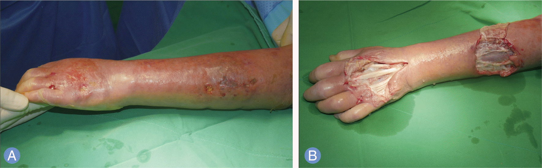 | Fig. 1.
(A) Preoperative photo. Large painful ulcerations and widespread cellulitis originated from intravenous line in the left forearm. (B) Intraoperative photo shows massive granulomatous tissues around extensor tendons and muscles. |
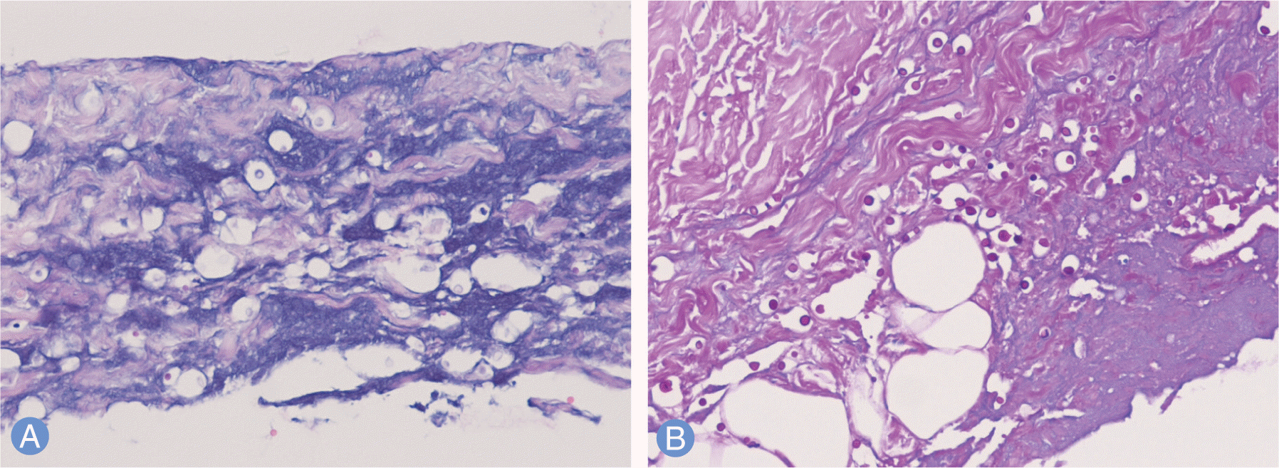 | Fig. 2.
(A) Histopathologic examination shows multiple round spores in necrotic tissue (H&E stain, ×200). (B) PAS stain shows reddish round spores (PAS stain, ×200). |
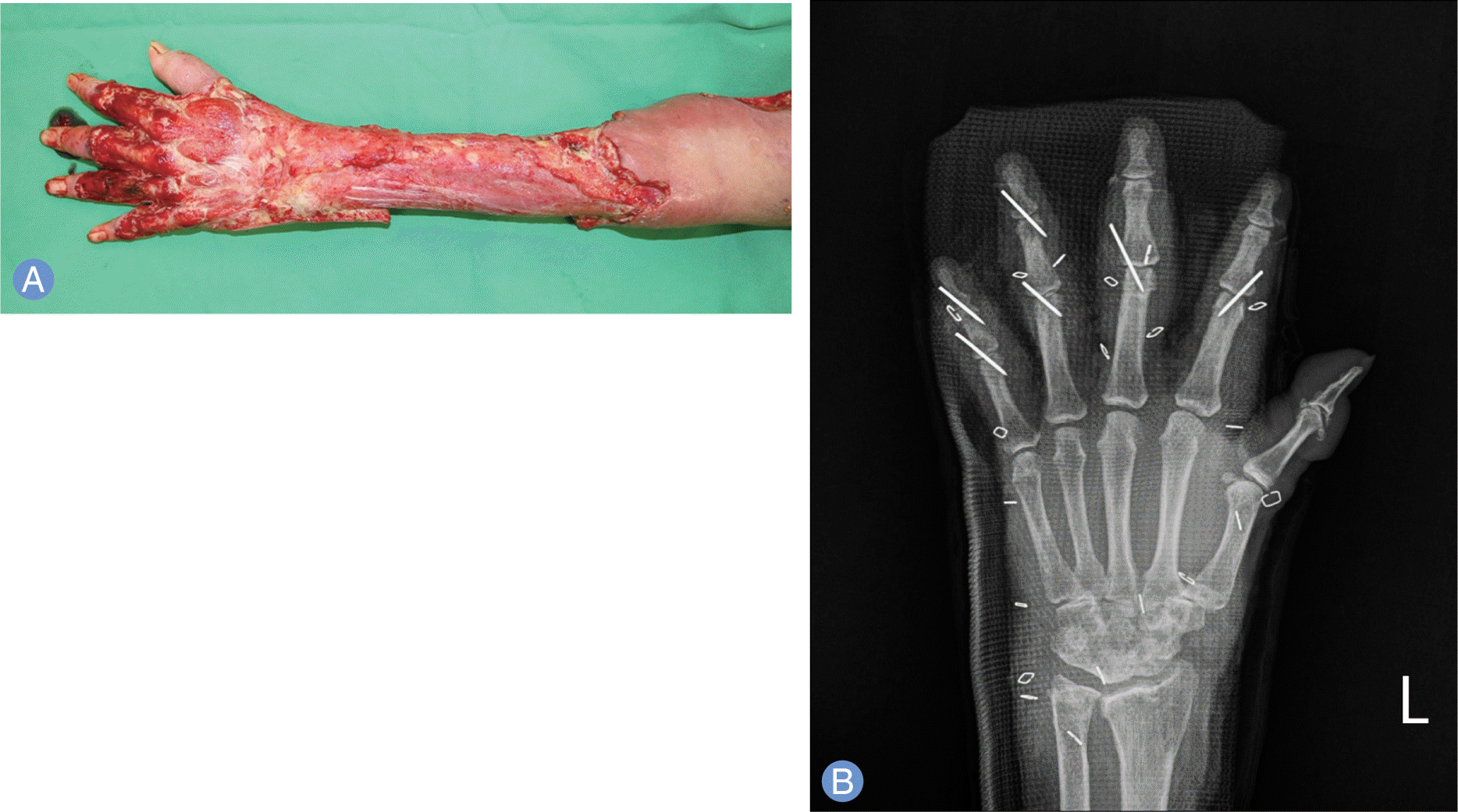 | Fig. 3.
(A) Intraoperative photo shows that skin and subcutaneous tissues were removed extensively above the fascias of extensor muscles due to progressive necrosis (one month after photographing Fig. 1). (B) Postoperative simple radiograph. After the second to fifth extensor tendons were removed, the interphalangeal joints were temporarily fixed with the Kirschner wires. |
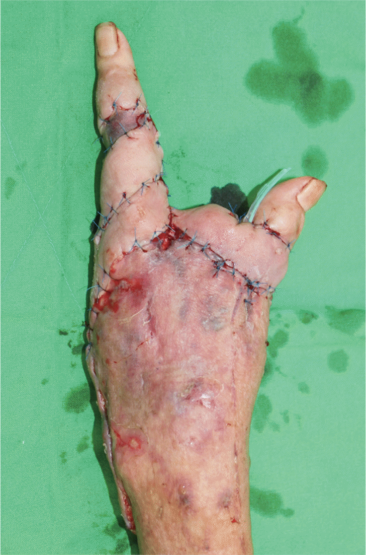 | Fig. 4.Two months after orthopedic surgery, second ray was also amputated for wound healing. Local advancement flap was performed immediately. |
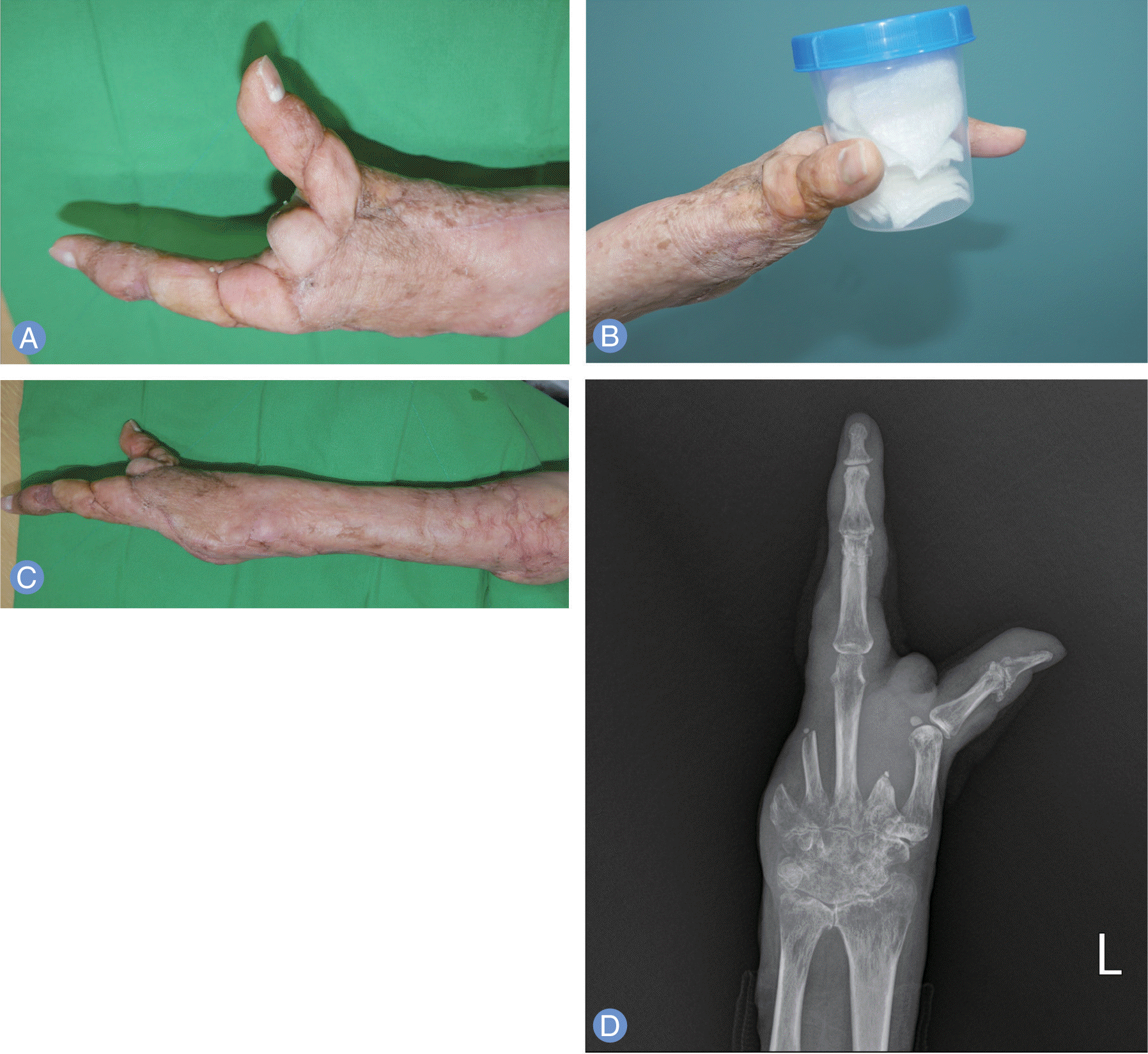 | Fig. 5.Eight months after orthopedic surgery. (A) The wound left hand was well healed, and the opposition of the thumb was possible. (B) Preservation of these 2 digits allowed to allow grasping large objects. (C) The wound of left forearm was also well healed. (D) Simple radiograph shows that second, fourth, and fifth rays were amputated. |
 XML Download